

Labeled vertebrae diagram with structure and functions explained
The concept of Vertebrae Diagram is essential in biology and helps explain real-world biological processes and exam-level questions effectively. This topic is important for understanding the structure and function of the human backbone, aiding students in learning to draw, label, and identify parts of the vertebral column for exams and practical applications.
Understanding Vertebrae Diagram
Vertebrae diagram refers to a labeled anatomical drawing of the human vertebral column. The vertebrae diagram visually displays the 33 bones that make up the spine, grouped into five regions: cervical, thoracic, lumbar, sacral, and coccygeal. This concept is important in areas like vertebral column anatomy, backbone diagram practice, and understanding functions of the vertebrae.
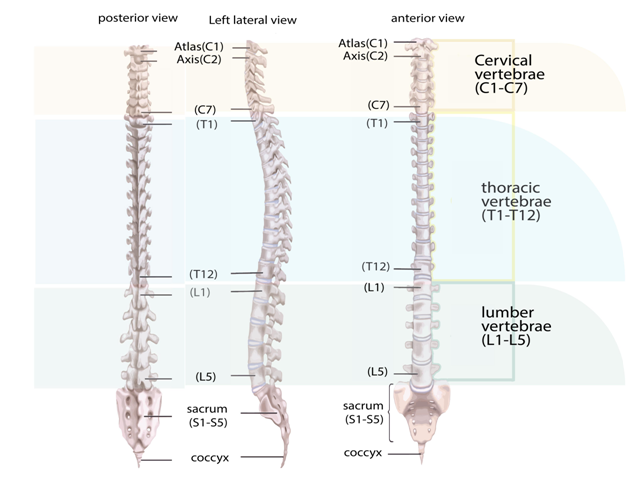
Types and Structure of Vertebrae: The Five Regions
- Cervical Vertebrae (7): Found in the neck region, labeled C1–C7. C1 (atlas) and C2 (axis) are special for head movement.
- Thoracic Vertebrae (12): Upper back; labeled T1–T12. Connect to ribs and form the chest's bony structure.
- Lumbar Vertebrae (5): Lower back; labeled L1–L5. Largest vertebrae for supporting body weight.
- Sacral Vertebrae (5, fused): Fused into a single bone (sacrum), connects spine to pelvis.
- Coccygeal Vertebrae (4, fused): Fused to form the tailbone (coccyx), supports ligament attachments.
Key Features in a Vertebrae Diagram
- Vertebral body: The large, weight-bearing part at the front.
- Vertebral arch: Forms the canal for the spinal cord.
- Spinous process: The projection you feel down your back.
- Transverse processes: Lateral projections for muscle attachment.
- Articular processes: Form joints with adjacent vertebrae.
Vertebrae Table – Numbering and Regions
| Region | Label | Number of Vertebrae | Key Features |
|---|---|---|---|
| Cervical | C1–C7 | 7 | Small, flexible, supports head |
| Thoracic | T1–T12 | 12 | Attached to ribs, heart-shaped body |
| Lumbar | L1–L5 | 5 | Largest, kidney-shaped body |
| Sacral | S1–S5 | 5 (fused) | Triangular, fused bone (sacrum) |
| Coccygeal | Co1–Co4 | 4 (fused) | Tailbone, small and fused (coccyx) |
Labeling and Drawing a Vertebrae Diagram
When drawing or using a vertebrae diagram in exams, make sure to label each region and indicate the numbers (e.g., C1 for the first cervical). Practice with both labeled and unlabeled sketches to improve memory retention and accuracy for quick revision.
Functions of Different Vertebrae Regions
- Cervical: Supports the head and allows neck movement.
- Thoracic: Protects organs and attaches to ribs.
- Lumbar: Bears body weight and provides flexibility in the lower back.
- Sacral: Connects spine to pelvis, supports hip bones.
- Coccygeal: Attachment point for ligaments and muscles of the pelvic floor.
Practice Questions
- What are the five main regions in a vertebrae diagram?
- Draw and label a simple vertebrae diagram showing all regions.
- What is the function of the lumbar vertebrae?
- How does the structure of the cervical vertebrae differ from thoracic vertebrae?
Common Mistakes to Avoid
- Mixing up the order or number of vertebrae in each region.
- Forgetting to label key regions like sacrum and coccyx in the vertebrae diagram.
- Omitting important features like spinous processes and foramina.
Real-World Applications
The concept of vertebrae diagram is used in fields like medicine, physiotherapy, anatomy, and sports science. It helps in understanding back pain, spinal injuries, and surgical planning. Vedantu helps students relate such topics to practical examples in daily life, developing awareness about body posture, movement, and health issues.
Page Summary
In this article, we explored vertebrae diagram, how to label its regions and bones, its practical significance, and key exam points. Practice drawing and labeling diagrams to improve recall and answer accuracy. To learn more and build confidence, keep practicing with Vedantu resources.
Further Learning – Internal Links
- Vertebrates and Invertebrates – Learn the differences in basic body plans for better context.
- Human Body and Its Movements – Discover how the vertebral column allows flexible movement.
- Vertebral Column – Dive deeper into the anatomical and functional details.
- Bones of Leg – Compare the backbone to other major skeletal bones.
- Central Nervous System – See the vertebrae's key role in protecting the spinal cord.
- Human Skeletal System – Overview of all major bones, perfect for revision.
- Difference Between Vertebrates and Invertebrates – Clarifies concepts often confused with vertebrae topics.
- Locomotion and Movement MCQ – Practice MCQs on related biology topics.
- Vertebrae – Study every type in detail for strong diagram answers.
- Nervous System – Understand how the vertebrae and spinal cord interact.
- Vertebrate System – Connect skeletal, nervous, and muscular systems for a complete understanding.


FAQs on Vertebrae Diagram and Anatomy of a Typical Vertebra
1. What is a vertebrae diagram?
A vertebrae diagram is a labeled illustration that shows the structure and parts of a single vertebra or the entire vertebral column. It typically highlights key anatomical components used in Biology and Anatomy studies.
- Shows the vertebral body, vertebral arch, and spinous process
- Labels openings like the vertebral foramen
- May include regions such as cervical, thoracic, and lumbar vertebrae
2. What are the main parts labeled in a typical vertebra diagram?
The main parts labeled in a typical vertebra diagram are the vertebral body, vertebral arch, and various processes.
- Vertebral body – anterior, weight-bearing portion
- Vertebral arch – forms the posterior part surrounding the spinal cord
- Spinous process – posterior projection for muscle attachment
- Transverse processes – lateral projections
- Vertebral foramen – opening for the spinal cord
3. What is the function of the vertebrae in the human body?
The primary function of the vertebrae is to protect the spinal cord and support the body’s weight. Each vertebra contributes to the overall role of the vertebral column.
- Protects the spinal cord within the vertebral canal
- Supports the head and trunk
- Allows flexibility and movement
- Provides attachment sites for muscles and ligaments
4. How many vertebrae are there in the human spine?
The human spine typically consists of 33 vertebrae in total. These are grouped into specific regions of the vertebral column.
- 7 cervical vertebrae
- 12 thoracic vertebrae
- 5 lumbar vertebrae
- 5 fused sacral vertebrae
- 4 fused coccygeal vertebrae
5. What is the difference between cervical, thoracic, and lumbar vertebrae?
The main difference between cervical, thoracic, and lumbar vertebrae lies in their location, size, and function.
- Cervical vertebrae (neck): small, allow head movement, have transverse foramina
- Thoracic vertebrae (chest): articulate with ribs
- Lumbar vertebrae (lower back): largest, bear maximum body weight
6. What is the vertebral foramen in a vertebra diagram?
The vertebral foramen is the central opening in each vertebra through which the spinal cord passes. It is formed by the vertebral body and vertebral arch.
- Aligns with other foramina to form the vertebral canal
- Protects the delicate spinal cord
- Allows passage of blood vessels and nerves
7. What is the role of the intervertebral discs between vertebrae?
The intervertebral discs act as shock absorbers between adjacent vertebrae. They are fibrocartilaginous structures located between the vertebral bodies.
- Reduce friction during movement
- Absorb mechanical shock
- Allow flexibility of the spine
8. How does the vertebral column protect the spinal cord?
The vertebral column protects the spinal cord by enclosing it within a bony canal. This protection is achieved structurally.
- Each vertebra has a vertebral foramen
- Stacked vertebrae form the vertebral canal
- Strong ligaments and muscles add support
9. What are spinous and transverse processes in a vertebra?
The spinous process and transverse processes are bony projections from a vertebra that serve as muscle and ligament attachment points.
- Spinous process: single posterior projection, palpable along the back
- Transverse processes: two lateral projections on each side
- Provide leverage for muscle movement
10. Why is studying a vertebrae diagram important in Biology?
Studying a vertebrae diagram is important because it helps learners understand spinal anatomy, structure, and function. It supports learning in human anatomy and physiology.
- Clarifies the arrangement of the vertebral column
- Helps identify labeled parts in exams
- Improves understanding of posture and spinal disorders


