

Labeled Diagram of Brain and Functions of Each Part
The concept of diagram of brain is essential in biology and helps explain real-world biological processes and exam-level questions effectively. Understanding and drawing a well-labeled brain diagram prepares students for board exams, gives clarity about brain parts and functions, and is a fundamental topic in nervous system studies.
Understanding Diagram of Brain
Diagram of brain refers to a labeled illustration or drawing that shows all the main parts of the human brain, such as the cerebrum, cerebellum, brainstem, and the different lobes. This concept is important in areas like brain anatomy, brain parts and functions, and visual aids for class revision. A clear brain diagram helps students identify regions, understand their functions, and label them accurately in exams.
Main Parts of the Brain
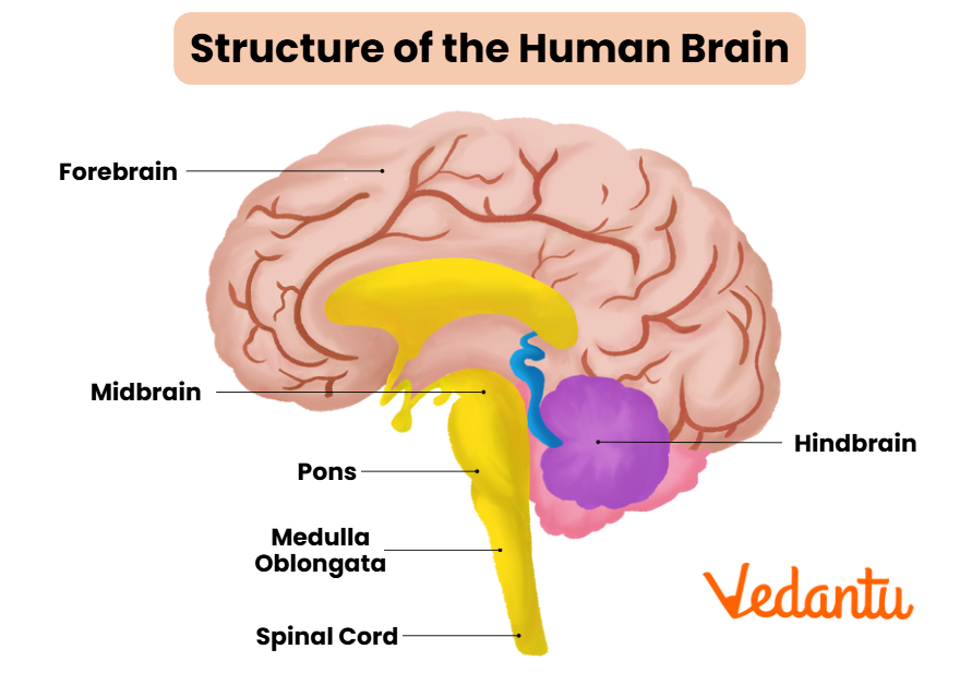
The human brain is mainly divided into three key parts:
- Forebrain (Cerebrum, Thalamus, Hypothalamus)
- Midbrain (Tectum, Tegmentum, Cerebral peduncles)
- Hindbrain (Cerebellum, Pons, Medulla oblongata)
Each of these regions consists of smaller parts that control important functions such as movement, balance, learning, sensory perception, and vital processes like breathing. A labeled diagram of the brain is frequently asked in board exams and is a must-know for students of Class 10 and 12.
Lobes of the Brain and Their Functions
The cerebrum—the largest part of the brain—is further divided into four lobes, each with distinct functions. A diagram of brain lobes and functions helps clarify their roles:
| Lobe | Main Function |
|---|---|
| Frontal Lobe | Speech, reasoning, planning, motor functions |
| Parietal Lobe | Sensory perception, spatial orientation, body awareness |
| Occipital Lobe | Visual processing |
| Temporal Lobe | Hearing, recognition, processing of auditory stimuli |
Brain Stem and Ventricles
The brain stem consists of the midbrain, pons, and medulla oblongata. It connects the brain with the spinal cord and controls breathing, heartbeat, and reflexes. The ventricles are fluid-filled spaces inside the brain that protect it by circulating cerebrospinal fluid (CSF).
Easy Brain Diagram for Kids and Beginners
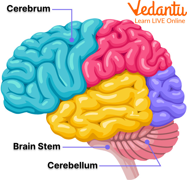
For younger students, a simple brain drawing includes the major sections—cerebrum, cerebellum, and brainstem. Use big labels and simple outlines for easy remembering. Vedantu recommends drawing step by step:
- Draw an oval for the cerebrum.
- Add a small rounded portion beneath it for the cerebellum.
- Draw the stem below for the brainstem.
- Label all parts clearly.
Quick Revision Table – Parts of the Brain
| Part | Location | Function |
|---|---|---|
| Cerebrum | Top, largest region | Controls voluntary actions, thinking, memory |
| Cerebellum | Back, below cerebrum | Balance and coordination |
| Brainstem | Base, connects to spinal cord | Breathing, heartbeat, reflexes |
Common Mistakes to Avoid
- Confusing cerebellum with cerebrum when labeling.
- Missing out on labeling smaller parts like pons and medulla.
- Not connecting each label to its function in the diagram.
Real-World Applications
The knowledge of diagram of brain helps in medicine, psychology, and understanding diseases and injuries. For example, doctors use MRI brain diagrams to diagnose stroke or tumor locations. Students using Vedantu resources gain a practical view of how understanding the brain supports learning, health, and everyday safety.
In this article, we explored diagram of brain, its labeled parts, their functions, lobes, and tips for drawing and revision. Proper practice helps in perfect labeling and understanding. To learn more about the human nervous system or specific brain parts, continue exploring with Vedantu.
Related Reading
- Human Brain – Deep dive on brain anatomy and structure for class revision.
- Parts of the Brain – Explains each brain part with diagrams for exam reference.
- Cerebrum – Focuses on cerebrum’s structure, location, and functions.
- Cerebellum – Detailed look at the cerebellum, a key diagram label.
- Brain Tumour Symptoms – Links brain anatomy to real-life health.
- Central Nervous System – Connects brain diagrams to the rest of the nervous system.
- Difference Between Brain and Spinal Cord – Clarifies confusion about brain vs spinal cord diagrams.
- Nervous System – Explains brain’s connection to nerves and body function.
- Neuron – Shows how neurons are organized in the brain’s structure.


FAQs on Diagram of Brain with Labeled Structures and Functions
1. What is a diagram of the brain?
A diagram of the brain is a labeled visual representation that shows the major parts and structures of the human brain and their positions. It helps learners understand brain anatomy by clearly marking key regions such as:
- Cerebrum
- Cerebellum
- Brainstem
- Lobes of the brain (frontal, parietal, temporal, occipital)
Brain diagrams are commonly used in biology to explain structure, function, and coordination of the central nervous system.
2. What are the main parts shown in a brain diagram?
The main parts shown in a typical human brain diagram are the cerebrum, cerebellum, and brainstem. These major regions include:
- Cerebrum – largest part responsible for thinking, memory, and voluntary actions
- Cerebellum – controls balance and coordination
- Brainstem – connects the brain to the spinal cord and regulates vital functions
Some detailed diagrams also label internal structures like the thalamus, hypothalamus, and corpus callosum.
3. What are the four lobes of the brain in a labeled diagram?
The four lobes of the brain shown in a labeled diagram are the frontal, parietal, temporal, and occipital lobes. Each lobe has a specific function:
- Frontal lobe – reasoning, decision-making, and voluntary movement
- Parietal lobe – sensory perception (touch, temperature, pain)
- Temporal lobe – hearing and memory
- Occipital lobe – vision and visual processing
These lobes are clearly separated by grooves called sulci in most brain structure diagrams.
4. What is the function of the cerebrum in the brain diagram?
The cerebrum is responsible for higher mental functions, voluntary movements, and sensory interpretation. It performs several important roles:
- Controls voluntary muscle actions
- Processes sensory information
- Enables thinking, memory, and intelligence
- Regulates speech and emotions
In most brain diagrams, the cerebrum appears as the largest, uppermost part divided into two hemispheres.
5. What is the function of the cerebellum in a brain diagram?
The cerebellum coordinates balance, posture, and smooth muscle movements. It plays a key role in:
- Maintaining body equilibrium
- Coordinating voluntary movements
- Ensuring precise motor control
In a labeled brain diagram, the cerebellum is located below the cerebrum and behind the brainstem.
6. What is the brainstem and what does it do?
The brainstem is the lower part of the brain that connects it to the spinal cord and controls vital life functions. It regulates:
- Breathing
- Heartbeat
- Blood pressure
- Swallowing and reflexes
A brainstem diagram usually shows three parts: midbrain, pons, and medulla oblongata.
7. What is the difference between the cerebrum and cerebellum?
The main difference between the cerebrum and cerebellum is that the cerebrum controls thinking and voluntary actions, while the cerebellum controls coordination and balance. Key differences include:
- Cerebrum – largest part; responsible for intelligence, memory, and sensation
- Cerebellum – smaller; responsible for posture and motor coordination
- Cerebrum is located at the top; cerebellum is located at the back and bottom
Both structures are clearly labeled in a standard human brain diagram.
8. What is the corpus callosum in a brain diagram?
The corpus callosum is a thick band of nerve fibers that connects the left and right cerebral hemispheres. Its main function is to:
- Allow communication between both hemispheres
- Coordinate sensory and motor information
In a sagittal section brain diagram, the corpus callosum appears as a curved structure located above the thalamus.
9. What does a sagittal section of the brain show?
A sagittal section of the brain shows a side view that divides the brain into left and right halves. It clearly displays internal structures such as:
- Corpus callosum
- Thalamus
- Hypothalamus
- Brainstem
- Cerebellum
This type of brain diagram is useful for understanding the internal anatomy of the human brain.
10. Why is a labeled diagram of the brain important for biology students?
A labeled diagram of the brain is important because it helps students visually identify brain structures and understand their functions clearly. It supports learning by:
- Improving memory through visual representation
- Clarifying the position of major brain parts
- Helping in exams that require labeling or explanation
- Connecting structure with function in the central nervous system
Brain diagrams make complex neuroanatomy easier to study and revise effectively.


