


What Is the Structure and Function of the Vertebral Column in NEET Biology?
The vertebral column, also called the backbone or spine, is a core part of the human skeletal system studied in Biology for NEET. It supports the body, protects the spinal cord, and plays roles in movement and flexibility. Understanding the vertebral column is crucial for NEET aspirants because it forms the foundation for several questions across anatomy and physiology. A clear concept of the vertebral column not only helps in answering direct questions but also supports learning related systems in human biology.
What is the Vertebral Column?
The vertebral column is a long, flexible, segmented bony structure running down the back. It is made up of several small bones called vertebrae, which are stacked one over the other. The column extends from the base of the skull to the pelvis. Its main function is to provide structural support, protect the spinal cord, allow movement, and serve as an attachment point for muscles and ribs. In human beings, the vertebral column is a defining feature of the axial skeleton and plays a central role in posture and locomotion.
Core Ideas and Fundamentals of the Vertebral Column
Basic Structure of the Vertebral Column
The vertebral column consists of a series of bones called vertebrae, which are joined together by intervertebral discs and ligaments. This arrangement provides both strength and flexibility. The vertebral column is divided into specific regions based on the location and type of vertebrae present.
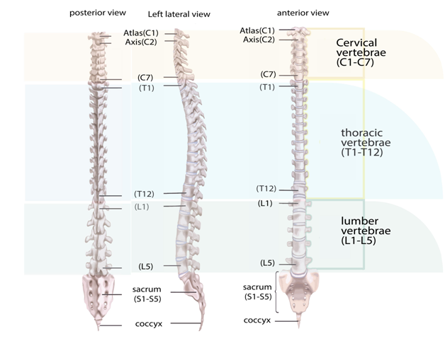
Regions and Number of Vertebrae
- Cervical (Neck Region) - 7 Vertebrae
- Thoracic (Chest Region) - 12 Vertebrae
- Lumbar (Lower Back) - 5 Vertebrae
- Sacral (Pelvic Region) - 5 Fused Vertebrae
- Coccygeal (Tailbone) - 4 Fused Vertebrae
Functions of the Vertebral Column
- Supports body weight and maintains posture
- Protects the spinal cord running inside the vertebral canal
- Allows flexibility and movement (bending, twisting)
- Serves as a point of attachment for muscles and ribs
Important Sub-Concepts Related to the Vertebral Column
Structure of a Typical Vertebra
A typical vertebra consists of a vertebral body, vertebral arch, vertebral foramen, and various processes (spinous, transverse, and articular). The vertebral foramen forms a canal for the spinal cord. The processes provide points of muscle and ligament attachment.
Intervertebral Discs
Between adjacent vertebrae (except between some fused vertebrae), there are intervertebral discs made of cartilage. These discs act as cushions, absorb shock, and permit flexible movement of the spine.
Curvatures of the Vertebral Column
In adults, the vertebral column has four normal curves - cervical and lumbar curves are convex forward, while thoracic and sacral curves are convex backward. These curves increase the strength and flexibility of the spine.
Key Relationships, Principles, and Facts
- There are a total of 33 vertebrae in human beings, but due to fusion, only 26 are distinct in adults.
- The vertebral column encloses and protects the fragile spinal cord.
- Abnormalities like scoliosis (lateral curvature), kyphosis (increased thoracic curve), and lordosis (increased lumbar curve) can affect posture and health.
- Intervertebral discs help distribute body weight and provide flexibility.
Features, Functions, and Importance
- Segmented Structure: Allows limited independent movements, increasing overall flexibility and shock absorption.
- Protection: Safeguards the spinal cord, a vital pathway for nerves connecting the brain and body.
- Weight Bearing: Transmits body weight from head to pelvis and legs.
- Organ Attachment: Provides attachment for ribs, pelvic girdle, and muscles.
Table: Regions of the Vertebral Column and Their Features
| Region | Number of Vertebrae | Main Features |
|---|---|---|
| Cervical | 7 | Smallest, support head; first two (atlas and axis) allow head movement |
| Thoracic | 12 | Articulate with ribs, support chest cage |
| Lumbar | 5 | Largest, bear most weight, limited movement |
| Sacral | 5 (fused) | Form sacrum, connect spine to hips |
| Coccygeal | 4 (fused) | Form coccyx (tailbone), very little function in humans |
This table helps visualize differences between the regions of the vertebral column, which is useful for memory and quick NEET revision.
Why is the Vertebral Column Important for NEET?
Questions on the vertebral column often appear in NEET Biology due to its anatomical and functional significance. It is commonly tested in human physiology, anatomy, and applied clinical contexts (like injuries or congenital defects). Understanding the vertebral column is essential because:
- It forms a basic part of human skeletal structure, linking to muscles, nerves, and movement topics.
- A solid grasp helps in visualizing related systems and answering diagram-based or clinical scenario questions.
- Related facts, such as number and function of vertebrae or common abnormalities, are high-yield MCQ topics.
How to Study Vertebral Column Effectively for NEET
- Start by understanding the overall structure and regions (cervical, thoracic, lumbar, etc.).
- Memorize the number of vertebrae in each region using mnemonics if needed.
- Study diagrams, such as the vertebrae diagram, and practice drawing and labeling them.
- Learn the function and importance of intervertebral discs and curvatures.
- Solve past NEET MCQs involving the vertebral column and related abnormalities.
- Revise using tables and bullet lists for quick memory recall.
- Clarify doubts on confusing terms like atlas, axis, sacrum, and coccyx with the help of visual aids.
Common Mistakes Students Make Regarding the Vertebral Column
- Confusing the number and types of vertebrae in each region
- Mixing up the structure and function of atlas and axis vertebrae
- Ignoring structural differences between cervical, thoracic, and lumbar vertebrae
- Overlooking the role of intervertebral discs and curvatures in function and health
- Not practicing enough diagram-based questions
Quick Revision Points on the Vertebral Column
- Vertebral column - flexible rod made up of 33 vertebrae, 26 distinct in adults due to fusion.
- Regions: Cervical (7), Thoracic (12), Lumbar (5), Sacral (5 fused), Coccygeal (4 fused).
- Atlas (C1) and Axis (C2) are specialized cervical vertebrae for head movement.
- Intervertebral discs act as shock absorbers and provide flexibility.
- The vertebral column protects the spinal cord and supports the body.
- Normal curvatures increase strength and flexibility of the spine.
- Remember common abnormalities: scoliosis, kyphosis, and lordosis.
- Practice drawing and labeling the vertebrae diagram for fast recall.
FAQs on Vertebral Column: Essential Concepts for NEET Students
1. What is the vertebral column and what is its main function in the human body for NEET?
The vertebral column is the central supportive structure of the human skeleton, commonly known as the backbone or spine. It primarily functions to:
- Provide structural support to the body
- Protect the spinal cord, a major part of the nervous system
- Facilitate movement and flexibility
- Serve as an attachment point for muscles and ribs
2. How many vertebrae are present in the human vertebral column according to the NEET syllabus?
The human vertebral column typically consists of 33 vertebrae in total. These are categorized as:
- 7 cervical vertebrae
- 12 thoracic vertebrae
- 5 lumbar vertebrae
- 5 sacral vertebrae (fused into the sacrum)
- 4 coccygeal vertebrae (fused into the coccyx)
3. What are the main regions of the vertebral column in humans for NEET exam preparation?
The vertebral column is divided into five distinct regions based on location and structure:
- Cervical (neck region) – 7 vertebrae
- Thoracic (upper back) – 12 vertebrae
- Lumbar (lower back) – 5 vertebrae
- Sacral (sacrum) – 5 fused vertebrae
- Coccygeal (tailbone) – 4 fused vertebrae
4. What is the significance of intervertebral discs for NEET students?
Intervertebral discs are soft, cushion-like pads between adjacent vertebrae that absorb shocks and allow flexibility. Their main significance includes:
- Providing shock absorption during movement
- Preventing friction between vertebrae
- Helping the vertebral column bend and twist
5. How does the structure of the vertebral column contribute to its function in humans?
The vertebral column's structure ensures both stability and flexibility. Key contributions include:
- Curved alignment (cervical, thoracic, lumbar curves) for balance and shock absorption
- Interlocking vertebrae provide strength and movement
- Intervertebral discs add flexibility and protection
- Attachment points for muscles and ligaments
6. Name the bones that form the vertebral column. (Scraped)
The vertebral column is made up of vertebrae arranged in five groups:
- 7 cervical vertebrae
- 12 thoracic vertebrae
- 5 lumbar vertebrae
- 5 sacral vertebrae (fused)
- 4 coccygeal vertebrae (fused)
7. What is the difference between cervical, thoracic, and lumbar vertebrae?
Cervical, thoracic, and lumbar vertebrae have unique characteristics based on location:
- Cervical vertebrae: Smallest, with foramen in transverse processes
- Thoracic vertebrae: Medium-sized, attached to ribs
- Lumbar vertebrae: Largest, strong bodies for weight-bearing
8. Which part of the vertebral column supports the skull?
The cervical region of the vertebral column, especially the first cervical vertebra (atlas), directly supports the skull. It enables:
- Head support and upright posture
- Neck movement and flexibility
9. What is the function of the sacrum and coccyx in the vertebral column?
The sacrum and coccyx are the fused, lower sections of the vertebral column. Their functions include:
- Sacrum: Connects the spine to the pelvic girdle, stabilizing pelvis
- Coccyx: Provides attachment for ligaments and muscles
10. Why is the vertebral column called the backbone? (Scraped)
The vertebral column is commonly called the backbone because it forms the central axis and main support for the body’s framework. It:
- Runs along the back from skull to pelvis
- Supports the body's weight and posture
11. How is the vertebral column adapted to protect the spinal cord for NEET?
The vertebral column protects the spinal cord by enclosing it within the vertebral canal, a bony tunnel formed by stacked vertebral arches. Adaptations include:
- Bony protection against injury
- Shock absorption through discs and curves
- Flexibility without compromising safety
12. List the functions of the vertebral column. (Scraped)
The vertebral column serves several essential functions:
- Supports the head and trunk
- Protects the spinal cord
- Provides attachment for ribs and muscles
- Enables flexible movement of the body