

Structure and Functions Explained in a Labeled Liver Diagram
The concept of liver diagram is essential in biology and helps explain real-world biological processes and exam-level questions effectively. A good understanding of the liver diagram is important for students preparing for CBSE, ICSE, and other board exams, since labeling and drawing this structure is a common requirement.
Understanding Liver Diagram
Liver diagram refers to a visual representation that shows the anatomical structure, position, and main lobes of the liver within the human body. This concept is important in areas like human anatomy, digestive system biology, and exam preparation. The liver plays a significant role in digestion, detoxification, and metabolism. Recognizing its anatomy through diagrams helps students identify different parts such as lobes, blood vessels, and its orientation inside the abdomen.
Location of Liver in the Human Body
The liver is the largest gland in the human body and is located in the upper right portion of the abdomen, just beneath the diaphragm and above the stomach. It is mainly on the right side but extends towards the left. Understanding the location of liver in body is vital for correct labeling and diagram drawing in biology exams.
Features of a Labeled Liver Diagram
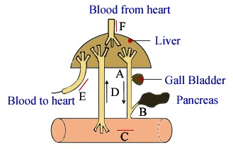
A liver diagram labeled clearly marks the main anatomical features. For exam purposes, remember to show:
- Right lobe (larger in size)
- Left lobe (smaller in size)
- Falciform ligament (divides the lobes)
- Gallbladder (under right lobe, stores bile)
- Major blood vessels: hepatic artery, portal vein, hepatic vein
- Bile duct
These parts are frequently asked in board and competitive exams. Practice drawing and labeling them for clear answers!
Simple and Unlabeled Liver Diagrams for Practice
For revision, students benefit from simple or liver diagram unlabeled sketches. Start with the basic shape—a wedge or prism with a rounded right side and a pointed left side. Practice with unlabeled drawings helps test your memory and labeling accuracy.
- Draw the outline (wedge shape)
- Divide into right and left lobes
- Add gallbladder under the right lobe
- Sketch blood vessels
Step-by-step drawing enhances your confidence during quick exam revision.
Anatomy and Lobes of the Liver
The liver is divided into lobes and segments. For exam diagrams, focus on:
- Right lobe (largest, to the right of the falciform ligament)
- Left lobe (smaller, to the left of the ligament)
- Other segments: caudate and quadrate (may be labeled for extra marks)
The hepatic artery, portal vein, and bile duct are often drawn near the lower or posterior edge, showing their relationships to the lobes.
Relationship of Liver to Other Organs
A detailed liver diagram with ribs or posterior views sometimes shows the liver in relation to:
- Ribcage (protects upper part of the liver)
- Stomach (left and slightly below liver)
- Right kidney and diaphragm (behind and above the liver)
- Gallbladder (under the right lobe)
This helps you understand its positioning during practical or theory questions.
Main Functions Shown in a Liver Diagram
The liver has several key functions, often summarized in revision tables:
| Function | Description |
|---|---|
| Bile production | Helps digest fats in the intestine |
| Detoxification | Removes toxins from blood |
| Metabolism | Regulates glucose, fats, and proteins |
| Storage | Stores vitamins, iron, and glycogen |
| Blood filtration | Removes old blood cells |
| Clotting factor synthesis | Produces important proteins for clotting |
Learn these functions well for quick revision. For a full list, see liver functions on Vedantu.
Practice Questions
- Draw and label a liver diagram as asked in exams.
- List the main functions of the liver shown in the diagram.
- Where is the liver located in the human body?
- Why are blood vessels important to label in a liver diagram?
Common Mistakes to Avoid
- Mixing up the right and left lobes (right is always larger).
- Forgetting to add and label the gallbladder or bile duct.
- Omitting key blood vessels (portal vein, hepatic artery, hepatic vein)
- Confusing the liver’s position (mainly on the right, but crosses midline to the left)
Real-World Applications
The concept of liver diagram is used in fields like medicine, medical imaging, and surgery. Understanding its structure helps doctors detect diseases (like hepatitis or cirrhosis), plan surgeries, and explain treatments. Students who master diagram-based questions with Vedantu gain clarity not only in exams, but also for future careers in health sciences and biotechnology.
In this article, we explored liver diagram, its key structures, drawing tips, main functions, and practice points for exam revision. To learn more about the anatomy and role of the liver, or to strengthen your biology concepts, refer to Liver and Human Digestive System on Vedantu for further reading and practice.
- Liver – Detailed structure and functions
- Human Digestive System – Connection of liver in digestion
- Bile – Importance in fat digestion
- Rectum – Relation to abdominal organ layout
- Kidneys – Comparative positioning to liver
- Digestive System Diagram – Integrated diagram practice
- Human Body Anatomy – Full map of organ positions
- Peristalsis – Liver’s role in digestive movement
- Human Excretory System – Excretory functions of liver


FAQs on Liver Diagram and Labeled Anatomy of the Human Liver
1. What is a liver diagram?
A liver diagram is a labeled illustration that shows the structure, position, and major parts of the liver in the human body. It helps students understand the anatomical features and internal organization of the liver. A typical liver diagram includes:
- The right and left lobes
- The gallbladder
- The hepatic artery and portal vein
- The bile duct
- The functional unit called the hepatic lobule
2. Where is the liver located in the human body?
The liver is located in the upper right side of the abdominal cavity, just below the diaphragm. It lies mainly in the right hypochondriac region and partly in the epigastric region. Key location features include:
- Below the diaphragm
- Above the stomach and intestines
- Protected by the lower ribs
3. What are the main parts labeled in a liver diagram?
The main parts labeled in a liver diagram include its lobes, blood vessels, and bile ducts. Common labeled structures are:
- Right lobe (largest lobe)
- Left lobe
- Caudate lobe and quadrate lobe
- Hepatic artery (supplies oxygenated blood)
- Hepatic portal vein (brings nutrient-rich blood)
- Hepatic vein (drains blood to the inferior vena cava)
- Bile duct and gallbladder
4. What is the function of the liver in the human body?
The liver performs vital functions including metabolism, detoxification, and bile production. Its major functions include:
- Producing bile to help digest fats
- Storing glycogen, vitamins, and minerals
- Detoxifying harmful substances and drugs
- Producing plasma proteins like albumin
- Regulating blood glucose levels
5. What is a hepatic lobule in a liver diagram?
A hepatic lobule is the structural and functional unit of the liver shown in microscopic liver diagrams. It is typically hexagonal in shape and consists of:
- A central vein at the center
- Radiating plates of hepatocytes
- Sinusoids (blood capillaries)
- A portal triad at each corner (hepatic artery, portal vein, bile duct)
6. What is the difference between the right lobe and left lobe of the liver?
The right lobe of the liver is larger and performs more metabolic activity than the smaller left lobe. Key differences include:
- The right lobe occupies most of the upper right abdomen
- The left lobe extends toward the left side of the body
- They are separated by the falciform ligament
7. What blood vessels are shown in a liver diagram?
A liver diagram shows three major blood vessels: the hepatic artery, portal vein, and hepatic vein. Their roles include:
- Hepatic artery – supplies oxygen-rich blood from the heart
- Hepatic portal vein – brings nutrient-rich blood from the digestive organs
- Hepatic vein – carries filtered blood to the inferior vena cava
8. How does bile flow in the liver according to a liver diagram?
Bile flows from hepatocytes to the bile ducts and then to the gallbladder or small intestine. The step-by-step flow is:
- Produced by hepatocytes
- Collected in bile canaliculi
- Drains into small bile ducts
- Moves to the common hepatic duct
- Stored in the gallbladder or released into the duodenum
9. Why is the liver called a vital organ?
The liver is called a vital organ because it performs essential life-sustaining metabolic and detoxification functions. Without a functioning liver:
- Toxins would accumulate in the blood
- Digestion of fats would be impaired due to lack of bile
- Blood protein production would decrease
- Blood sugar regulation would fail
10. How do you label a simple liver diagram for exams?
To label a simple liver diagram for exams, identify and mark the major visible structures clearly and correctly. Common exam labels include:
- Right lobe
- Left lobe
- Gallbladder
- Hepatic artery
- Portal vein
- Bile duct


