

Labeled uterus diagram structure layers parts and reproductive functions
The concept of uterus diagram is essential in biology and helps explain real-world biological processes and exam-level questions effectively. Understanding a uterus diagram helps students visualize the female reproductive system, know important terminology, and perform well in board exams and NEET-level tests.
Understanding Uterus Diagram
Uterus diagram refers to a labelled or unlabelled graphical representation of the uterus, highlighting its shape, location in the female body, and anatomical parts such as the fundus, body, cervix, and ligaments. This concept is important in areas like human reproductive biology, exam preparation (labelling), and understanding pregnancy physiology. Uterus diagrams help clarify spatial relationships with adjacent organs (urinary bladder, rectum), pathways such as the fallopian tubes, and support structures like ligaments.
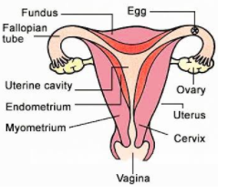
Main Parts Seen in a Uterus Diagram
- Fundus: The dome-shaped upper part above the openings of the fallopian tubes.
- Body (Corpus): The main central portion where implantation occurs.
- Cervix: Lower constricted region connecting the uterus to the vagina, divided into internal and external os.
- Isthmus: Narrow segment between body and cervix.
- Ligaments: Broad, round, ovarian, cardinal, and uterosacral ligaments provide support.
- Endometrial lining: Inner mucous membrane, important in menstruation and pregnancy.
- Myometrium: Thick muscular middle layer responsible for contractions.
Uterus Structure and Function
- Shape & Position: Pear-shaped, situated between the urinary bladder and rectum.
- Zones: Fundus (top), body (middle), isthmus (narrowing), cervix (neck).
- Wall Layers: Serous perimetrium (outer), myometrium (muscle), endometrium (lining).
- Blood & Nerves: Supplied by uterine artery; innervated by uterovaginal plexus.
- Ligaments: Broad, round, uterosacral, ovarian, and cardinal ligaments anchor the uterus.
Functions of the Uterus
- Site for implantation of embryo during pregnancy.
- Supports, nourishes, and protects the developing fetus.
- Contracts to help expel the fetus during childbirth.
- Regenerates endometrial lining during each menstrual cycle.
- Plays a role in menstruation, labour, and postpartum involution.
Variations and Clinical Aspects of Uterus Diagrams
- Pregnancy: The uterus enlarges, and diagrams show the growing cavity and fetal position.
- With IUD: Diagrams indicate the placement of intrauterine devices for contraception.
- Position Variations: Anteverted (normal), retroverted, anteflexed, and retroflexed positions.
- For Kids/Exams: Simple versions use fewer labels for easy recall and neat diagramming.
- Ligament Attachments: Medical diagrams highlight surrounding support structures.
How to Draw and Label a Uterus Diagram for Exams
- Draw a central pear-shaped outline.
- Mark the fundus (top), body (center), and cervix (bottom narrowing).
- Show fallopian tube openings on each top side.
- Add internal cavity, endometrial lining, and muscle layers if required.
- Label support ligaments around the outline (broad, round, ovarian, etc.).
- Neatly label each part outside the diagram with straight lines.
- Keep diagram symmetric and labels clear for board scoring.
Common Mistakes to Avoid
- Mixing up uterus and ovary diagrams.
- Incorrect label placement (especially ligaments and cervix/os).
- Ignoring changes during pregnancy or failing to mention functions.
- Missing out layers (endometrium, myometrium) in diagrams when asked.
Real-World Applications
The concept of uterus diagram is used in medicine (gynaecology, obstetrics), health education, clinical diagnosis, fertility treatments, and illustrating concepts in textbooks. Vedantu helps students relate such topics to real-life medical applications, understand birth control methods, or explain pregnancy stages in simple diagrams.
In this article, we explored uterus diagram, its key processes, real-life significance, and how to solve questions based on it. To learn more and build confidence, keep practicing with Vedantu resources and work on more labelled diagrams for exams.
Related Vedantu Internal Links
- Female Reproductive System
- Human Reproductive System
- Uterus and Development of Placenta
- Implantation in Human
- Ovary (Plant)
- Rectum
- Hormones
- Ovum
- Male Reproductive System
- Neuron Diagram
- Labeled Diagram of Human Ear


FAQs on Uterus Diagram with Labeled Structure and Functions
1. What is the uterus in the female reproductive system?
The uterus is a hollow, muscular organ in the female reproductive system where implantation and development of the embryo and fetus occur.
- It is located in the pelvic cavity between the bladder and rectum.
- It plays a central role in menstruation, pregnancy, and childbirth.
- It connects to the vagina through the cervix and to the ovaries via the fallopian tubes.
2. What are the main parts labeled in a uterus diagram?
A standard uterus diagram typically labels the fundus, body, cervix, and uterine layers.
- Fundus – the upper rounded portion.
- Body (corpus) – the main central part.
- Cervix – the lower narrow opening into the vagina.
- Endometrium, myometrium, and perimetrium – the three uterine layers.
3. What are the three layers of the uterus?
The three layers of the uterus are the endometrium, myometrium, and perimetrium.
- Endometrium – the inner lining that thickens and sheds during the menstrual cycle.
- Myometrium – the thick muscular middle layer responsible for uterine contractions.
- Perimetrium – the outer protective serous layer.
4. What is the function of the uterus?
The main function of the uterus is to support implantation, nourish the developing embryo, and enable childbirth.
- Receives the fertilized egg for implantation.
- Forms part of the placenta during pregnancy.
- Contracts during labor to deliver the baby.
- Sheds its lining during menstruation if fertilization does not occur.
5. What does the endometrium do in the uterus?
The endometrium is the inner lining of the uterus that thickens to prepare for pregnancy and sheds during menstruation if fertilization does not occur.
- Responds to hormones like estrogen and progesterone.
- Provides nutrients to the early embryo.
- Breaks down and is expelled as menstrual flow in each cycle.
6. Where is the uterus located in the human body?
The uterus is located in the pelvic cavity, between the urinary bladder and the rectum.
- It lies above the vagina.
- It is normally tilted slightly forward, a position called anteverted uterus.
- Its position allows connection to the ovaries through the fallopian tubes.
7. What is the difference between the uterus and the cervix?
The uterus is the main muscular organ for fetal development, while the cervix is its lower narrow opening that connects to the vagina.
- The uterus includes the fundus and body.
- The cervix controls the passage of sperm and menstrual flow.
- During childbirth, the cervix dilates to allow delivery.
8. How does the uterus change during pregnancy?
During pregnancy, the uterus enlarges significantly to accommodate and nourish the growing fetus.
- The myometrium expands and thickens.
- The endometrium forms part of the placenta.
- Its size increases from a small pear-shaped organ to one that fills much of the abdominal cavity.
9. What is the role of the myometrium in childbirth?
The myometrium produces strong rhythmic contractions that help expel the baby during childbirth.
- It is composed of smooth muscle fibers.
- Hormones like oxytocin stimulate contractions.
- These contractions dilate the cervix and push the fetus out.
10. Why is a uterus diagram important for biology students?
A uterus diagram is important because it helps students visually understand the structure, layers, and reproductive functions of the organ.
- Clarifies the position of the fundus, body, and cervix.
- Helps identify uterine layers and their roles.
- Supports understanding of menstruation, fertilization, and pregnancy processes.


