

Labelled TMV Diagram Explanation of Structure and Components
The concept of TMV diagram is essential in biology and helps explain real-world biological processes and exam-level questions effectively. Understanding the structure of Tobacco Mosaic Virus is important for students preparing for board and competitive exams, especially when labeling diagrams or describing virus features is required.
Understanding TMV Diagram
TMV diagram refers to the visual representation of the Tobacco Mosaic Virus (TMV) structure. TMV is a plant virus that mainly infects tobacco, tomatoes, potatoes, and other members of the Solanaceae family. This concept is important in areas like virus structure, plant pathology, and molecular biology. TMV diagram is widely used in NCERT, Class 11 Biology, and various entrance exams to test knowledge of viral morphology and labeling skills.
Features of TMV Structure
A well-drawn TMV diagram should clearly indicate the main structural features. Below are the key features you must know and label:
- TMV is a rod-shaped virus approximately 300 nm long and 18 nm in diameter.
- It has a protein coat called the capsid, made up of about 2130 identical protein subunits.
- The genetic material is a single-stranded RNA located inside the capsid.
- The capsid forms a helical structure around the RNA chain.
- Each helix turn has about 16.3 capsomeres.
- The RNA consists of around 6395 nucleotides and is coiled tightly inside the protein shell.
- The structure displays helical symmetry and inherent chirality.
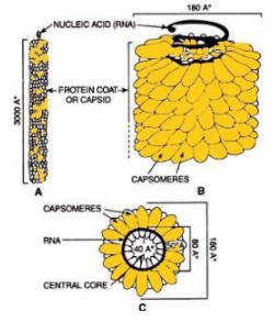
Labeled TMV Diagram Explanation
The labeled TMV diagram should indicate the protein capsid, RNA core, and the helical arrangement of proteins. Label the following parts clearly:
- Capsid or protein coat
- Single-stranded RNA (ssRNA)
- Helical structure
- Capsomeres (protein subunits)
Good labeling and neat diagram are essential for scoring full marks in practical or board exams. Use arrows for pointing out each part, as shown in standard TMV diagrams in textbooks or PDFs.
TMV Diagram in Exams
In board exams and competitive tests, questions often ask you to draw and label the TMV diagram or explain its main features. Tips to score high marks:
- Draw to scale, maintain proportions.
- Label each part clearly and legibly.
- Create neat, bold lines for main structures.
- Highlight the difference between RNA and protein coat.
- Revise from TMV diagram PDFs and check for correct spellings.
Download TMV Diagram PDFs
For fast revision and convenience, download labeled TMV diagram PDFs for practice. Hindi versions and blank unlabeled diagrams are helpful for quick self-tests and last-minute revision. Many students prefer digital notes or printable sheets for study.
Common Mistakes to Avoid
- Confusing TMV diagram with diagrams of other plant viruses or bacteria.
- Missing labels or incorrect placements of RNA and capsid parts.
- Forgetting to show the helical symmetry or not drawing the full rod-shaped outline.
- Writing 'DNA' instead of 'RNA' inside TMV.
Real-World Applications
The TMV diagram is essential not only for exams, but also for understanding viral infections in plants. TMV has industrial and research uses in biotechnology and nanotechnology. Knowing its structure helps in genetic engineering and plant pathogen management. Vedantu makes these concepts easy to grasp for all students.
Practice Questions
- Draw and label a neat diagram of TMV as per NCERT standards.
- What are the main features of a TMV diagram?
- How do you differentiate TMV structure from that of bacteria?
- Explain the role of the protein coat in the TMV structure.
- Why is RNA important in the TMV diagram?
In this article, we explored TMV diagram, its key processes, real-life significance, and how to solve questions based on it. To learn more and build confidence, keep practicing with Vedantu.
Further Reading
- Virus – Basic virus structure, essential for understanding TMV and other plant viruses.
- Differences between Virus and Bacteria – Clarifies TMV’s classification and resolves common doubts.
- Plant Cell Structure and Function – Explains how TMV infects plant cells and its impact on plant physiology.
- Cell Structure and Function – Helpful to understand how TMV relates to general cell biology topics.
- Molecular Basis of Inheritance – Provides background on RNA, as seen in TMV core.
- Photosynthesis Process – Important because TMV affects photosynthetic tissues in plants.
- Fungi – Comparative study of major plant pathogens.
- Microorganisms – Puts TMV in the larger context of plant pathogens.
- Plant Tissue vs Animal Tissue – Useful for compare-and-contrast board questions about TMV impact.
- Cell Membrane – Explains how TMV enters plant cells, a frequent exam topic.


FAQs on TMV Diagram and Detailed Structure of Tobacco Mosaic Virus
1. What is a TMV diagram in biology?
A TMV diagram is a labeled representation of the structure of Tobacco Mosaic Virus (TMV), showing its helical protein coat and RNA core. It typically illustrates:
- The helical capsid made of protein subunits
- The central single-stranded RNA (ssRNA)
- The rod-shaped structure of the virus
2. What are the main parts labeled in a TMV diagram?
The main parts labeled in a TMV diagram are the protein coat and the RNA core. Key labeled components include:
- Capsid – protective protein covering
- Capsomeres – repeating protein subunits
- Single-stranded RNA – genetic material
- Helical structure – spiral arrangement of capsomeres around RNA
3. What is the structure of Tobacco Mosaic Virus (TMV)?
The Tobacco Mosaic Virus (TMV) has a rigid, rod-shaped helical structure composed of RNA and protein. Its structure includes:
- A central single-stranded RNA (ssRNA)
- About 2,130 identical capsomeres
- A helical capsid surrounding the RNA
4. Why is TMV described as a helical virus?
TMV is described as a helical virus because its capsid proteins are arranged in a spiral around its RNA. In this arrangement:
- Protein subunits wind around the RNA molecule
- The structure forms a cylindrical rod
- The RNA lies in a groove inside the helix
5. What is the function of the capsid in the TMV diagram?
The capsid in the TMV diagram functions as a protective protein coat that surrounds and safeguards the viral RNA. Its main roles are:
- Protecting genetic material from damage
- Helping in virus attachment to host cells
- Maintaining the virus’s rigid rod shape
6. What type of genetic material does TMV contain?
TMV contains single-stranded RNA (ssRNA) as its genetic material. This RNA:
- Acts directly as messenger RNA (mRNA) in host cells
- Encodes viral proteins needed for replication
- Is enclosed within the helical capsid
7. How does TMV infect plant cells?
TMV infects plant cells by entering through wounds and releasing its RNA to replicate inside the host. The infection process involves:
- Entry through damaged plant tissue
- Uncoating of the capsid
- Release of viral RNA into the cytoplasm
- Replication using host ribosomes and enzymes
8. What is the difference between TMV and bacteriophage structure?
The main difference between TMV and a bacteriophage is that TMV has a simple helical rod shape, while bacteriophages have a complex head-tail structure. Key differences include:
- TMV: Helical capsid, ssRNA, rod-shaped, infects plants
- Bacteriophage: Icosahedral head, tail fibers, usually DNA, infects bacteria
9. Why is the TMV diagram important in biology exams?
The TMV diagram is important in biology exams because it demonstrates the classic example of a helical virus structure. Students are often asked to:
- Draw and label the helical capsid and RNA core
- Explain viral symmetry
- Describe structure-function relationships in viruses
10. Who discovered Tobacco Mosaic Virus (TMV)?
Tobacco Mosaic Virus (TMV) was first discovered by Dmitri Ivanovsky in 1892. Later contributions include:
- Martinus Beijerinck – recognized it as a contagious living fluid (virus)
- Wendell Stanley – crystallized TMV in 1935


