

Chromosome diagram definition structure and labeled parts in detail
The concept of chromosome diagram is essential in biology and helps explain real-world biological processes and exam-level questions effectively. A labeled chromosome diagram clearly shows the structure, anatomy, and major parts of a chromosome, making it easier for students to understand genetic inheritance, mutation, and cell division. This topic is frequently tested in board exams and is vital for Class 10, 11, and 12 Biology students.
Understanding Chromosome Diagram
Chromosome diagram refers to a detailed, labeled drawing that illustrates the structure of a chromosome inside the cell nucleus. This concept is important in areas like chromosome structure & function, genetic inheritance, and cell division processes. Chromosome diagrams help visualize key features like the centromere, chromatids, telomeres, and arms, all of which play vital roles during mitosis and meiosis.
Key Parts of a Chromosome Diagram
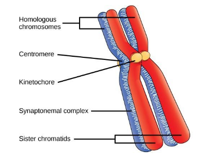
A chromosome diagram usually shows these major parts:
- Chromatids: Two identical halves of a chromosome, visible during cell division.
- Centromere: The constricted region joining the two chromatids and dividing the chromosome into two arms.
- Arms (p and q): The shorter arm is called 'p', and the longer arm is 'q'.
- Telomere: The terminal end portions of the chromosome protecting it from fusion with other chromosomes.
- Kinetochore: A disc-shaped protein on the centromere where spindle fibers attach during cell division.
- Satellite: Sometimes found at the end of a chromosome, connected by a thin filament.
Types of Chromosome Based on Centromere Position
The position of the centromere gives chromosomes their characteristic shapes. Here’s a helpful table to understand chromosome types better:
Types of Chromosome Diagram
| Type | Centromere Position | Shape |
|---|---|---|
| Metacentric | Centromere in the middle | V-shaped, arms equal length |
| Submetacentric | Centromere slightly off center | J or L-shaped, arms unequal |
| Acrocentric | Centromere close to one end | Rod-like, one very short arm |
| Telocentric | Centromere at the terminal end | I-shaped, arms on one side only |
How to Draw & Label a Chromosome Diagram
Drawing a chromosome diagram correctly is crucial for exam marks. Here’s a simple step-by-step guide:
- Draw an X-shape (for replicated chromosome) or a rod for single chromatid.
- Mark the constriction (centromere) joining the two arms.
- Label the 'p' (short) and 'q' (long) arms on either side of the centromere.
- Point out telomeres at both ends.
- Label chromatids and, if relevant, mark the position of alleles or genes.
- Add the kinetochore on the centromere, if needed for class 11/12.
Chromosome vs. Chromatin vs. DNA Diagram
Students often get confused between chromosome, chromatin, and DNA in diagrams:
| Component | Appearance | When Visible |
|---|---|---|
| Chromosome | Condensed X-shaped | During cell division |
| Chromatin | Loose, thread-like | In interphase (resting cell) |
| DNA | Double helix, molecular view | At all times (invisible with light microscope) |
Class-wise Chromosome Diagrams for Exams
Board exams for classes 10, 11, and 12 frequently require students to draw and label a chromosome diagram. Class 10 students may be asked for simpler labeling (centromere, arms), while class 11/12 might require alleles, satellite, and kinetochore. Practicing different diagrams improves accuracy and exam marks.
Common Mistakes to Avoid
- Confusing chromosome diagram with chromatin or DNA structures.
- Missing key labels such as the centromere or mislabeling arms.
- Not showing both chromatids in metaphase diagrams.
Real-World Applications
The concept of chromosome diagram is used in genetics, medicine, and biotechnology. Clear chromosome diagrams help in understanding genetic diseases (like Down’s syndrome), analyzing karyotypes, and during advanced genetics research. Vedantu helps students relate these diagrams to real class experiments and practical biology.
Practice Questions
- What are the main parts labeled in a standard chromosome diagram?
- Explain the differences between metacentric and acrocentric chromosomes with diagrams.
- How is the position of the centromere shown in a chromosome diagram?
- Draw and label a chromosome diagram showing genes and alleles.
Explore Related Concepts
- Chromosome
- Difference Between Gene and Chromosome
- Cell Structure and Function
- Nucleus
- DNA Structure
- Chromatin
- Cell Cycle
- Differences Between Mitosis and Meiosis
In this article, we explored chromosome diagram, its key processes, real-life significance, and how to solve questions based on it. To learn more and build confidence, keep practicing with Vedantu’s structured biology resources and diagrams.


FAQs on Chromosome Diagram Explained with Structure and Labels
1. What is a chromosome diagram?
A chromosome diagram is a labeled representation that shows the structure and parts of a chromosome. It typically illustrates key components such as:
- Chromatids – two identical halves of a duplicated chromosome
- Centromere – the constricted region joining sister chromatids
- Telomeres – protective end caps of the chromosome
- p arm (short arm) and q arm (long arm)
Chromosome diagrams are commonly used in biology to understand chromosome structure, cell division, and genetic organization.
2. What are the main parts of a chromosome in a diagram?
The main parts of a chromosome shown in a diagram are the sister chromatids, centromere, telomeres, and the p and q arms. These parts include:
- Sister chromatids – identical DNA copies formed after replication
- Centromere – attachment site for spindle fibers during cell division
- Telomeres – repetitive DNA sequences protecting chromosome ends
- p arm – short arm of the chromosome
- q arm – long arm of the chromosome
These labeled parts help students understand chromosome structure and function during mitosis and meiosis.
3. What is the function of the centromere in a chromosome diagram?
The centromere is the region of a chromosome that holds sister chromatids together and allows their separation during cell division. Its key functions include:
- Serving as the attachment site for spindle fibers
- Ensuring equal distribution of chromosomes in mitosis and meiosis
- Maintaining chromosome stability
In a chromosome diagram, the centromere appears as a constricted or pinched region.
4. What is the difference between a chromatid and a chromosome?
A chromosome is a complete DNA structure, while a chromatid is one of two identical copies of a replicated chromosome. The difference can be understood as:
- Before DNA replication: one chromosome consists of a single chromatid
- After DNA replication: one chromosome consists of two sister chromatids joined at the centromere
Chromosome diagrams often show duplicated chromosomes as X-shaped structures made of two chromatids.
5. Why are telomeres important in a chromosome diagram?
The telomeres are protective DNA sequences at the ends of chromosomes that prevent genetic damage. Their importance includes:
- Protecting chromosome ends from deterioration
- Preventing fusion with other chromosomes
- Maintaining stability during DNA replication
In a chromosome diagram, telomeres are shown at both ends of each chromatid.
6. How does a chromosome diagram help in understanding mitosis and meiosis?
A chromosome diagram helps explain how chromosomes replicate and separate during mitosis and meiosis. It shows:
- Formation of sister chromatids after DNA replication
- Attachment of spindle fibers at the centromere
- Separation of chromatids into daughter cells
This visual representation makes it easier to understand chromosome movement and genetic distribution during cell division.
7. What do the p arm and q arm represent in a chromosome diagram?
The p arm and q arm represent the short and long sections of a chromosome separated by the centromere. Specifically:
- p arm – the shorter arm (“p” stands for petite)
- q arm – the longer arm
These labels are used in genetics to describe gene locations and chromosomal abnormalities.
8. What is the structure of a chromosome made of?
A chromosome is made of DNA tightly coiled around histone proteins forming chromatin. Its structural organization includes:
- DNA double helix carrying genetic information
- Histones that package DNA into nucleosomes
- Condensed chromatin fibers forming visible chromosomes during cell division
Chromosome diagrams often represent the condensed form seen during mitosis.
9. How many chromosomes are shown in a human chromosome diagram?
A human chromosome diagram typically shows 46 chromosomes arranged in 23 pairs. These include:
- 22 pairs of autosomes
- 1 pair of sex chromosomes (XX in females, XY in males)
Such an arrangement is called a karyotype, which helps in studying genetic disorders and chromosomal variations.
10. What is the difference between chromatin and chromosome in a diagram?
The main difference is that chromatin is the uncondensed DNA-protein complex, while a chromosome is the condensed form visible during cell division. The distinction includes:
- Chromatin – loosely packed, present during interphase
- Chromosome – tightly coiled and visible during mitosis or meiosis
In diagrams, chromatin appears as thread-like material, whereas chromosomes appear as distinct rod- or X-shaped structures.


