


Download Class 11 Biology NCERT Solutions PDF
Biology Class 11 builds the foundation for important topics like cell structure, plant physiology, human physiology, and biological classification. Many students find diagrams, definitions, and long answers difficult to understand. Our Class 11 Biology NCERT solutions are created to simplify every concept with clear explanations and step by step answers. The NCERT solutions for Class 11 Biology are fully aligned with the latest Class 11 Biology CBSE syllabus for the academic year 2026-27. Each answer follows the correct exam pattern so students can learn how to write structured and scoring responses. Whether you are revising theory or preparing for tests, these solutions help you strengthen your basics and improve accuracy. Each chapter of NCERT Solutions for Class 11 Biology can be downloaded in a PDF format by clicking on the “Download PDF” button at the top of each chapter page given below. Access the Biology Class 11 NCERT PDF and study anytime to stay confident and exam ready.
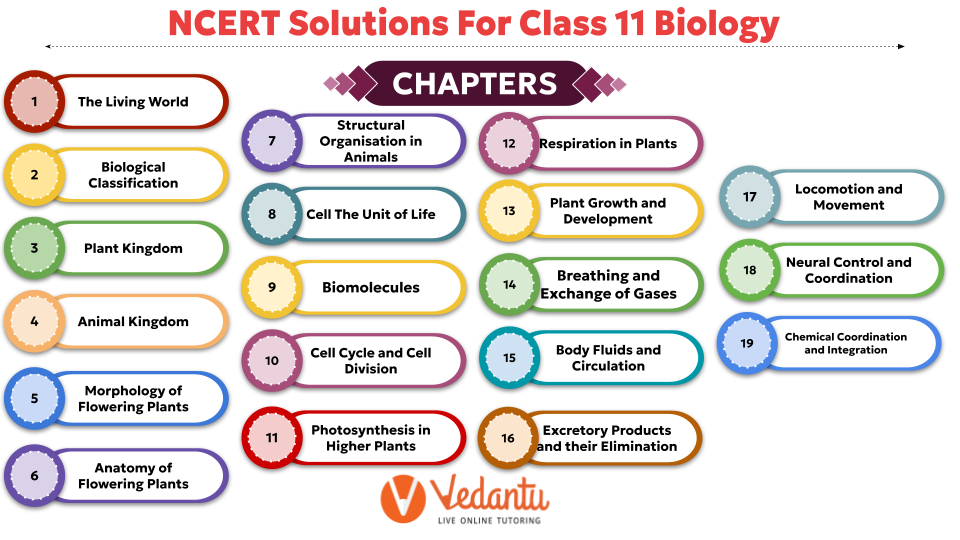
 Table of Content
The Following chapters have been removed from the NCERT Class 11 Biology textbook 2026-27.
Transport in Plants
Mineral Nutrition
Digestion and Absorption

FAQs on NCERT Solutions For Class 11 Biology - 2026-27
1. Are the NCERT Solutions for Class 11 Biology aligned with the latest CBSE 2026-27 syllabus?
Yes, the NCERT Solutions for Class 11 Biology are meticulously updated to conform to the latest CBSE curriculum for the 2026-27 academic year. This ensures every solution corresponds to the current textbook exercises and learning objectives, providing you with the most relevant and accurate study material.
2. How can I effectively use the NCERT Solutions for Class 11 Biology to prepare for my exams?
To effectively prepare, first attempt to solve the textbook exercises on your own. Then, use the NCERT Solutions to:
Verify your answers and understand the correct step-by-step methodology.
Identify any gaps in your conceptual understanding.
Learn the precise way to frame answers, especially for diagram-based and long-answer questions, to score maximum marks.
3. Are the NCERT textbook and its solutions sufficient to score well in the Class 11 Biology final exam?
The NCERT textbook is the foundation for the Class 11 Biology exam. Using the NCERT Solutions helps master all the core concepts and question types from the prescribed syllabus. While they are comprehensive, supplementing your study with practice papers and revision notes can enhance your performance and confidence.
4. Are NCERT Solutions available for all chapters in the Class 11 Biology syllabus for 2026-27?
Yes, complete and detailed NCERT Solutions are provided for all 19 chapters across the five units of the Class 11 Biology syllabus, as prescribed by CBSE for the 2026-27 session. This includes solutions for units like Diversity in the Living World, Structural Organisation in Plants and Animals, Cell Structure and Function, Plant Physiology, and Human Physiology.
5. How do the NCERT Solutions for Class 11 Biology help in clearing doubts?
The solutions provide detailed, step-by-step explanations for every question in the textbook. When you are stuck on a difficult question, the solution breaks down the complex biological concept into simpler parts. This methodical approach not only gives you the answer but also clarifies the underlying principle, effectively resolving your doubts.
6. Why is following the step-by-step method in NCERT Solutions important for Biology?
Following the step-by-step method is crucial because it helps you understand the logical flow of a biological process or concept. In exams, marks are often awarded for specific steps and keywords. This approach teaches you how to structure your answers correctly, ensuring you don't miss critical stages in explanations, such as the phases of meiosis or the mechanism of enzyme action, thereby helping you score full marks.
7. How can I use these solutions to answer questions I haven't seen before?
The NCERT Solutions don't just give answers; they teach a problem-solving approach. By regularly practising with them, you learn how to break down questions, identify the key biological principle being tested, and structure a logical answer. This skill is transferable, allowing you to confidently tackle unfamiliar or application-based (HOTS) questions in your exams.
8. How do the solutions for diagram-based questions improve understanding?
The solutions for diagram-based questions are vital as they explain not just 'what' to label but 'why' each label is significant. They connect anatomical structures to their physiological functions, for example, explaining the role of each part of the nephron in urine formation. This transforms diagrams from mere illustrations into powerful tools for understanding complex biological systems.
9. Beyond just checking answers, how can I use these solutions for self-assessment?
For effective self-assessment, compare your entire answer-writing process with the solution. Ask yourself:
Did I use the correct biological terminology?
Was my explanation as clear and concise as the solution?
Did I miss any crucial steps in my explanation of a process?
This detailed comparison helps pinpoint specific weak areas for targeted revision.