


Biology Notes for Chapter 16 Excretory Products and Their Elimination Class 11 - FREE PDF Download
Class 11 Excretory Products and Their Elimination Notes are prepared to simplify the essential processes of excretion in humans and other organisms for students. These notes focus on key topics such as the structure and functioning of the human excretory system, the formation of urine, the role of nephrons, and the regulation of kidney function. With clear explanations, diagrams, and summaries, students can easily grasp complex concepts like osmoregulation and the role of hormones in regulating excretory processes. Class 11 Biology Notes help students enhance their understanding and excel in their CBSE exams by focusing on concise content and illustrative examples.
 Table of Content
Download the FREE Excretory Products And Their Elimination Class 11 NCERT Notes PDF from Vedantu, updated according to the latest CBSE Class 11 Biology Syllabus, to improve your learning and preparation for the exam.
Section–A (1 Mark Questions)
1. Write the name of uricotelic organisms.
Ans. Birds and insects excrete nitrogenous wastes as uric acid hence are uricotelic animals.
2. What are podocytes?
Ans. The epithelial cells of Bowman’s capsule called podocytes.
3. What is the pH of urine?
Ans. Urine is slightly acidic with the pH-6.0.
4. Which is the shared terminal duct of the reproductive and urinary system in the human male?
Ans. The shared terminal duct of the reproductive and urinary system in the human male is urethra.
5. Which exact of part of nephron is directly influenced by ADH?
Ans. The exact part of the nephron that is directly influenced by ADH is the distal convoluted tubule and collecting duct.
Section–B (2 Mark Questions)
6. Refer the given figure of the nephron and label correctly the parts a to f.
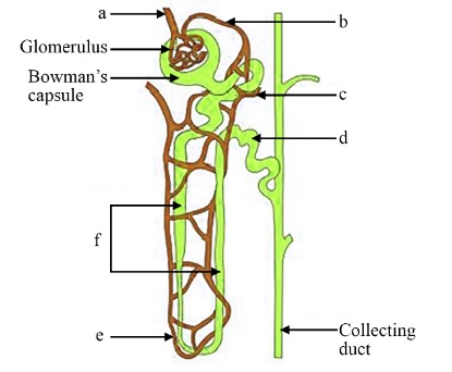
Ans. a-afferent arteriole, b-efferent arteriole, c-PCT, d-DCT, e-vasa recta, f-Henle’s loop,
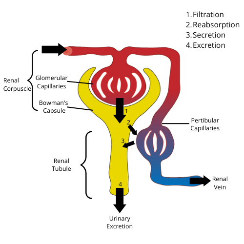
7. What is the correct sequence of processes involved in urine formation?
Ans. Three processes involved in urine formation are: glomerular filtration, reabsorption and secretion. Filtration of blood by the glomerulus is called glomerular filtration, absorption of selected materials from the filtrate into the blood of the peritubular capillaries or vasa recta is called reabsorption and excretion of additional wastes from the bloodstream into the filtrate is called secretion.
8. Complete the following:
(i) Urinary excretion = – tubular reabsorption + tubular secretion
(ii) Dialysis fluid = Plasma–
Ans. (i) Urinary excretion = Glomerular filtration – tubular reabsorption + tubular secretion
(ii) Dialysis fluid = Plasma – nitrogenous wastes
9. Write the significance of the sebaceous gland?
Ans. Sebaceous glands are involved in the removal of substances, such as squalene, cholesterol, triglycerides, wax, and esters via sebum that provides a greasy covering to the skin.
10. Where does the selective reabsorption of glomerular filtrate take place?
Ans. Selective reabsorption involves the reuptake of useful substances from the filtrate and occurs in the convoluted tubules (proximal and distal). The tubular epithelial cells in different segments of nephron perform this either by active or passive mechanisms.
11. Mention the substances that exit from the tubules in order to maintain a concentration gradient in the medullary interstitium.
Ans. The concentration gradient in medullary interstitium is established primarily by renal tubules of loop of Henle and the blood vessels surrounding them (vasa recta) in a process called counter current exchange. The substances that exit from tubules for maintenance of such gradients are mainly sodium chloride (NaCl), water and urea (containing H+, K+ and NH3).
Various harmful substances are formed in the body as a result of different metabolic reactions. It contains urea, uric acid, ammonia, carbon dioxide, water, and ions, among other things.
The three major types of nitrogenous wastes found in higher animals are Urea, ammonia, and uric acid. Ammonia is one of the most dangerous nitrogenous waste out of these.
Ammonotelic animals are those that expel ammonia as nitrogenous waste, such as most bony fishes and aquatic amphibians. Ammonia is excreted through diffusion. Ureotelic organisms are those that excrete urea as nitrogenous waste. Mammalian, amphibian, and other ureotelic organisms are examples.
Uricotelic organisms are those that excrete uric acid as nitrogenous waste, such as reptiles and birds.
Excretory Organs Are Found In A Wide Range Of Organisms
The excretory structures of different organisms differ. For excretion, amoeba and paramecium have contractile vacuoles. In sponges, the excretory system is known as the canal system. Sponges excrete through a canal system. The excretory function of coelenteron is found in Hydra. Platyhelminthes contain flame cells. Annelids, such as earthworms, contain nephridia. Prawns have excretory glands that are green in color. In insects, malpighian tubules form the excretory system.
The Excretory System Of Humans
The human excretory system consists of two kidneys, two ureters, a urinary bladder, and a urethra.
The kidneys are bean-shaped and are located in the abdominal cavity. The right kidney is slightly lower in the body than the left kidney. The inner surface of the kidney is concave, while the outer surface is convex. Hilum is a notch that can be found near the center of the inner concave surface of the kidney. The ureter, blood vessels, and nerves all enter through the hilum. The renal pelvis is a funnel-shaped space inside the hilum with projections known as calyces. The kidneys are divided into two sections: the outer and inner kidneys. The outer part of the kidney is called the cortex, and the inner part is known as medulla. The medulla is divided into medullary pyramids, which are conical masses. The column of Bertini is an extension of the renal cortex that separates the pyramids.
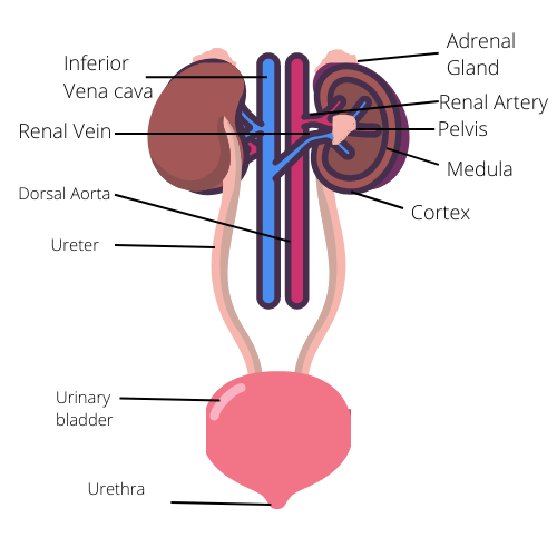
Labeled Diagram of Human Excretory System
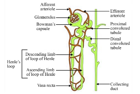
The structural and functional unit of the kidneys is the nephron. Two major parts of the nephron are the renal corpuscle and the renal tubule.
A tuft of capillaries forms glomerulus. The afferent arteriole transports blood into the glomerulus, while the afferent arteriole transports blood out of the glomerulus. Bowman's capsule is a cup-shaped structure that holds Glomerulus together. Bowman's capsule, along with the glomerulus, is referred to as the renal corpuscles or Malpighian body.
Labeled Diagram of Nephron of Human Kidney
The proximal convoluted tubule (PCT) is a highly coiled structure that is an extended tubular structure of the Malpighian body. Henle's loop is the next section of the tubule. Henle's loop is made up of two limbs, one is an ascending limb and the other one is a descending limb. The ascending limb extends into a distal convoluted tubule (DCT). The DCT then connects to the collecting duct.
There are two main types of nephrons which are - Cortical nephrons and medullary nephrons.
Cortical Nephrons: Cortical nephrons are formed when only a small portion of their loop of Henle is found in medulla because of its short length.
Medullary Nephrons: Medullary nephrons are formed when the loop of Henle becomes long and spreads into the medulla.
Urine Production
The three major steps in the production of urine are glomerular filtration, tubular reabsorption, and secretion.
Glomerular Filtration: When blood enters the glomerulus via an afferent arteriole, glomerular filtration begins. Water and nitrogenous waste enter the glomerulus and blood cells, while proteins exit through the efferent arteriole. The kidney filters approximately 1100 ml to 1200 ml of blood per minute on average. The glomerular capillary blood pressure causes blood to filter through three different layers. The first layer is the endothelium, which surrounds the glomerular blood vessels. The second layer is Bowman's capsule epithelium, with a basement membrane separating the two. Podocytes are the epithelial cells of the Bowman's capsule that are arranged in an intricate pattern to leave some minute spaces known as slit pores or filtration slits. The amount of filtrate produced by the kidneys per minute is referred to as the glomerular filtration rate.
Labeled Diagram Of Steps Involved In Formation Of Urine
Tubular reabsorption: Tubular reabsorption is the process of absorbing necessary molecules like glucose, amino acids, etc or ions like sodium ions, etc. Some substances are absorbed actively, while others are absorbed passively. Glucose and amino acids are actively absorbed, whereas water is passively absorbed.
Urine Secretion: The final step in the formation of urine is secretion. Potassium ions, hydrogen ions, and ammonia are released to maintain the ionic and acid balance of the body fluids.
Functions Of Tubules
Proximal Convoluted Tubules (PCT): Simple cuboidal brush border epithelium lines the proximal convoluted tubules (PCT). Such epithelium provides a larger surface area for reabsorption. In PCT, the majority of the electrolytes and water are reabsorbed. It contributes to the maintenance of the pH and ionic balance of body fluids through the secretion of hydrogen ions, potassium ions, and ammonium ions into the filtrate.
Henle’s Loop: Henle's loop is very helpful in maintaining the fluid's osmolarity. In the ascending limb, there is very little reabsorption. It is water-impermeable but electrolyte-permeable. The descending limb absorbs the majority of the water, concentrating the filtrate. The descending limb is nearly impermeable to all electrolytes. As a result, different parts of Henle's loop absorb differently.
Functions Of Tubules Of Nephron
Distal Convoluted Tubule (DCT): To maintain the fluid's ionic balance, the distal convoluted tubule absorbs water, sodium ions, and bicarbonate ions while excreting potassium ions and hydrogen ions.
Collecting Ducts: The collecting duct reabsorbs a large amount of water to concentrate the urine. The collecting duct also secretes hydrogen ions and potassium ions. It keeps the blood's ionic balance and pH stable.
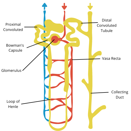
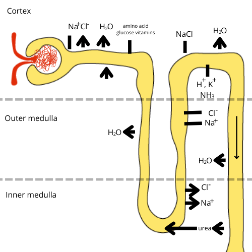
Mechanism of Concentration of Filtrate
Mammals, including humans, can produce concentrated urine to conserve water. This process mainly happens in the Henle’s loop and vasa recta within the kidneys. Here’s how it works:
Counter Current System: In Henle's loop, the flow of the filtrate (the liquid that will become urine) moves in opposite directions in its two limbs, creating a counter-current. Similarly, blood flows in opposite directions in the two limbs of the vasa recta, which is a network of capillaries around Henle's loop.
Osmolarity Gradient: This counter-current system helps create a difference in osmolarity (concentration of solutes) from the outer part (cortex) to the inner part (medulla) of the kidney. The osmolarity increases as it moves towards the medulla, going from 300 mOsmol/L in the cortex to 1200 mOsmol/L in the inner medulla. This gradient is created mostly by sodium chloride (NaCl) and urea.
NaCl and Urea Transport: Sodium chloride is transported by the ascending limb of Henle’s loop and exchanged with the descending limb of the vasa recta. Urea also enters the thin ascending limb of Henle’s loop and moves back to the kidney's medullary interstitium through the collecting tubule.
Counter Current Mechanism: This movement of substances between the loops and the vasa recta helps maintain a concentration gradient in the medullary area of the kidneys. This gradient makes it easier for water to leave the collecting tubules, leading to the concentration of the urine.
In simple terms, the counter-current mechanism allows the kidney to reabsorb water efficiently and produce urine that can be up to four times more concentrated than the original filtrate. This process is crucial for conserving water in the body.
Regulation Of Kidney Function
The hormones released by hypothalamus, juxtaglomerular apparatus (JGA), and heart are involved in kidney regulation. Any change in blood volume or ionic balance activates the body's osmoreceptors. Due to this, the hypothalamus produces an antidiuretic hormone (ADH)/ vasopressin. This aids in water reabsorption from the tubules. This increases blood volume and, via a negative feedback mechanism, turns off the osmoreceptors.
During a drop in glomerular blood pressure, the juxtaglomerular apparatus is activated. An enzyme called Renin is released by Juxtaglomerular cells that converts angiotensinogen in the blood to angiotensin I, which is then converted to angiotensin II. Because angiotensin II is a potent vasoconstrictor, it raises glomerular blood pressure. Angiotensin II also stimulates the adrenal cortex to produce aldosterone, which increases sodium ion and water reabsorption from the distal tubules. This procedure is known as the renin-angiotensin-aldosterone system (RAAS).
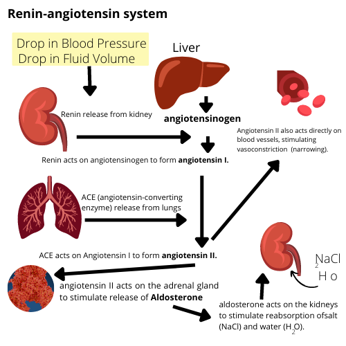
Renin-Angiotensin-Aldosterone System (RAAS) regulates kidney functions
A polypeptide hormone called atrial natriuretic factor (ANF) is produced by the heart which acts as a vasodilator and thus lowers the blood pressure. It functions as a negative feedback mechanism for the renin-angiotensin system.
Micturition: Micturition is the process of excretion or release of urine outside from the body. It is controlled by the CNS via a variety of neural mechanisms.
Other Organs in Excretion: The lungs, kidneys, liver, and skin work together to eliminate wastes such as carbon dioxide, toxic substances, urea, and so on.
Excretory System Dysfunctions:
Uremia: It is the accumulation of urea in the blood as a result of kidney failure.
Acute renal failure: It occurs when one or both the kidneys fail to filtrate the urine and are unable to function for a variety of reasons.
Renal calculi: Insoluble kidney stones caused by the accumulation of insoluble crystals such as oxalates.
Glomerulonephritis: It is an inflammation of the Glomerulus.
In cases of kidney failure, hemodialysis can be used to remove excess urea from the blood. Blood is removed from the body via cellophane tubules and dialyzed against an isotonic liquid to remove wastes before being pumped back into the body.
Students can study sitting from anywhere with the help of our notes of Ch 16 Bio Class 11, which are available to be downloaded in PDF Format. It makes learning highly flexible for students to access these notes even without an internet connection and on any device. Students who are more comfortable studying from a hard copy can print out these Class 11 Biology Ch 16 notes as per their convenience.
5 Important Topics of Biology Class 11 Chapter 16 you shouldn’t Miss!
Importance of Excretory Products and Their Elimination Class 11 Notes
Class 11 Excretory Products And Their Elimination Notes break down the complex processes involved in the human excretory system, such as urine formation and the regulation of kidney function, making them easier to understand.
Important processes like the countercurrent mechanism in Henle’s loop are explained with clear diagrams, helping visual learners grasp difficult concepts quickly.
Class 11 Biology Chapter 16 Notes PDF focuses on the key topics that are frequently asked in exams, helping students to prepare efficiently and score well.
Concise summaries and bullet points help in better retention of information, making revision faster and more effective before exams.
By focusing on both human and comparative excretion in animals, Class 11 Excretory Products And Their Elimination Notes provides an overall view of the topic, which helps in understanding the broader biological significance of excretion.
Tips for Learning the Class 11 Biology Chapter 16 Excretory Products and Their Elimination
Understand the structure and function of the kidneys, ureters, bladder, and urethra.
Focus on the three key processes of urine formation: filtration, reabsorption, and secretion.
Use diagrams to grasp the counter-current mechanism in Henle’s loop and vasa recta.
Draw and label diagrams of the nephron and kidney to reinforce learning.
Create a chart comparing excretory products (ammonia, urea, uric acid) in different animals.
Write bullet-point summaries after each study session for quick revision.
Practice NCERT and previous exam questions to assess understanding.
Revise regularly using flashcards or short notes to retain key concepts.
Conclusion
Excretory Products And Their Elimination Notes explain how the human body removes waste products to maintain balance and health. The chapter covers the structure and function of the excretory system, the process of urine formation, and how the body regulates water and salts. It also highlights the role of kidneys and different excretory products in animals. By understanding these concepts, students can better appreciate the importance of excretion in maintaining homeostasis, making it a crucial chapter for both exams and overall biological knowledge.
Related Study Materials for Class 11 Biology Chapter 16 Excretory Products And Their Elimination
Students can also download additional study materials provided by Vedantu for Biology Class 11, Chapter 16–
Revision Notes Links for Class 11 Biology
Important Study Materials for Class 11 Biology
FAQs on CBSE Notes Class 11 Biology Chapter 16 - Excretory Products and Their Elimination - 2026-27
1. What are the key points students should revise for Chapter 16 Excretory Products and Their Elimination in Class 11 Biology?
For quick revision, students should focus on:
- Structure of the human excretory system, including the kidneys, nephrons, and their parts
- Major steps of urine formation: glomerular filtration, tubular reabsorption, and tubular secretion
- Counter-current mechanism in Henle’s loop and vasa recta
- Hormonal regulation of kidney function, including roles of ADH and aldosterone
- Types and significance of excretory products (ammonia, urea, uric acid)
2. How can concept maps help in revising Excretory Products and Their Elimination for Class 11?
Concept maps help by visually linking major topics such as excretory organs, processes of urine formation, regulation, and disorders. Creating a concept map allows students to see connections between concepts, making last-minute revision more effective and helping to recall information during exams.
3. What is the best order to revise the core topics in this chapter for maximum retention?
For effective revision, follow this order:
- Start with excretory products and their types
- Study comparative excretory structures in various organisms
- Move to human excretory system structure (including labeled diagrams)
- Revise urine formation steps in detail
- Understand the counter-current mechanism
- Review hormonal and neural regulation
- Finish with common excretory system disorders and function of accessory excretory organs
4. How can bullet-point summaries aid in revising important concepts of Chapter 16?
Bullet-point summaries break down complex processes into concise steps and definitions. This format makes it easier to review, spot key terms, and remember critical facts for the CBSE exam. Use bullet-point notes especially for metabolic pathways, hormone action, and disease mechanisms.
5. What are the most common revision mistakes made by students for Excretory Products and Their Elimination?
Common mistakes include:
- Neglecting labeled diagrams of the nephron and human kidney
- Memorising without understanding the sequence of urine formation
- Overlooking the counter-current mechanism and its importance
- Confusing roles of ADH and aldosterone in regulation
- Not differentiating between ammonia, urea, and uric acid excretion in various animals
6. Why is the counter-current mechanism essential in the context of Class 11 Biology revision notes?
The counter-current mechanism in the kidney enables the body to produce concentrated urine, conserving water—an important survival adaptation. It is often tested in exams and reflects integration of structural and functional understanding, making it crucial for both quick revision and conceptual clarity.
7. How do the revision notes connect the excretory system with overall body homeostasis?
Revision notes highlight that the excretory system removes metabolic wastes and regulates water, salt, and pH balance. By controlling these factors, kidneys and accessory excretory organs help maintain homeostasis, ensuring a stable internal environment necessary for cellular functions.
8. What revision strategies are most effective for mastering the regulation of kidney function?
Effective strategies include:
- Drawing flowcharts for hormonal regulation (ADH, RAAS, ANF)
- Listing each hormone’s role in bullet form
- Solving previous years’ short-answer questions on regulatory mechanisms
- Using mnemonics for sequence of enzyme/hormone actions
9. How should students approach revising excretory system disorders for exams?
Categorise disorders (e.g., glomerulonephritis, uremia, renal calculi) and write short notes on their causes, symptoms, and treatment. Use tables or charts to summarise for rapid review before tests.
10. What types of quick self-tests can reinforce learning from revision notes in this chapter?
After revision, test yourself by:
- Labeling diagrams from memory
- Writing step-wise processes for urine formation
- Comparing types of nitrogenous wastes with examples
- Explaining the impact of a blocked ADH pathway in one or two lines