


Neural Control and Coordination Class 11 Extra Questions and Answers Free PDF Download
Very Short Answer Questions. (1 Mark)
1. How does an impulse travel across a synapse?
Ans. The impulse travels across a synapse from axons to the cell body and dendrites to the next neuron.
2. How many pairs of cranial nerves are present in a man?
Ans. 12 pairs of cranial nerves are present in man.
3. What is saltatory conduction?
Ans. Saltatory conduction refers to a type of conduction of nerve impulse by myelinated nerve fiber, wherein action potential jumps from one node of Ranvier to the other one.
4. Name the band of nerve fibers that joins the two cerebral hemispheres in mammals.
Ans. Corpus callosum is the band of nerve fibers that joins the two cerebral hemispheres in mammals.
5. What is the threshold stimulus for nerve cells?
Ans. The minimum strength of a stimulus required to start the depolarization of neurons is called threshold stimulus.
6. What is a compound eye?
Ans. In insects, the eye is composed of independent visual elements called ommatidia. These types of eyes are referred to as compound eyes.
7. What types of neurons are found in the dorsal root of the spinal nerve?
Ans. Sensory neurons are found in the dorsal root of the spinal nerve.
8. What is the basic unit of the neural system?
Ans. Neurons are the basic unit of the neural system.
9. Why is blind spot devoid of the ability for vision?
Ans. Blindspot has no photoreceptor cells – rods or cones hence it is devoid of the ability for vision.
10. Name the fluid present in the membranous labyrinth.
Ans. Endolymph is the fluid present in the membranous labyrinth.
11. Name the area of the retina where only cones are densely packed.
Ans. The fovea is the area of the retina where only cones are densely packed.
12. Name the innermost menning of the brain.
Ans. Piamater is the innermost menning of the brain.
13. To which part of the brain communication and memory are associated?
Ans: Cerebrum is the part of the brain where communication and memory are associated.
14. Name the bundle of fibers that connect two cerebral hemispheres in human beings.
Ans. The Corpus callosum is the bundle of fibers that connect two cerebral hemispheres in human beings.
15. Name the photopigment present in the rod cells.
Ans. Rhodopsin is the photopigment present in rod cells.
16. Why can impulses flow only in one direction?
Ans. Impulses flow only in one direction because each synapse allows the impulse to cross it in a single direction.
17. Where is the hypothalamus located in the brain?
Ans. Hypothalamus is located at the base of the thalamus in the brain.
Short Answer Questions (2 Marks)
1. What is a reflex?
Ans. Reflex is defined as an involuntary action that is performed by muscle under the direction of the spinal cord as a response to the stimulus. Since it is an automatic response to a stimulus hence it is not under any conscious control.
Example: Respiration, peristalsis, secretion of saliva in the mouth, etc.
2. What happens when the membrane of a nerve cell carries out a sodium pump?
Ans. When a membrane carries a sodium pump, it carries three sodium ions from
axoplasm to the cell exterior:
It transfers two potassium ions exchanged from the ECF to the cell interior.
The exterior is positively charged.
3. What are the events that take place at the point of stimulation of the axon?
Ans. The events that take place at the point of stimulation of axon are-
Membrane permeability changes; it becomes freely permeable to Na+ ions.
The rapid inflow of Na+ ions occurs and the axoplasm becomes positively charged while the exterior becomes negatively charged. This is known as the depolarized state and the potential difference across the membrane is the action potential.
The current flows through axoplasm from the depolarized region, to the next polarised region and through ECF from the polarised region to the depolarised region.
4. Give parts of the neuron.
Ans. The neuron is a microscopic structure made up of three parts:
Cell body – It contains cytoplasm with cell organelles and some granular bodies called Nissl’s granules.
Dendrites – These are the short fibers that branch repeatedly and project out of the cell body. They transmit impulses towards the cell body (cyton).
Axon – It is a long fiber with branched distal ends. Each branch terminates into a bulb-like structure known as a synaptic knob.
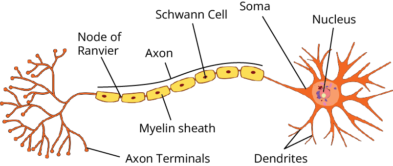
Structure of Neuron
5. Describe the role and location of the ciliary body in the human eye.
Ans. The choroid becomes thick where the cornea and the sclera meet and it is called the ciliary body.
The function of the ciliary body is that it continues in front of the lens to form an opaque structure called the iris.
6. What is the mosaic vision?
Ans. A type of vision that is found in insects due to the compound eye. In such a type of vision, a complete image of the object as seen by the compound eye is formed by a number of small lineages each of which is contributed by an ommatidium. Such an image formed by many bits of images is called a mosaic image and the vision is the mosaic image vision.
7. Where does cerebrospinal fluid occur in our body? Mention two if its function.
Ans. Cerebrospinal fluid is found in the subarachnoid space between arachnids and
The parameter of the meninges around the brain and spinal cord and also in the cavities of the brain.
Its functions are:
1) It protects the brain and spinal cord by acting as a cushion to absorb shocks.
2) It helps in removing harmful metabolites, drugs, etc. away from the brain.
8. What is the chemical and difference between rods and cones?
Ans. The difference between rods and cones are listed below:
9. Why are gray matter and white matter contained in the human nervous system named so?
Ans. Gray matter contains spindle, pyramidal, cell bodies with grayish brown appearance and hence named as gray matter.
The white matter contains innumerable myelinated axons, a large amount of myelin gives it tissue an opaque white appearance, therefore, called white matter.
10. Fill in the blanks in the different columns A to D:
Ans. The blanks are:
(a) To collect sound waves
(b) Eustachian tube
(c) Colour vision
(d) Iris
Short Answer Question (3 Marks)
1. Differentiate between dorsal spinal roots and ventral spinal roots.
Ans. The difference between dorsal spinal roots and ventral spinal roots are listed below:
2. Describe the human neural system.
Ans. The human neural system is divided into two parts:
1) Central Neural system (CNS) – It includes the brain and spinal cord and is the site of information processing and control.
2) Peripheral neural system (PNS) – PNS consists of all nerves of the body associated with the CNS. The nerve fibers of PNS are of two types i.e. afferent and efferent fibers.
(a) Afferent nerve fibers are responsible for transmitting impulses from tissues or organs to the CNS.
(b) Efferent nerve fibers are responsible for transmitting impulses from CNS to concerned peripheral tissues or organs.
PNS is further divided into –
(1) Somatic neural system which relays impulse from CNS to the skeletal muscles.
(2) Autonomic neural system transmits impulses from CNS to the involuntary organs as well as the smooth muscles of the body. It is divided into two parts -
a) Sympathetic neural system
b) Parasympathetic neural system
3. Why do giant squids have very thick nerve fiber?
Ans. The velocity of a nerve impulse in a nerve fiber depends on its myelinated and also on the thickness of the fibers. The impulses travel faster in thicker nerve fibers and as the giant squids are large-sized aquatic animals, they have thick nerve fibers.
4. Where are synaptic vesicles found? Name their chemical contents? What is the function of these contents?
Ans. Synaptic vesicles are found in the bulbous expansion called synaptic knob, at the nerve terminal. Each of the synaptic vesicles contains as many as 10,000 molecules of a neurotransmitter substance which is responsible for the transmission of nerve impulses across the synapse.
When a wave of depolarization reaches the presynaptic membrane, the voltage-gated calcium channels concentrated at the synapse open, and the Ca++ ions diffuse into a terminal from the surrounding fluid.
The Ca++ ions stimulate synaptic vesicles to move to the terminal membrane, fuse with it and then rupture by exocytosis into the cleft.
This neurotransmitter diffuses across the synapse and stimulates the membrane of the next neuron.
5. Give the location and function in the human eye, of the following –
(i) Cornea (ii) Iris (iii) Vitreous humor
Ans. 1) Cornea is the dome-shaped part of the sclera which is transparent and curved. It refracts light towards the retina.
2) Iris is the colored (pigmented) at the front and formed by choroid. It encloses the pupil and the iris contains ciliary muscles which regulate the size of the pupil and controls the amount of light.
3) Vitreous humor is present in the posterior chamber of the eye. It helps in shaping the eye and also supports the retina and the lens. It also refracts light rays.
6. Why are nerve impulses conducted more rapidly in myelinated nerve fiber than in a non – myelinated one? Explain.
Ans. In a myelinated nerve fiber, lipid-rich myelin acts as an insulator, and depolarization occurs in nodes of Ranvier wherein the myelin sheath is absent. Since the action potential jumps from one node to another, conduction becomes faster and such a type of conduction is called saltatory conduction.
In a non–myelinated fiber, this depolarization occurs all along its length thus slowing down the conduction.
7. Observe the diagram given right and answer the following questions:
(i) Label the parts A and B
(ii) Give the function of C and D.
(iii) Name the layers which wrap this organ.
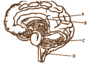
Parts of Brain
Ans. The different parts of the given figure are:
(i) A: Cerebrum; B: Corpus callosum
(ii) C: Balancing of body and maintain posture; D: Vomiting, coughing, breathing, salivation, or any other correct answer (any one).
(iii) Piameter, arachnoid and duramater.
Long Answer Questions (5 Marks)
1. Draw a labeled diagram to show the structural view of the human ear in the sectional view.
Ans. The diagram to show the structural view of the human ear in the sectional view is below:
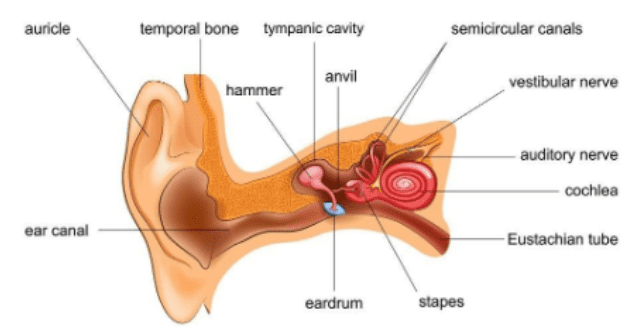
Anatomy of Ear
2. What is meant by the resting membrane potential of a neuron? How do ion channels and sodium-potassium pumps contribute to the resting potential?
Ans. The resting membrane potential is the electrical potential difference across the membranes of a resting neuron is called resting membrane potential.
The membrane is polarized with a negative interior and a positively charged exterior.
The permeability of the membrane to K+ ions is greater than its permeability to the Na+ ions.
Negatively charged protein molecules can cross the membrane.
The sodium pump transports three Na+ ions to the exterior, while in exchange only 2K+ ions come inside.
Hence, the surface carries a positive charge, which the interior negatively charged.
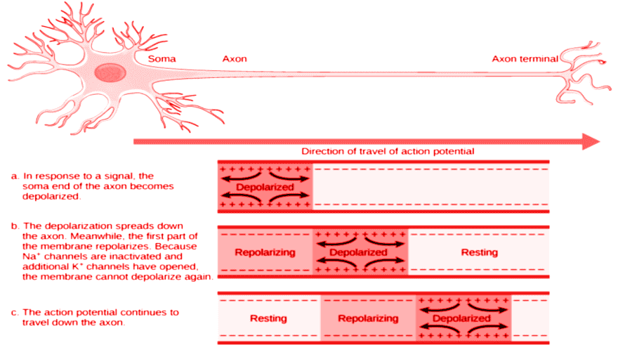
Transmission of Electrical Impluse Thorugh Neuron
3. Reflex arc. Taking one example, describe the functioning of the various components of a spinal.
Ans. A reflex arc is the specific neural pathway from stimulus to reflex.
The components of reflex arc are-
(1) Receptors– These are the organs/tissues which receive stimulus and send it as an impulse.
(2) Sensory or afferent nerves – These are neurons that conduct the impulse from the receptor to the central Nervous system (spinal cord)
(3) Relay or intermediate neurons – These are neurons that conduct impulses from the afferent neurons to the efferent neurons.
(4) Effectors/motor neurons – These neurons conduct impulses from the spinal cord/relay neurons to the effectors’ organ concerned.
(5) Effectors – It is the organ/tissue or gland that functions accordingly.
Related Study Materials for Class 11 Biology Chapter 18
CBSE Class 11 Biology Chapter-wise Important Questions
CBSE Class 11 Biology Chapter-wise Important Questions and Answers cover topics from all other chapters, helping students prepare thoroughly by focusing on key topics for easier revision.
Additional Study Materials for Class 11 Biology

FAQs on Important Questions For Class 11 Biology Chapter 18 Neural Control and Coordination - 2026-27
1. What are the most important topics to prepare from the CBSE Class 11 Biology Chapter 18 – Neural Control and Coordination for the 2026-27 exam?
- Structure and function of neurons
- Transmission of nerve impulses (including saltatory conduction)
- Reflex action and reflex arc
- Central and peripheral nervous systems
- Autonomic nervous system—sympathetic vs parasympathetic
- Sensory organs (eye and ear anatomy, mechanism of vision and hearing)
- Human brain regions and associated functions
- Differences between rods and cones
2. Which types of questions are frequently asked in CBSE Class 11 Biology exams from Neural Control and Coordination?
- Short answer questions on neural structure and synapse function
- Diagram-based questions (neuron, reflex arc, human eye/ear)
- Differences (e.g., between CNS and PNS, rods and cones, dorsal and ventral roots)
- Mechanism-focused HOTS (e.g., conduction of impulse, reflex action steps, saltatory conduction)
- Application: identifying neural defects, predicting outcomes of nerve damage
3. What is saltatory conduction, and why is it advantageous in myelinated nerve fibres? (CBSE 2026–27, 3-mark)
- Saltatory conduction is the process where action potentials jump from one node of Ranvier to the next in a myelinated nerve fibre.
- This sharply increases the speed of impulse transmission by skipping insulated segments.
- It is energy efficient and ensures rapid responses in higher organisms.
4. How does the structure of the human eye support the mechanism of vision? (CBSE Conceptual FUQ)
- The cornea and lens refract light onto the retina for focused images.
- The iris controls light entry for optimal vision in varying brightness.
- Photoreceptor cells (rods and cones) in the retina transform light into nerve impulses.
- Nerve signals then travel via the optic nerve to the visual cortex for processing.
5. Compare the structures and functions of the central and peripheral nervous systems. Why is this distinction important?
- Central nervous system (CNS): Made up of the brain and spinal cord; processes, interprets, and stores information; coordinates all body functions.
- Peripheral nervous system (PNS): All nerves outside CNS; relays messages to and from CNS; includes somatic and autonomic systems.
- This distinction is vital because CNS damage leads to loss of control/coordination, while PNS injury affects sensory/motor pathways.
6. What is a reflex arc and how does it ensure a fast response? (CBSE 2026–27, HOTS/FUQ)
- A reflex arc is a neural pathway that mediates an automatic response to a stimulus.
- It involves a receptor, sensory neuron, relay neuron, motor neuron, and effector.
- By bypassing the brain for simple decisions, reflex arcs enable immediate protective actions, such as withdrawing from a hot object.
7. Why are impulses conducted only in one direction at synapses? (Common conceptual FUQ)
- At a synapse, only the presynaptic neuron contains neurotransmitter vesicles and releases chemical signals into the synaptic cleft.
- Postsynaptic neurons possess receptors for these chemicals.
- This chemical arrangement ensures impulses propagate in one direction only.
8. List common misconceptions students have about the autonomic nervous system.
- Confusing voluntary and involuntary control: Autonomic nervous system (ANS) regulates involuntary actions only.
- Assuming sympathetic and parasympathetic always work oppositely—They often do, but can sometimes both be active (e.g., sexual response).
- Mistaking all automatic responses as ANS-mediated: Some reflexes are mediated via the CNS only.
9. How do rods and cones function differently in the human retina? (Expected 2-mark)
- Rods are sensitive to dim light and are responsible for black-and-white vision; they contain rhodopsin pigment.
- Cones function in bright light and enable color vision; they contain iodopsin pigment.
- This distinction allows humans to see in both daylight and low-light conditions.
10. What are the roles of cerebrospinal fluid (CSF) in the nervous system? (Frequently asked, CBSE 2026–27)
- Protects the brain and spinal cord by absorbing shocks
- Provides nutrients to nervous tissue
- Removes metabolic wastes and toxins
11. Explain the marking scheme and typical question distribution for Chapter 18 in the CBSE Class 11 Biology Board Exam 2026–27.
- Chapter 18 usually contributes 2 to 5 marks through a mix of 1-mark very short, 2/3-mark short, and 5-mark long questions.
- High-weightage topics: neural pathways, reflex mechanisms, sensory organ functions.
- Unit 5 (Human Physiology) as a whole accounts for ~18 marks, so mastering this chapter boosts your overall score.
12. How does damage to the corpus callosum affect body coordination? (Conceptual FUQ)
- The corpus callosum connects the two cerebral hemispheres.
- Damage impairs communication between right and left brain sides, affecting motor coordination, problem-solving, and sensory integration.
13. What events occur during a nerve impulse transmission at the axon terminal? (Expected HOTS, CBSE 2026–27)
- Arrival of action potential at axon terminal opens voltage-gated calcium channels.
- Calcium influx triggers synaptic vesicle fusion with the membrane.
- Neurotransmitter is released into the synaptic cleft, binding receptors on the next neuron.
- This generates a new action potential in the postsynaptic neuron (one-way flow).
14. How can students avoid common errors in diagram-based questions from Neural Control and Coordination?
- Label all parts clearly and accurately (e.g., neuron, synapse, reflex arc, eye, ear)
- Include direction of impulse flow
- Ensure anatomical proportions are reasonable and key segments present (e.g., node of Ranvier, lens, cochlea)
- Practice CBSE previous years' and sample diagrams for best results
15. Why is threshold stimulus important in neural function? (High-order expected FUQ)
- Threshold stimulus is the minimum intensity required to depolarize a neuron and initiate an action potential.
- Ensures neurons only respond to sufficiently strong signals, preventing accidental firing due to random fluctuations.