


Biology Notes for Chapter 6 Anatomy of Flowering Plants Class 11 - FREE PDF Download
Class 11 Biology Chapter 6 Notes make learning about the internal structure of flowering plants easier for students. Anatomy of Flowering Plants Class 11 notes cover important topics like plant tissues, roots, stems, and secondary growth. These notes simplify complex ideas into easy-to-understand points with clear explanations and summaries. Key facts are highlighted to help students quickly grasp and remember essential information. Examples and diagrams are provided to further enhance understanding. Class 11 Biology Notes are ideal for quick revision and exam preparation, helping students confidently understand the main concepts and perform well in exams.
 Table of Content
Download the FREE PDF for Anatomy of Flowering Plants Notes Class 11, prepared by experts at Vedantu and updated according to the latest CBSE Class 11 Biology Syllabus, to make your study sessions more effective and organised.
The Tissues
Tissues in plants vary depending on their location, which affects their structure and function. Based on structure and location, there are three types of tissue systems: the epidermal tissue system, the ground or fundamental tissue system, and the vascular or conducting tissue system.
A tissue is a collection of cells that share a common origin and function.
A plant is made up of a variety of tissues.
Meristematic and permanent tissues are the two types of tissues. This is founded on affecting the cells' ability to divide.
Meristematic tissue cells can divide, whereas permanent tissue cells do not split any farther.
Epidermal Tissue System
The epidermal tissue system forms the outermost covering of the plant body, consisting of epidermal cells, stomata, and appendages like trichomes and hairs.
The epidermis is a single-layered, compact arrangement of elongated cells with a waxy cuticle to prevent water loss (cuticle is absent in roots).
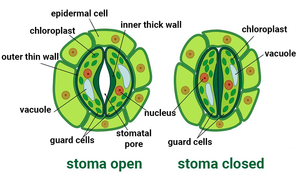
Stomata are structures on the leaf epidermis that regulate transpiration and gas exchange. They consist of two guard cells that enclose a stomatal pore.
Guard cells are bean-shaped in most plants and dumb-bell-shaped in grasses, with thickened inner walls. They contain chloroplasts to control stomatal opening and closing.
Some nearby epidermal cells, called subsidiary cells, assist the guard cells, forming the stomatal apparatus.
Root hairs are unicellular extensions of epidermal cells that aid in water and mineral absorption.
Trichomes are the multicellular hairs on the stem, which can be branched, unbranched, soft, stiff, or secretory. They help reduce water loss through transpiration.
The Ground Tissue System
The ground tissue includes all tissues except the epidermis and vascular bundles. It is made up of simple tissues like parenchyma, collenchyma, and sclerenchyma. Parenchyma cells are commonly found in the cortex, pericycle, pith, and medullary rays of primary stems and roots. In leaves, the ground tissue is made of thin-walled cells containing chloroplasts, known as mesophyll.
The Vascular Tissue System
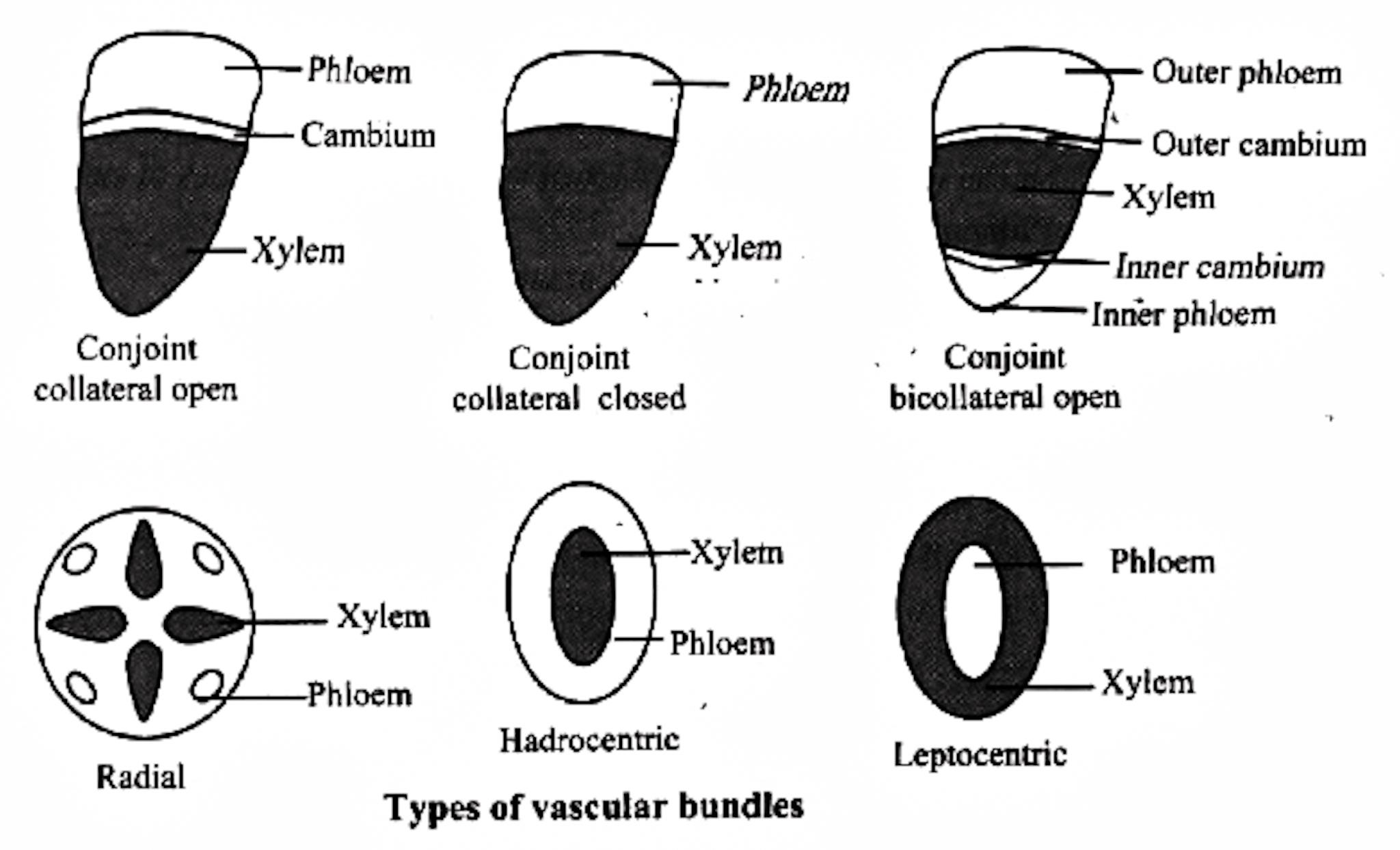
The vascular system is made up of complex tissues: xylem and phloem, which together form vascular bundles. In dicot stems, a cambium layer between the xylem and phloem allows for the formation of secondary tissues, making them open vascular bundles. In monocots, there is no cambium, so they are called closed vascular bundles. In a radial arrangement, the xylem and phloem are positioned alternately along different radii, as seen in the roots. In conjoint vascular bundles, the xylem and phloem are located along the same radius, commonly found in stems and leaves, with the phloem positioned on the outer side of the xylem.
Anatomy of Dicotyledonous and Monocotyledonous Plants
The anatomy of dicotyledonous plants includes vascular bundles arranged in a ring, with cambium present for secondary growth. In monocotyledonous plants, vascular bundles are scattered, lacking cambium, and do not exhibit secondary growth.
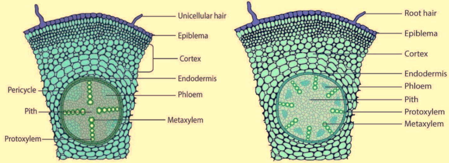
Dicotyledonous Root
The outermost layer is the epiblema, which has unicellular root hairs.
The cortex consists of thin-walled parenchyma cells with intercellular spaces.
The innermost layer of the cortex is the endodermis, made of barrel-shaped cells with Casparian strips of suberin.
Pericycle, located next to the endodermis, contains thick-walled parenchyma cells and initiates lateral roots and vascular cambium.
The pith is either small or absent.
Conjunctive tissue, made of parenchymatous cells, lies between the xylem and phloem.
There are usually 2-4 patches of xylem and phloem and a cambium ring forms between them.
The stele consists of the pericycle, vascular bundles, and pith.
Monocotyledonous Root
The anatomy of a monocot root is similar to that of a dicot root, with structures like the epidermis, cortex, endodermis, pericycle, vascular bundles, and pith.
However, monocot roots typically have more than six xylem bundles (polyarch) and a large, well-developed pith. Unlike dicot roots, monocot roots do not undergo secondary growth.
Dicotyledonous Stem
In a transverse section of a young dicotyledonous stem, the outer epidermis is covered by a thin cuticle, sometimes with trichomes and stomata. Below it, the cortex has three layers: the hypodermis of collenchymatous cells for strength, parenchymatous cells with spaces, and the innermost endodermis rich in starch (starch sheath). The pericycle has semi-lunar sclerenchymatous patches, and parenchymatous cells between vascular bundles form medullary rays. The vascular bundles, arranged in a ring, are conjoint, and open, with an endarch protoxylem. The centre is filled with parenchymatous cells, forming the pith.
Monocotyledonous Stem
The monocot stem has a sclerenchymatous hypodermis, numerous scattered vascular bundles, each enclosed by a sclerenchymatous bundle sheath, and a large parenchymatous ground tissue. The vascular bundles are conjoint and closed, with smaller bundles near the periphery and larger ones centrally. Phloem parenchyma is absent, and water-containing cavities are present within the vascular bundles.
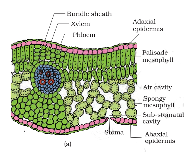
Dorsiventral(Dicotyledonous) Leaf
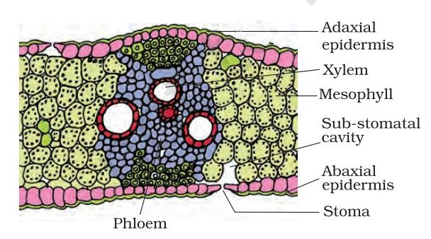
A vertical section of a dorsiventral leaf reveals three main parts: the epidermis, mesophyll, and vascular system. The epidermis, covering both sides of the leaf, has more stomata on the lower surface. The mesophyll, between the epidermal layers, contains chloroplasts and is made of palisade parenchyma (elongated cells) and spongy parenchyma (loosely arranged with air spaces). The vascular system, in the veins and midrib, has bundles surrounded by thick-walled sheath cells, varying in size.
Isobilateral (Monocotyledonous) Leaf
The anatomy of isobilateral leaves resembles dorsiventral leaves but differs in key ways. Isobilateral leaves have stomata on both surfaces and lack differentiation between palisade and spongy parenchyma. In grasses, bulliform cells on the adaxial surface help regulate water loss by causing the leaf to curl when flaccid and spread when turgid. Monocot leaves show parallel venation with similarly sized vascular bundles.
5 Important Topics of Class 11 Chapter 6 You Shouldn’t Miss!
Importance of Anatomy of Flowering Plants Class 11 Notes
Helps in understanding the internal structure of plants, which is important for studying plant biology in detail.
Simplifies complex topics, making it easier for students to understand and remember key concepts about plant tissues and their functions.
Provides a clear understanding of the different tissues and cells involved in the growth and development of plants.
Includes useful diagrams and explanations, which make it easier to visualise and understand plant anatomy.
Improves exam preparation by organising important information in a simple format, helping students do better in assessments.
Lays a strong foundation for future studies in botany and related fields by covering fundamental concepts.
Tips for Learning the Class 11 Biology Chapter 6 Anatomy of Flowering Plants
Divide the chapter into smaller sections, such as plant tissues, root anatomy, and stem anatomy, and focus on each section separately.
Study diagrams of plant tissues and structures to better understand their functions. Drawing these diagrams yourself can also help reinforce what you've learned.
Create flashcards for key terms like xylem, phloem, cambium, and others to help with quick revision.
Write short summaries of each section in your own words to make sure you understand the concepts clearly.
Solve questions related to the anatomy of plants to test your understanding and get familiar with exam-style questions.
Conclusion
The Anatomy of Flowering Plants Class 11 Notes make it easier to understand important ideas like plant tissues, their structure, and how they function. These notes break down difficult topics into simple points, and the use of helpful diagrams makes learning and remembering details easier. Going over these notes regularly will improve your understanding and help you perform better in exams. The notes not only simplify the content but also guide you through challenging topics. By using these notes consistently, you can strengthen your knowledge of plant anatomy and perform well in your studies.
Related Study Materials for Class 11 Biology Chapter 6 Anatomy of Flowering Plants
Students can also download additional study materials provided by Vedantu for Biology Class 11, Chapter 6–
Chapter-wise Class 11 Biology Notes PDF Download
Related Study Materials Links for Class 11 Biology
FAQs on Anatomy of Flowering Plants Class 11 Biology Chapter 6 CBSE Notes - 2026-27
1. How can I use these notes for a quick revision of the Anatomy of Flowering Plants chapter?
These notes are structured for efficient revision. They condense key concepts into easy-to-digest points, diagrams, and summaries. For a fast review, focus on the highlighted terms and flowcharts to quickly recall the main topics before an exam.
2. What are the most important concepts to focus on in these revision notes for Class 11 Biology, Chapter 6?
When using these notes for revision, you should pay special attention to the following key areas:
- The structure and types of meristematic and permanent tissues.
- Key differences between dicot and monocot anatomy (root, stem, and leaf).
- The complete process of secondary growth.
- The components and functions of the vascular bundles, specifically xylem and phloem.
3. How do these notes help in remembering the differences between dicot and monocot anatomy?
The notes provide clear, side-by-side comparisons, often using tables and labelled diagrams. This format helps you quickly see and remember the distinct features of dicot and monocot roots, stems, and leaves, which is a frequently asked topic in exams.
4. Is the topic of secondary growth simplified in these revision notes?
Yes, complex topics like secondary growth are broken down into simple, sequential steps. The notes clearly explain the roles of the vascular cambium and cork cambium, making it easier to understand how a plant stem increases in girth.
5. Why is it helpful to revise the different tissue systems together?
Revising the three tissue systems (epidermal, ground, and vascular) provides a complete framework for the chapter. It helps you understand how various tissues are organised and function together in a plant, making your revision more logical and less about memorising isolated facts.
6. How can I use these notes to avoid confusing xylem and phloem during revision?
The notes clearly list the distinct components and functions of each tissue. To avoid confusion, focus on the summary points that compare xylem (transports water and minerals) with phloem (transports food). This direct comparison is an effective revision technique.
7. How do the diagrams in these notes aid in exam preparation?
The diagrams offer a visual summary of complex structures, such as the cross-section of a root or a leaf. Revising with these diagrams enhances memory retention and is essential for answering questions that require drawing or labelling, which often carry significant marks.
8. Are these Class 11 Biology revision notes updated for the current academic session?
Yes, these notes are fully aligned with the CBSE syllabus for the 2026-27 academic year. They cover all the necessary topics from the Anatomy of Flowering Plants chapter, ensuring your revision is complete and relevant to your exams.