


Biology Notes for Chapter 7 Structural Organisation in Animals Class 11 - FREE PDF Download
Multicellular organisms' cells are organised into tissues to perform various functions.
Tissue: A collection of similar cells and intercellular substances that work together to perform a specific function.
All complex animals are made up of just four basic tissue types.
Organ: A collection of similar and dissimilar tissues in a living organism that have been organised and adapted to perform a common specific function, such as the heart, lung, kidney, or stomach.
Organ System: A collection of organs that work together to perform a single function. Each has a specific function in the body and is made up of specific tissues.
Organ and Organ System
The body's basic tissue types organise in various ways to form organs. In multicellular organisms, a group of such organs will then associate with one another to form organ systems.
The organisation of the body into tissues, organs, and organ systems is necessary for the body to function more efficiently. It also aids in the better coordination of the activities of the millions of cells that comprise an organism. Every organ in our bodies is made up of one or more types of tissues. For example, our heart is made up of all four types of tissues: epithelial, connective, muscular, and neural.
The complexity of organs and organ systems exhibits a discernible trend, which is referred to as an evolutionary trend.
Frog
Frogs are a type of amphibious vertebrate. • They are members of the Phylum Chordata's class Amphibia and can live on both land and in freshwater.
Rana tigrina is the most common Indian species.
They are cold-blooded, also known as poikilotherms.
Poikilotherms: These are organisms whose body temperature changes in response to their surroundings. As a result, they do not maintain a constant body temperature.
Camouflage: The ability to change the colour of their skin in order to blend in with their surroundings and hide from their enemies.
Mimicry: An organism's protective coloration that blends with the environment and camouflages it from the enemy.
Aestivation: An organism's dormant state during the summer. This assists the organism in surviving the summer's extreme heat. Summer sleep is another name for it. At this stage, the organism's metabolic and physiological activities are at their lowest.
Hibernation: An organism's dormant state during the winter. This assists the organism in surviving the bitter cold of winter. Winter sleep is another name for it. At this stage, the organism's metabolic and physiological activities are minimal.
Morphology:
Because of the presence of mucus, the skin is smooth and slippery. This aids in keeping the skin moist at all times.
The dorsal side of the body is olive green with dark irregular spots. The ventral side is a light yellow colour.
The frog does not consume water. Instead, it absorbs it through the skin.
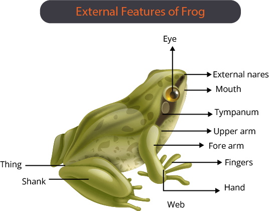
A frog's body is divided into two parts: the head and the trunk.
There is no neck or tail.
Above the mouth, there is a pair of nostrils.
The eyes are dilated and covered by a nictitating membrane.
Nictitating membrane: In birds, reptiles, and some mammals, it is a whitish or translucent membrane that forms an inner eyelid.
Tympanum: A membranous structure that represents the ear on either side of the eye. It is capable of receiving sound signals.
The forelimbs and hindlimbs aid in walking, swimming, leaping, and burrowing.
The hind limbs are larger and more muscular than the forelimbs. They all end in five digits.
Forelimbs: Smaller and less muscular than hind limbs. They all have four digits at the end.
Webbed digits on the feet aid in swimming.
Frogs have two sexes.
Male frogs are distinguished by the presence of sound-producing vocal sacs and a copulatory pad on the first forelimb digit.
Anatomy
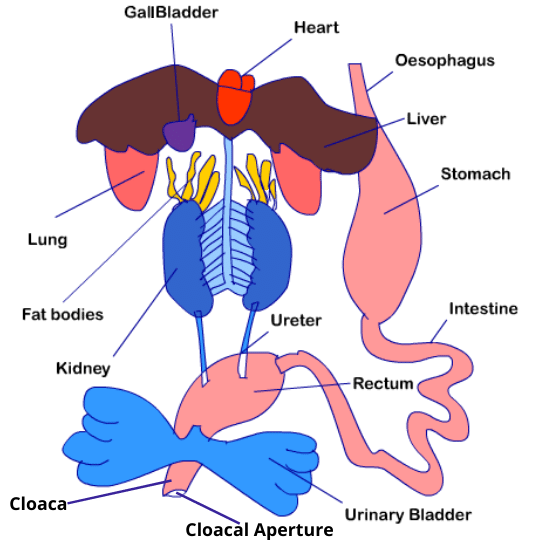
Digestive System:
It is made up of an alimentary canal as well as digestive glands.
Because frogs are carnivores, the alimentary canal is short, and the intestine is shorter.
The mouth opens into the buccal cavity, which leads to the oesophagus via the pharynx.
The oesophagus is a narrow tube. It connects to the stomach. The stomach is followed by the intestine. The intestine opens into the rectum, which then opens into the outside world via the cloaca.
Cloaca: a common chamber at the digestive tract's end. It is used in vertebrates (except most mammals) and certain invertebrates to release excretory and genital products.
Pancreas: A digestive gland that produces pancreatic juice containing digestive enzymes.
Bilobed tongue: It aids in food capture.
Stomach: This organ secretes gastric juice and HCl. This aids in the digestion of food.
Chyme: Food that has been partially digested and is formed in the stomach.
Chyme travels from the stomach to the duodenum.
The duodenum is the first section of the intestine. Through a common bile duct, it receives bile from the gallbladder and pancreatic juice from the pancreas. Pancreatic juice digests proteins and carbohydrates while bile emulsifies fats.
The intestine is where the final digestion takes place.
The numerous villi and microvilli absorb digested food.
Microvilli: These are the numerous finger-like projections that protrude from the intestine's inner wall. They broaden the surface area available for absorption.
Undigested solid waste enters the rectum and exits through the cloaca.
Respiration in Water:
In water, they exhibit cutaneous respiration, in which the skin functions as an aquatic respiratory organ.
Diffusion exchanges dissolved oxygen in water through the skin.
Respiration on Land:
They exhibit pulmonary respiration.
Respiratory organs include the buccal cavity, skin, and lungs.
The lungs are a pair of elongated, pink sac-like structures located in the upper part of the trunk (thorax).
The nostrils allow air into the buccal cavity. It then travels to the lungs.
Respiration occurs through the skin during aestivation and hibernation.
Circulatory System:
It is well developed, with a closed type circulatory system and a lymphatic system.
The blood vascular system consists of the heart, blood vessels, and blood itself.
Lymph, lymph channels, and lymph nodes comprise the lymphatic system.
The heart is a muscular structure located in the upper cavity of the body. It is made up of three chambers: two atria and one ventricle. The pericardium is a membrane that surrounds the heart.
The right atrium is joined by a triangular structure known as the sinus venosus.
The vena cava, or major veins, supply blood to the heart.
The ventricle gives way to the conus arteriosus. It is a sac-like structure found on the heart's ventral side.
The arteries (arterial system) transport blood from the heart to all parts of the body. The veins (venous system) collect blood from various parts of the body and transport it to the heart.
Hepatic portal system: A venous connection between the liver and the intestine.
Renal portal system: A specialised venous connection that connects the kidney to the lower parts of the body.
Blood: Blood is made up of plasma and cells. RBC (red blood cells) or erythrocytes, WBC (white blood cells) or leucocytes, and platelets are examples of blood cells.
RBCs are nucleated and contain the pigment haemoglobin, which is red in colour.
Lymph is not the same as blood. It is devoid of RBCs and contains few proteins.
During circulation, the blood transports nutrients, gases, and water to their respective sites.
Blood circulation is caused by the pumping action of the muscular heart.
Excretory System:
It is the organ system in charge of eliminating nitrogenous wastes from the body.
It is made up of two kidneys, ureters, a cloaca, and a urinary bladder.
Kidneys are red, bean-like structures found in the back of the body on either side of the vertebral column.
The nephron is the kidney's structural and functional unit. There are numerous nephrons or uriniferous tubules in each kidney.
Ureters are tubular structures that develop from the kidney. They are urogenital ducts that lead to the cloaca.
In females, the oviduct and ureters open independently into the cloaca.
Urinary bladder: It has thin walls and is located ventral to the rectum. The rectum connects to the cloaca as well.
Ureotelic: Animals that excrete nitrogenous waste as urea. A ureotelic animal is a frog.
Control and Co-ordination:
In frogs, it is highly evolved, and it includes both the neural system and the endocrine glands.
Endocrine glands: Secretory tissues that release various chemicals known as hormones, which cause chemical coordination in animals. Pituitary, thyroid, parathyroid, thymus, pineal body, pancreatic islets, adrenals, and gonads are the most important endocrine glands in frogs.
The nervous system is divided into three parts: the central nervous system, the peripheral nervous system, and the autonomic nervous system.
The brain gives rise to ten pairs of cranial nerves.
The brain is protected by a bony structure known as the cranium or brain box.
The brain is divided into three sections: the forebrain, the midbrain, and the hindbrain.
The forebrain is made up of the olfactory lobes, paired cerebral hemispheres, and an unpaired diencephalon.
The midbrain is distinguished by a pair of optic lobes.
The cerebellum and the medulla oblongata make up the hindbrain. The medulla oblongata exits the foramen magnum and enters the spinal cord. The vertebral column protects the spinal cord.
Organs of sense: sensory papillae
Organs of taste: taste buds
Organs of smell: nasal epithelium
Organs of vision: eyes
Hearing organs: tympanum
Internal ears and eyes are well-organised structures.
The rest (sensory papillae, taste buds, nasal epithelium) are cellular aggregations that form around nerve endings.
Eyes: A pair of structures located in the orbit of the skull. They are basic eyes with a single unit.
Ears: There are no external ears. Externally, the tympanum can be seen. It is the hearing organ as well as the organ of balance or equilibrium.
Reproduction:
They have well-organised reproductive systems for both males and females.
The male reproductive system consists of a pair of yellowish ovoid testes.
They attach to the upper part of the kidney via the mesorchium, a double fold of the peritoneum.
The serous membrane lining the cavity of the abdomen and covering the abdominal organs is known as the peritoneum.
Mesorchium: The peritoneum fold that connects the testis to the upper part of the kidney.
Vasa efferentia: They are 10-12 in number and originate in the testes. They enter the kidneys from the side and exit through Bidder's canal.
Bidder's canal: Bidder's canal is found inside the kidney of a frog. It receives sperm from the testes via several vasa efferentia. It connects to the urinogenital duct and enters the cloaca.
Cloaca: A small median chamber that transports faeces, urine, and sperms to the outside.
Female Reproductive System: It is made up of two ovaries located near the kidneys.
The ovaries and the kidneys have no functional relationship.
Oviduct: A tube that connects the ovary and the cloaca. It opens independently into the cloaca.
A mature female can lay between 2500 and 3000 eggs at a time.
Fertilisation is external and takes place in water.
The frog life cycle exhibits complete metamorphosis, which includes a larval stage.
Tadpole: A frog's larval stage. After metamorphosis, it grows into an adult.
Frogs are beneficial to humans because they eat insects and protect crops. Frogs maintain ecological balance in the ecosystem because they serve as an important link in the food chain and food web.
Man eats the muscular legs of frogs in some countries.
Important Points to Remember
Cells of multicellular organisms
Tissue, Organ and Organ System
Biology Class 11 chapter 7 Notes will also include information about neural tissue, organ and organ system.
Frog, its morphology, anatomy, respiration in water, respiration on the land digestive system, circulatory system, excretory system, control and coordination, reproduction including the male reproductive system and female reproductive system. Students will also learn about tadpoles and the importance of frogs.
Structural Organisation in Animals Basic Subjective Questions - Class 11 Revision Notes
Section–A (1 Mark Questions)
1. State the number of segments in earthworms which are covered by a prominent dark band or clitellum.
Ans. Three segments of segments of earthworm are covered with clitellum. They are 14th, 15th and 16th segment.
2. Where are sclerites present in cockroach?
Ans. Sclerites are the chitinous plates that forms the exoskeleton of cockroach. Sclerites are present in all body segment.
3. A muscle fibre tapers at both ends and does not show striations. Name the muscle fibre.
Ans. Smooth muscles are the muscle fibres that tapers at both the ends and does not show striations.
4. What is the difference between cutaneous and pulmonary respiration?
Ans. Cutaneous respiration takes place through skin and pulmonary respiration takes place through lungs.
5. Name any two cell junctions found in tissues.
Ans. Gap junction and adhering junction.
Section–B (2 Mark Questions)
6. What are the cellular components of blood?
Ans. The cellular components of blood are: Red blood cells, white blood cells and platelets.
7. Write a short note on cardiac muscles.
Ans. Cardiac muscles are the muscles which are present only in heart. They are faintly striated muscles. They are branched. They have centrally placed nucleus. They have intercalated disc. These muscles are involuntary.
8. Where do you find Malpighian tubules?
Ans. In insects, at the junction of midgut and hindgut is present another ring of 100-150 yellow-coloured thin filamentous Malpighian tubules. They help in removal of excretory products from haemolymph.
9. Stratified epithelial cells have limited role in secretion. Justify their role in our skin.
Ans. Stratified epithelial cells are present where the outermost cell becomes hard and dead. They play a major role in protection but they have limited role in secretion. At some locations, the stratified epithelium has keratin on the outermost layer which makes the layer waterproof.
10. Why are neurons called excitable cells? Mention special features of the membrane of the neuron?
Ans. Membranes of neurons are in the polarized state. Different ions interact with membrane to change the polarization and thus neurons become excited.
The ability to become polarized or depolarized is a special feature necessary for the transmission of nerve impulse.
11. Name the different types of nephridia present in earthworms on the basis of location? What is the function of nephridia?
Ans. There are three different types of nephridia in earthworms. They are:
Pharyngeal nephridia
Septal nephridia
Integumentary nephridia
Nephridia play an important role in excretion in earthworms.
5 Important Topics of Class 11 Chapter 7 You Shouldn’t Miss!
Importance of Structural Organisation in Animals Class 11 Notes
The Class 11 Biology notes on Structural Organisation in Animals are crucial for grasping how different animal tissues and organ systems work.
They simplify complex concepts, breaking down the functions and structures of various tissues and organs.
With clear explanations, diagrams, and key facts, these notes make it easier to understand and remember essential details.
They are ideal for quick revision and exam preparation, helping students confidently tackle questions on animal anatomy and physiology.
Tips for Learning the Class 11 Biology Chapter 7 Structural Organisation in Animals
Focus on the main tissue types and organ systems in animals, including their functions and structures.
Study diagrams to visualize the anatomy and organization of different tissues and organs.
Create concise notes summarizing each organ system and its components.
Regularly revisit your notes to reinforce your understanding and retention.
Test yourself with practice questions to check your grasp of the material.
Relate different tissues and organs to their functions to see how they work together in the animal body.
Conclusion
Structural Organisation in Animals Class 11 Notes make understanding key concepts like animal tissues, organs, and body systems much easier. These notes break down complex ideas into simple, easy-to-follow points and include helpful diagrams to make learning and remembering details more straightforward. Regularly reviewing these notes will strengthen your grasp of the subject and help you do well in exams. By using these notes often, you'll get a clearer picture of how animals are structured and function, which will support your study and exam preparation.
Related Study Materials for Class 11 Biology Chapter 7 Structural Organisation in Animals
Students can also download additional study materials provided by Vedantu for Biology Class 11, Chapter 7–
Chapter-wise Class 11 Biology Notes PDF Download
Related Study Materials Links for Class 11 Biology
FAQs on Structural Organisation in Animals Class 11 Biology Chapter 7 CBSE Notes - 2026-27
1. How can I use these revision notes for 'Structural Organisation in Animals' effectively before an exam?
Start by reviewing the summary for each major topic, like animal tissues and the organ systems of a frog. Use the key points and diagrams to quickly refresh your memory on the main concepts. Focus on any tables that differentiate between tissue types, as this is great for quick comparison and recall.
2. What are the main topics covered in these Class 11 Biology Chapter 7 notes?
These notes provide a concise summary of the entire chapter, focusing on the core concepts for your exams. Key areas include:
- The four primary types of animal tissues: Epithelial, Connective, Muscular, and Neural.
- The concept of organs and organ systems.
- The detailed morphology and anatomy of the frog as a representative example.
3. How do these notes help clarify the difference between the various types of connective tissues?
The notes simplify this complex topic by grouping connective tissues into clear categories: loose, dense, and specialised. They use bullet points to highlight the unique features, cell types, and functions of each type, such as bone, cartilage, blood, and adipose tissue, making them easier to compare and remember during revision.
4. While revising with these notes, why is it important to understand the frog's anatomy?
Studying the frog's anatomy is crucial because its organ systems are very similar to those of other vertebrates, including humans. These notes help you revise how the digestive, circulatory, respiratory, and nervous systems work together. Understanding this example helps build a strong foundation for more complex biology topics.
5. Do these notes include diagrams for revising the organ systems?
Yes, the notes include simplified and clearly labelled diagrams of important structures, such as the different types of epithelial tissues and the digestive system of a frog. These visuals are designed to make revision faster and help you recall key anatomical details during your exam.
6. How do these notes explain the connection between a tissue's structure and its function?
The notes explain this by providing direct examples. For instance, they show how the cube-like cells of cuboidal epithelium are ideal for secretion in glands, while the strong fibres in dense connective tissues (like tendons) are perfect for connecting muscles to bones. This helps you understand the 'why' behind the structures, not just the 'what'.
7. What is the most efficient way to revise the four primary animal tissues using these notes?
The most effective method is to use the summary tables and bullet points. First, revise the main function of each primary tissue—Epithelial for covering, Connective for support, Muscular for movement, and Neural for control. Then, review the sub-types detailed in the notes for a complete and quick overview.