


Biology Notes for Chapter 17 Locomotion And Movement Class 11 - FREE PDF Download
Section–A (1 Mark Questions)
1. What do you mean by isotropic and anisotropic bands?
Ans. The light bands contain actin filaments and is called I-band or Isotropic band, whereas the dark band called ‘A’ or Anisotropic band contains myosin filaments.
2. What is locomotion? How is locomotion different from the movement?
Ans. Locomotion is the displacement of a body from one place to another. E.g., running, walking etc. On the contrary, movement is the displacement of a body or a part of the body from its original position. E.g., movement of hand.
3. What is the significance of locomotion in animals?
Ans. In animals, locomotion plays an important role in helping them to move from one place to another. Animals move for many reasons to support their living such as food, shelter, mate and defense.
4. Name the different types of movement exhibited by the cells of the human body.
Ans. Amoeboid movement, flagellar movement, ciliary movement and muscular movement.
5. Name the movable bone of the skull.
Ans. Mandible is the only movable bone of the skull.
Section–B (2 Mark Questions)
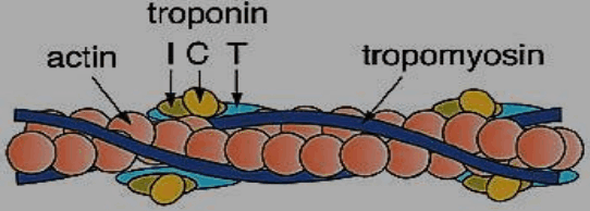
6. Label the different components of actin filament in the diagram given below.
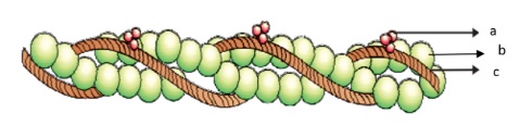
Ans. a-Troponin, b- Actin, c-Tropomyosin
7. Write a short note on gout.
Ans. Gout occurs due to a defect in purine metabolism that causes an excess of uric acid and its salts. In gout, uric acid is raided in the blood. When crystals of uric acid salts accumulate in the joints it causes gouty arthritis. The excess of urates can form stones in the kidneys. Treatment with certain drugs can increase the excretion of urates.
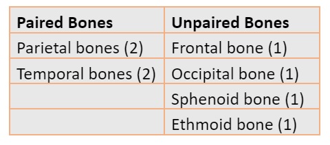
8. Name all the bones that form cranium.
Ans. Cranium is called the brain box. It is made up of eight bones. These bones are
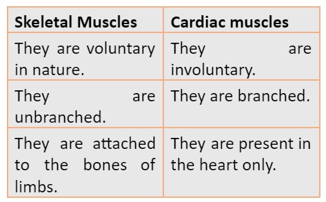
9. How are skeletal muscles different from cardiac muscles?
Ans.
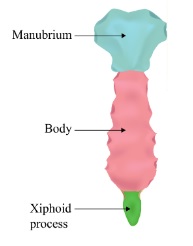
10. Draw a well labelled diagram of sternum.
Ans.
11. Write down the function of sternum.
Ans. Some of the functions of sternum are:
It protects the internal organs in the thoracic region.
It also provides the surface for muscle attachment.
The sternum helps in the respiratory mechanism.
It protects the internal organs in the thoracic region.
It also provides the surface for muscle attachment.
The sternum helps in the respiratory mechanism.
PDF Summary - Class 11 Biology Locomotion and Movement Notes (Chapter 17)
Locomotion and Movement
Movement is defined as the movement of living organisms from one place to another; if the movement causes a change in location or position, it is called locomotion; such as walking, climbing, running, etc.
Kinds of Movement
There are three kinds of movement which are ciliary, amoeboid, and muscular.
Ciliary Movement
This type of movement occurs in those organs which are covered with ciliated epithelium. It helps to capture dust particles that are inhaled during breathing and also helps to move the egg from the fallopian tube into the uterus.
Amoeboid Movement
This type of movement can be seen in some immune cells, such as macrophages and white blood cells. It can also be seen in amoeba moving through pseudopods.
Amoeboid movement in Amoeba
Muscular Movement
Muscle movement is seen in the tongue, chin, limbs, etc. The muscles, bones, and nervous system are all involved in locomotion.
Muscle
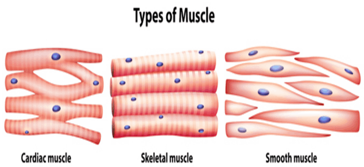
Muscle or muscle tissue is essentially mesoderm. It is an organization of cells that is involved in body movement. Skeletal muscle, smooth muscle and cardiac muscle are the three main muscle types.
Skeletal muscles are voluntary muscles that mean these muscles are under the control of our will and under the control of the somatic nervous system. They are striated muscles because of the characteristic striations present on them. These muscles are attached to the bones through tendons and are involved in keeping the body in a particular posture and performing different body movements.
Smooth muscles lack striations and are also called visceral muscles. These muscles control involuntary body movements. They are located in the walls of both the digestive tract and reproductive tract.
Cardiac muscles are the heart muscles that help the heart contract and relax rhythmically. These muscles are involuntary in nature and also have cross stripes with branching patterns.
Types of Muscles
Structure of Muscle
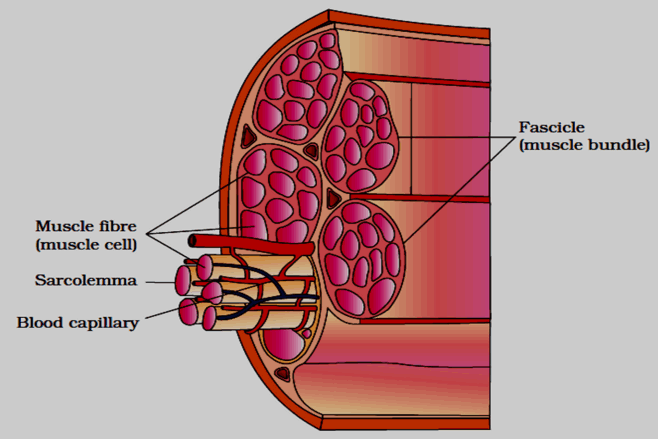
Several muscle bundles in skeletal muscle form a fascicle. Each muscle bundle is composed of several muscle fibers. The plasma membrane arranged on the muscle fibers is called sarcolemma. The muscle membrane or sarcolemma surrounds the sarcoplasm. There are several nuclei in muscle fibers which are called syncytium. The endoplasmic reticulum present in the muscle fibers is called the sarcoplasmic reticulum. The calcium ions stored in the sarcoplasmic reticulum participate in the contraction of muscles. Muscle fibers contain parallel strands called myofibrils of myofilaments. The epimysium is the fibrous tissue that is present around the skeletal muscle.
Structure of the Muscle
The characteristic stripes of skeletal muscle are due to the presence of two kinds of proteins which are actin and myosin. The light stripes are also called isotropic bands and have action. But the dark stripes are called anisotropic bands that have myosin protein. Filaments that are thin are called actin filaments, and thick filaments are called myosin filaments.
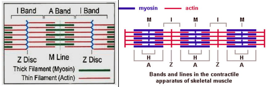
Structure of Sarcomere
There is an elastic fiber called the Z-line present in the centre of each actin stripe. The part of myofibrils between two consecutive Z-lines is called a sarcomere. The sarcomere is called the functional unit of muscle contraction.
Structure of Contractile Protein
The two main contractile proteins are actin and myosin. The monomer unit of actin is called G-actin or globular actin. The polymer of G-actin forms F-actin or F-filament. Two of the F-filaments twist around each other to form actin molecules. The protein tropomyosin surrounds the F-actin. Another protein named troponin is evenly distributed in tropomyosin.
The monomeric unit of myosin is called meromyosin. Each of the meromyosins is composed of two parts: a spherical head and a long tail. The spherical head has ATPase activity and actin-binding sites.
Structure of Contractile Protein
Muscle Contraction Mechanism
The muscle contraction is explained with the help of the sliding filament theory. During the contraction of muscles, the fine fibres slide over the thick fibres. When the signal is transmitted from the central nervous system to the motor neurons, muscle contraction begins. The neuromuscular junction is where motor neurons and muscle fibres come together. The release of neurotransmitters such as acetylcholine at the neuromuscular junction will generate action potentials in the muscle plasma membrane.
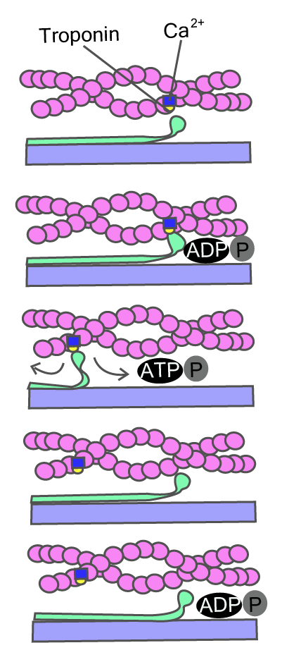
Sliding Filament Theory
The action potential causes the release of calcium ions from the sarcoplasmic reticulum into the sarcoplasm. The increase in calcium levels causes the binding of calcium ions to troponin present on actin filaments. This shows the active myosin binding sites. The ATPase activity of myosin exposes sites that allow cross-bridging between actin and myosin. It causes sarcomere shortening that leads to muscle contraction. Then the calcium ions are pumped back to the sarcoplasmic reticulum. This process hides the actin filaments by returning the muscle to its original position.
Formation of Lactic Acid in Muscles
When the muscles are reactivated, such as during exercise or running, the anaerobic breakdown of glycogen in the muscles leads to the accumulation of lactic acid in the muscles, which leads to muscle pain and fatigue.
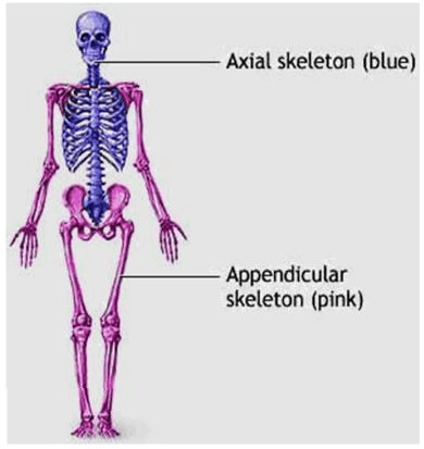
Skeletal System
The skeletal system comprises bones and cartilage. It helps the body to move. Due to the presence of calcium salts, bones are hard, and due to the presence of chondroitin sulfate, cartilage is flexible. A person consists of 206 bones and a small amount of cartilage. The skeletal system consists of two parts: the axial skeletal system and the appendicular skeletal system.
1. Axial Skeletal System
There are a total of 80 bones present in the axial skeletal system including the skull, sternum, vertebral column, and ribs.
Axial Skeletal System
Skull is composed of 22 facial and cranial bones. The cranial bones are a total of 8 in number which protects the brain. The facial area is composed of 14 bones, forming the front part of the skull. The U-shaped hyoid bone is located at the bottom of the mouth. Each middle ear is composed of three small bones: the malleus, incus, and stapes. These are collectively called ear ossicles.
The Vertical column or spine is made up of 33 vertebrae. The vertebral column extends from the skull base and makes the basic structure of the trunk. Each of the vertebrae has a hollow central part called the neural tube through which the spinal cord passes.
The first vertebra is called the atlas and is connected to the occipital condyle. The spine or vertebral column is divided into 7 cervicals, 12 thoracic, 5 lumbar, 1 sacral, and 1 coccyx beginning from the skull. The number of cervical vertebrae in mammals is preserved.
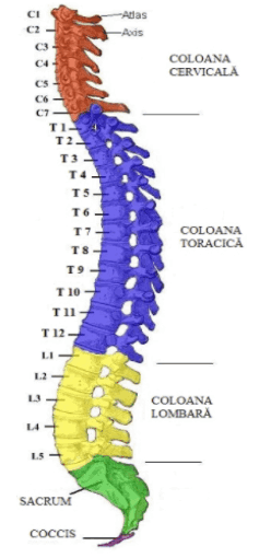
Vertebral Column
Ribs: True ribs include the first seven pairs of ribs. They are called true ribs because they are attached to the sternum directly. The eighth, ninth, and tenth ribs pairs are connected to the seventh pair of ribs instead of directly connected to the sternum. Therefore, these are called false ribs. The eleventh and twelfth pairs are the last two pairs of ribs that are not directly connected to the sternum, they are called floating ribs. The thoracic vertebrae, ribs, and sternum together make up the rib cage.
The sternum is a flat bone located at the midline of the chest. Twelve pairs of ribs are connected to the breastbone or sternum.
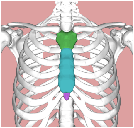
Sternum
2. Appendicular Skeletal System
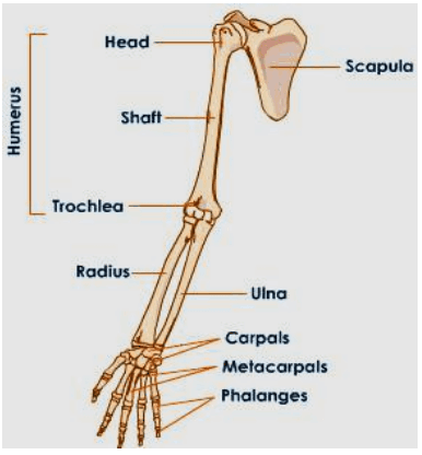
The Appendicular Skeletal System is made up of limb bones and girdles. Each limb has 30 bones.
Forelimb Bones: The bones of the front leg or arm or forelimb are the humerus, radius and ulna, wrist (8 carpal bones), and metacarpal bone (5 palm bones), and phalanges (14 digit bones).
Hindlimb Bones
Hindlimb Bones: There are several bones present in the hind leg or limb which are the femur, the thigh bone (the longest bone), tibia and fibula, and 7 tarsals (the ankle bones), 5 metatarsals, and 14 phalanges. The cup-shaped bones present on the knees are called the patella.
Hindlimb Bones
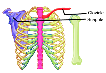
Pectoral girdle has two bones, the collarbone/clavicle, and the scapula. The scapula has a cavity called the glenoid, which forms a hinge in the form of a ball and socket joint with the humerus and is connected to the bones of the forelimbs.
Bones of Pelvic Girdle
The pelvic girdle has a cup-shaped cavity called the acetabulum, which forms a spherical connection as a ball and socket joint with the femur that is connected with the bones of the hind leg, and the thigh muscles are connected with the pelvic girdle.
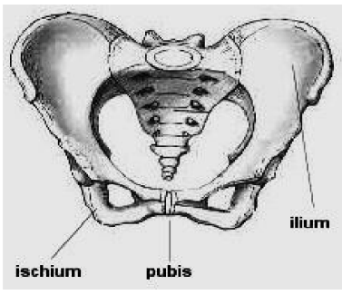
Bones of Pelvic Girdle
Joints
Joints are connections between bones or between bones and cartilage. They are important for locomotion because they act as fulcrums for the force exerted by muscles to induce movement. There exist three important types of joints:
1. Synovial Joints: There is a characteristic fluid-filled synovial cavity between the two bones, which allows more flexibility and more movement. For example-hinge joints (knee and elbow joints), ball and socket joints (hip and shoulder joints), pivot joints (neck), etc.
2. Fibrous Joints: The bones are connected by dense fibrous tissue forming sutures. They are motionless and can be seen in the joints between the flat bones of the skull.
3. Cartilaginous Joints: Cartilage exists and helps to connect two bones. Such joints are partially movable and located between the vertebrae.
Disorders Related to Muscular and Skeletal Systems
Myasthenia Gravis: This disease affects neuromuscular nodes, causing skeletal muscle fatigue, weak and paralyzed. Myasthenia gravis is an autoimmune disease.
Muscular Dystrophy: It is a genetic disease that causes progressive destruction of skeletal muscles.
Tetany: This leads to low levels of calcium ions in body fluids, which leads to rapid muscle spasms.
Arthritis: Arthritis is the inflammation of joints.
Gout: This condition is caused by the accumulation of uric acid crystals in the joints, which can cause inflammation of the joints.
Osteoporosis: The bone mass is reduced, which increases the risk of fractures. It is related to age and is usually related to decreased estrogen levels.
5 Important Topics of Biology Class 11 Chapter 17 You Shouldn’t Miss!
Importance of Class 11 Biology Chapter Locomotion And Movement Notes
Learning about movement helps set the stage for understanding more complex biology topics later on.
Locomotion And Movement Class 11 Notes PDF Download explains how our bones and muscles work, which is important for knowing how our body moves and is useful in physical activities.
Understanding how we move can help in treating injuries and doing physical therapy.
These notes make it easier to do lab work by explaining how muscles contract, how bones are structured, and how joints work.
They help with studying for exams by summarising key ideas and making it easier to remember important information.
Class 11 Locomotion And Movement Notes break down complicated ideas about muscles and bones, making them easier to understand.
Learning about movement connects to real-life situations, like improving sports performance and dealing with movement-related health issues.
Tips for Learning the Class 11 Biology Chapter 17 Locomotion and Movement
Study and draw diagrams of the skeletal and muscular systems to understand bone structures, joints, and muscle arrangements better.
Familiarise yourself with important terms like actin, myosin, and neuromuscular junction to grasp how muscles work and contract.
Connect concepts to everyday activities or sports to see how they apply in real-life scenarios, making the information more relatable and memorable.
Create summary notes or flashcards for each topic, including types of locomotion, muscle types, and joint functions, to reinforce key points.
Regularly practise labelling and drawing diagrams of muscles, bones, and joints to improve your understanding and recall.
Regularly revisit your notes and summaries to keep the information fresh in your mind and make it easier to recall during exams.
Conclusion
Locomotion And Movement Class 11 Notes provide essential insights into how organisms, especially humans, move. By understanding the structure and function of muscles, bones, and joints, students can grasp how movement is achieved. The chapter also explains various types of locomotion and the mechanisms behind muscle contraction. Understanding these concepts not only helps in exams but also connects to real-life applications, such as improving physical fitness and understanding movement-related health issues. Overall, Locomotion And Movement Short Notes offer a clear and simplified overview of complex biological processes.
Related Study Materials for Class 11 Biology Chapter 17 Locomotion And Movement
Students can also download additional study materials provided by Vedantu for Biology Class 11, Chapter 17–
Revision Notes Chapter wise Links for Class 11 Biology
Related Study Materials Links for Class 11 Biology
FAQs on CBSE Notes Class 11 Biology Chapter 17 - Locomotion And Movement - 2026-27
1. What are the key concepts summarised in the Locomotion and Movement Class 11 Revision Notes?
The Locomotion and Movement Class 11 Notes provide concise summaries of mechanisms of movement, types of muscles, human skeletal system, mechanism of muscle contraction, structure and function of joints, and common disorders such as arthritis and muscular dystrophy. These notes are structured for quick revision of all important NCERT and CBSE syllabus points.
2. How do revision notes help students prepare effectively for Class 11 Biology exams?
Revision notes are designed to help you quickly revisit essential concepts, organise your study sessions, and reinforce learning. They highlight key terms, provide chapter summaries, and include diagrams or tables for easy recall, which improves both speed and retention during exams.
3. What is the difference between isotropic and anisotropic bands as explained in the chapter notes?
Isotropic (I) bands in muscle fibers are the lighter regions containing actin filaments, while anisotropic (A) bands are the darker bands containing myosin filaments. This structural arrangement helps in understanding skeletal muscle contraction as described in the revision notes for Class 11 Biology.
4. Which types of movement are highlighted in the Class 11 Locomotion and Movement revision notes?
The main types of movement summarised are:
- Amoeboid movement (seen in white blood cells and amoeba)
- Ciliary movement (in respiratory tract and reproductive organs)
- Muscular movement (in body muscles such as limbs and tongue)
- Flagellar movement (in sperm cells)
5. How do revision notes summarise the structure of the human skeletal system?
The notes provide a summary of the axial skeleton (skull, vertebral column, ribs, sternum) and appendicular skeleton (bones of limbs and girdles), emphasising bone types, main functions, and joint classification for efficient last-minute study.
6. What is the sliding filament theory, and why is it important for quick revision?
The sliding filament theory explains the mechanism of muscle contraction: actin and myosin filaments slide past each other, powered by ATP and regulated by calcium ions. Knowing this process helps in understanding muscle movement questions frequently asked in CBSE Class 11 Biology exams.
7. Which disorders are concisely included in the Locomotion and Movement Class 11 revision notes?
The notes summarise important disorders, including:
- Arthritis – inflammation of joints
- Muscular dystrophy – progressive muscle weakness
- Osteoporosis – loss of bone density
- Gout – uric acid crystal accumulation in joints
- Myasthenia gravis – neuromuscular weakness
8. How are diagrams and labelled structures incorporated in revision notes for this chapter?
Revision notes for Class 11 Locomotion and Movement often include labelled diagrams of sarcomere, actin-myosin filaments, human skeleton, and joint types to enhance visual learning and support quick, effective recall before exams.
9. What are some effective revision tips specific to this chapter according to the notes?
Recommended revision strategies include:
- Drawing and practising diagrams of muscle structure and joints
- Making concept maps of different types of movement and muscle fibers
- Using flashcards for key terms and differences
- Regularly reviewing summaries for each topic
10. Why is it important to distinguish between movement and locomotion in Class 11 Biology revision?
Understanding the difference is essential because movement refers to any change in position of an organism or its body parts, while locomotion involves movement resulting in a change of location. This distinction is frequently tested and forms a basis for understanding higher concepts in physiology and animal behaviour.
11. In what ways do Class 11 Locomotion and Movement revision notes connect to real-life applications?
The notes relate concepts like muscle contraction and joint health to everyday activities such as sports, injury treatment, physiotherapy, and understanding movement-related disorders, making the subject relevant beyond exam preparation.
12. How should students use the Locomotion and Movement revision notes for last-minute exam revision?
Students should focus on key concepts, practice labelling diagrams, clarify differences and definitions, and quickly review highlighted points or summaries. Using the notes strategically in the final days before exams ensures better retention and confidence in attempting CBSE questions from Chapter 17.