


Class 11 Biology Chapter 6 Questions And Answers With Complete Solutions
Are you searching for NCERT solutions for class 11 biology chapter 6 anatomy of flowering plants? This chapter covers the internal structure of plants, including tissues, cells, and different plant parts. Class 11 biology chapter 6 ncert solutions help you understand complex topics like meristematic tissues, permanent tissues, and vascular bundles in simple terms.
 Table of Content
The solutions include:
- Detailed answers to all exercise questions about plant anatomy
- Clear explanations of meristematic and permanent tissues
- Step-by-step solutions for anatomy of flowering plants questions and answers class 11
- Simple diagrams and examples to understand tissue systems
Vedantu's Class 11 Biology NCERT Solutions make learning plant anatomy easy with clear explanations and proper examples. Each answer is written in simple language so students can understand every concept without confusion. Download the NCERT Solutions for class 11 biology chapter 6 PDF for free and study at your own pace!
1. State the position and role of various kinds of meristems.
Ans: Meristem refers to meristematic tissues which consist of actively dividing cells. Based on their position in the plant body, meristems are of three types. They are given below.
i. Apical Meristem: It is present at the apices of shoot and root and is accountable for the rise in length.
ii. Intercalary Meristem: It is present at the bases of leaves on top of the nodes or under the nodes and is accountable for the elongation of the organs.
iii. Lateral Meristem: It is present on the lateral side and is accountable for the rise in diameter or girth.
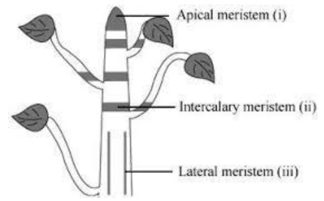
Fig.- Diagram showing different types of meristems.
2. Cork cambium forms tissues that form the cork. Do you agree with this statement? Explain.
Ans: Yes, I agree with this statement. Cork cambium (also called phellogen) cuts off cells both on its outer side and inner side. The cells cut off on the outer side form the cork (also called phellem ) and cells cut off on the inner side form the secondary cortex(also called phelloderm). The cells of the cork are dead while those of the secondary cortex is alive. Phelloderm, phellogen, and phellem are collectively known as periderm.
3. Describe the entire process of secondary development in the stems of woody angiosperms using a schematic representation. What is its importance?
Ans: The entire process is as follows:
Secondary growth refers to the formation of secondary tissues that leads to an increase in girth or width of dicot stems due to the activity of the cambium and cork cambium.
Secondary tissues are established by two kinds of lateral meristems, vascular cambium and cork cambium. The vascular cambium produces secondary vascular tissues whereas cork cambium forms periderm.
The vascular bundles in the dicot stem are conjoint, collateral, open, and are arranged in a ring. The cambium present among the phloem and xylem in vascular bundles is known as intrafascicular or fascicular cambium.
Besides this, some cells of medullary rays also turn into secondarily meristematic and this is known as interfascicular cambium. Both these cambia collectively constitute a complete cambial ring. This ring of vascular cambium divides the periclinal to cut off cells both on the inner side and outer side.
The cells cut off on the external side is the secondary phloem and the internal side is the secondary xylem.
The amount of secondary xylem cut-off is more than secondary phloem and therefore with the development of secondary tissue, a rise in diameter or girth occurs. The structure of the secondary xylem and secondary phloem is the same as that of the primary phloem and primary xylem.
With the rise in secondary tissue, the primary phloem and primary xylem get crushed. The ray initials of the vascular cambium ring are split by tangential divisions and add new cells. These new cells formed on both the sides of ray initials remain meristematic for some time and then distinguish into parenchymatous cells of rays.
The rays, produced by vascular cambium among the secondary phloem and secondary xylem, are known as secondary medullary rays. They are generally one to few layers in thickness and one to numerous layers in height.
The medullary rays form the radial system accountable for the radial conduction of solutes. They sustain the link between the cortex and pith. There is a significant difference in the activity of cambium with a change in season.
In spring, the activity of cambium is even greater, and therefore the wood elements are bigger in size with a wide lumen. The activity of the cambium is less in the autumn and the wood components are relatively small in size with the thin lumen.
Autumn wood and springwood of a year form the annual ring. (The age of a tree can be defined by calculating the annual rings. Numerous annual rings are consistent with the age of a tree.)
Phellogen cells(cork cambium) split on both the inner side and the outer side as well to form secondary tissues.
The secondary tissue developed on the inner side is known as the secondary cortex whereas the tissue developed on the outer side is known as cork.
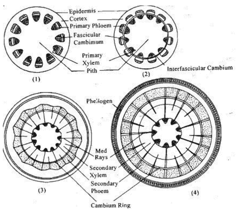
Fig.- Diagram indicating secondary growth in Dicot Stems (already in pdf)
The significance of secondary growth is as follows:
It adds to the girth of the plant thus provides support to the increasing weight of aerial parts due to growth.
It produces a corky bark around the tree trunk that protects the interior from abrasion, heat, cold, and infection.
It adds new vascular tissues for replacing old non-functioning one as well as for meeting increased demand for long-distance transport of sap and organic nutrients.
4. Draw images to sketch out the anatomical variation between:
a) Monocot root and dicot root
Ans: Variations between monocot root and dicot root are shown in the following picture and table.
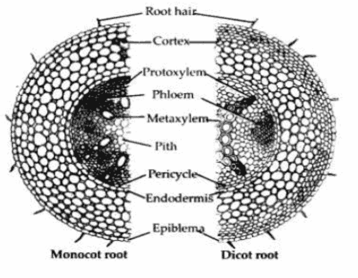
Fig.- Comparative pictures of dicot root and monocot root T.S.
b) Monocot stem and dicot stem
Ans: Variations between monocot and dicot stems are shown in the following illustration.
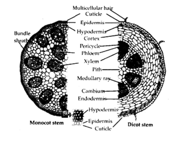
Fig.- Comparative pictures of dicot root and monocot stem T.S.
5. From your school garden, cut down a transverse section of the young stem of a plant and examine it by using the microscope. How would you determine whether it is a monocot stem or a dicot stem? Give reasons.
Ans: Looking through the microscope, I will check if the following features are observed.
Vascular bundles in the dicot stem are arranged in a ring whereas in monocot stem vascular bundles are scattered throughout the ground tissue.
Based on the arrangement of vascular bundles, it can be ascertained whether the young stem is dicot or monocot.
In addition to the indistinguishable ground tissue, sclerenchymatous hypodermis, spherical or egg-shaped vascular bundles with Y-shaped xylem are other distinguishing characteristics of monocot stem.
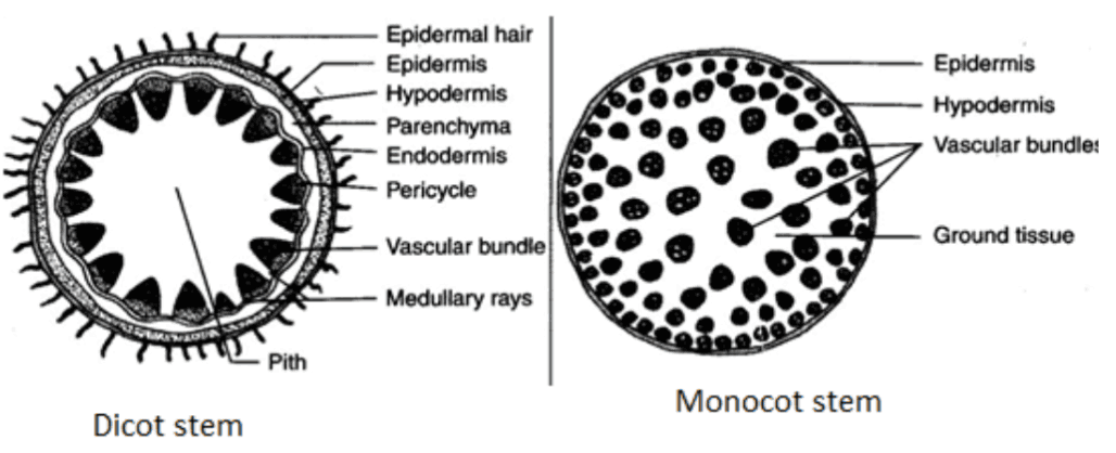
Fig: Arrangement of vascular bundles in dicot and monocot stems.
6. The transverse section of a plant material reveals the subsequent anatomical features – (a) the vascular bundles are conjoint, widely dispersed, and encircled by a sclerenchymatous bundle sheath, (b) phloem parenchyma is absent. What will you find it as?
Ans: The plant material is identified as a monocot stem
7. Why are phloem and xylem known as complex tissues?
Ans: Complex Tissues are made up of more than one type of cell and these work together as a unit. Xylem elements are responsible for the conduction of water and mineral salts from the roots to the other parts of the plant.
Xylem elements are highly lignified and dead except xylem parenchyma. It consists of:
Xylem vessels
Xylem tracheids
Xylem fibers
Xylem parenchyma
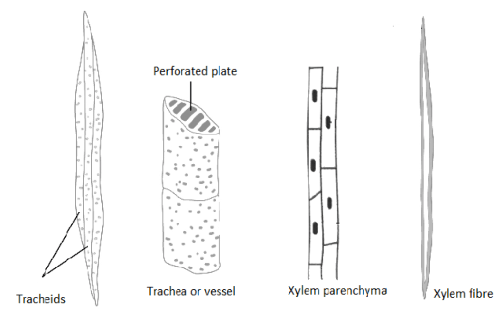
Fig: Complex tissue – Xylem
Phloem is a complex tissue associated with the translocation of food. Phloem elements are living except phloem fibers. It consists of:
Sieve tubes
Companion cells
Phloem fibers
Phloem parenchyma
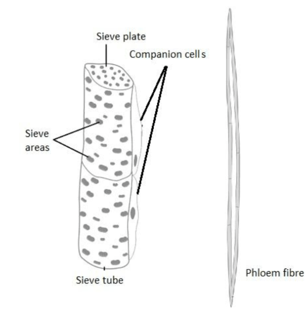
Fig: Complex tissue– Phloem.
8. What is the stomatal apparatus? With the help of diagrammatic representation describe the structure of stomata and label its parts.
Ans:
The stomatal apparatus comprises the following
A stoma –This is a small aperture or opening present in the epidermal cells of the leaf. This is called a stomatal aperture (singular- stoma, plural – stomata).
Two bean-shaped guard cells surrounding the stomatal aperture. ( It is to be noted that guard cells are dumbbell-shaped in monocots and bean-shaped in dicots.)
Subsidiary cells – These are specific epidermal cells in the vicinity of guard cells.
Function
Change in the turgidity or flaccidity of the guard cells is associated with stomatal opening and closure.
Stomata are essentially involved in gaseous exchange and transpiration.
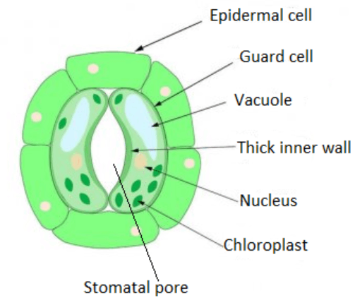
Fig.-Stomatal apparatus
9. Mention the three fundamental tissue systems in the flowering plants. Provide the tissue names under each system.
Ans:
The three basic tissue systems in flowering plants are the epidermal tissue system, ground tissue system, and vascular tissue system.
The epidermal tissue system comprises epidermal cells, stomata, trichomes, and hairs.
The ground tissue system is composed of the endodermis, cortex, pith, pericycle, and medullary rays, in the primary stems and roots.
In leaves, the ground tissue is comprised of thin-walled chloroplast-which contains the cells and is known as mesophyll.
The vascular tissue system is composed of complex tissues, the xylem, and the phloem.
10. In what way is the study of plant anatomy beneficial to us?
Ans:
The study of plant anatomy helps to understand structural adaptations in plants with respect to their different environmental conditions.
It also helps us in differentiating between monocots, dicots, and gymnosperms. This gives us an idea of the physiological state of the plants and so can be useful in crop improvement.
Internal structures also help us to predict the strength of wood and hence its utility for commercial activities.
Study of plant fibers such as jute, flax, and hemp, etc. may prove useful in their business-related exploitation.
11. What is periderm? How does periderm development occur in the dicot stems?
Ans: Periderm is a protective layer present outside the stem that replaces the epidermal layer in response to any injury or invasion of pathogens.
Phelloderm, phellogen, and phellem together constitute the periderm. Dicot stems produce phellogen or cork cambium in the external cortical cells. Phellogen cells split on both the inner side and the outer side to form secondary tissues. The secondary tissue produced on the inner side of the phellogen is known as the phelloderm or secondary cortex. On the outer side, phellogen produces phellem or cork.
12. Define the inner structure of a dorsiventral leaf by using a labeled diagram.
Ans:
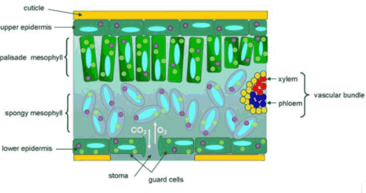
Fig: Internal Structure of a dorsiventral leaf.
Dorsiventral leaves are found in dicots. The significant anatomical characteristics of dorsiventral leaves are discussed below:
a) Upper Epidermis: This is usually the outermost single made of parenchymatous cells. The epidermal cells have sometimes outgrowths known as papillae, e.g., in Gladiolus. The epidermal cells are devoid of chloroplast and stomata are absent on the top epidermis.
b) Lower Epidermis: It is the same as the upper epidermis but here stomata are present. Chloroplasts are absent in the bottom epidermis also, except the guard cells of stomata.
c) Mesophyll: In between the lower and upper epidermis mesophyll tissues is present which can be split into two regions:
(i) Palisade Parenchyma: These are lengthened columnar cells without intercellular spaces. These contain chloroplast in them and are usually arranged in two layers.
(ii) Spongy Parenchyma: It is found below palisade parenchyma and is oval or spherical with intercellular spaces. They also contain chloroplasts but several chloroplasts are more in palisade parenchyma than spongy parenchyma.
d) Vascular Bundles: Vascular bundles are usually found at the boundary between the spongy and the palisade regions. The vascular bundle in the midrib area is the largest. Vascular bundles are collateral, conjoint, and closed. Each vascular bundle is encircled by a bundle sheath of parenchymatous cells. In the vascular bundle, phloem is found towards the lower epidermis and the xylem is present towards the top epidermis. Additionally, in the xylem, the protoxylem is towards the uppermost epidermis.
NCERT Solutions for Class 11 Biology Chapter 6 - Anatomy of Flowering Plants
Students who are studying in Class 11 need to get a detailed and coherent explanation of the chapter. They need a resource that can be a comprehensive guide to walk them through the different concepts given in the chapter. The best aspect is that the resource is available in the form of a free PDF that they can download and keep as an integral resource for studying. Also, the PDF has been drafted in a language that is easy to read and can be referred to get detailed information about the chapter. The students can study these NCERT Solutions for Class 11, Biology, Anatomy of Flowering Plants, to prepare the topics covered in this chapter for their exam.
NCERT Solutions for Class 11 Biology Chapter 6 Anatomy of Flowering Plants
The NCERT Solutions for Chapter 6 of Class 11 Biology explains the anatomy of a flower in detail. Anatomy refers to the detailed study of an organism's internal structure. So, when you study the anatomy of a plant, you need to study histology. Histology is the study of tissue's structure as well as the organization's structure. Also, by studying anatomy, you can get to know about the structural peculiarities of plants as well as the structural adaptation of the plants to different environments.
The chapter also explains the definition of a tissue. It is a group of cells that enjoy a common origin and perform common functions. The chapter further explains different types of plant tissues, such as apical meristem, lateral meristem, intercalary meristem, and parenchyma, collenchyma, sclerenchymas, and complex tissues.
Besides, the students can also get to know about the epidermal tissue system, which forms the outermost covering of the plant's body. The chapter also describes the secondary growth of a plant. The increase in the plant body's girth is called secondary growth. The tissues that are involved in the secondary growth of the plant are called cork cambium and vascular cambium.
NCERT Biology Class 11 Anatomy of Flowers: Weightage Marks
The Unit 2, Structural Organisation in Plants and Animals, of the NCERT Biology syllabus, covers the NCERT Solutions for Class 11 Biology, Chapter 6. The particular unit carries a weightage of 5 per cent of marks in the NEET exam and 12 per cent of marks in the final examination. The unit covers three chapters that carry an equal weightage in the examination.
Important Concepts Covered in Anatomy of Flowering Plants
In this chapter, students will learn about the anatomy of each part and its features in detail, from the seeds, parts of a monocot & dicot seed, its differentiation, different zones, and more. Given are the important topics covered in Anatomy of Flowering Plants:
Anatomy of Flowering Plants
Plants tissues
Meristematic tissues
Permanent tissue
Epidermal Tissue System
The ground Tissue System
The Vascular Tissue System
Dicotyledonous Root
Monocotyledonous Root
Dicotyledonous Stem
Monocotyledonous Stem
Dicotyledonous Leaf
Monocotyledonous Leaf
Secondary Growth
Benefits of NCERT Solutions for Class 11 Biology Chapter 6: Anatomy of Flowering Plants
Solutions are curated by experts at Vedantu and all are to the point.
Every solution is written in simple language which is very easy to understand.
The solutions will also be beneficial for students from the examination point of view for both board and competitive exams.
Labelled diagrams are provided to enable easy understanding of concepts in a better way.
The solutions are available in PDF format that can be downloaded absolutely free.
Study Materials for Class 11 Biology Chapter 6 Anatomy of Flowering Plants NCERT Solutions
Conclusion
The NCERT Solutions for Class 11 Biology Chapter 6 - Anatomy of Flowering Plants provided by Vedantu are an excellent resource for students to prepare for their exams. The solutions cover all the important topics in the chapter, including the internal structure of different plant organs, such as roots, stems, and leaves, and their functions and adaptations. The solutions are presented in a clear and concise manner, making it easy for students to understand the concepts. They also include diagrams and illustrations to help students visualize the different structures and processes. Vedantu provides additional study materials, such as notes, sample papers, and solutions to previous year question papers, to help students prepare for their exams. By using these resources, students can develop a deeper understanding of the anatomy of flowering plants and perform well in their exams.
NCERT Solutions for Class 11 Biology FREE PDF | Other Chapter-wise Links
Below are the other chapter-wise Links for the Solutions for Biology NCERT Class 11. You can download FREE PDFs of these chapter-wise solutions to familiarise yourself with the concepts.
Related Important Links for CBSE Class 11 Biology NCERT Solutions

FAQs on NCERT Solutions For Class 11 Biology Chapter 6 Anatomy Of Flowering Plants - 2026-27
1. What are NCERT Solutions for Class 11 Biology Chapter 6 – Anatomy of Flowering Plants?
NCERT Solutions for Class 11 Biology Chapter 6 provide stepwise answers to all textbook exercises as per the CBSE 2026–27 syllabus. They help students understand core concepts like plant tissues, secondary growth, and anatomical differences between monocots and dicots, following the NCERT methodology for exam preparation.
2. How are meristematic tissues classified in NCERT Class 11 Biology Chapter 6 Solutions?
According to NCERT Solutions, meristematic tissues are classified based on their location in the plant body into:
- Apical meristems (at root/shoot tips; increase length)
- Intercalary meristems (at leaf base/above nodes; elongate organs)
- Lateral meristems (on sides; increase girth/diameter)
3. Why are xylem and phloem called complex tissues in CBSE Class 11 Biology NCERT Solutions?
Xylem and phloem are called complex tissues because each is composed of multiple cell types working together for specific functions. Xylem transports water and minerals, while phloem translocates food. Their components include vessels, tracheids, fibers, and parenchyma (in xylem), as well as sieve tubes, companion cells, fibers, and parenchyma (in phloem).
4. What is the significance of secondary growth according to Class 11 Biology Chapter 6 NCERT Solutions?
Secondary growth, mainly found in dicot stems and roots, increases the girth of plants. This provides structural support, replaces old vascular tissue, and forms a protective corky layer (periderm), thereby safeguarding against pathogens, mechanical injury, and environmental stress, as explained in the official NCERT Solutions.
5. How can you distinguish between a monocot stem and a dicot stem using NCERT Solutions for Class 11 Biology Chapter 6?
The NCERT Solutions guide students to identify:
- Monocot stem: Vascular bundles are scattered, usually have bundle sheaths, phloem parenchyma is absent, and hypodermis is sclerenchymatous.
- Dicot stem: Vascular bundles arranged in a ring, open and collateral, bundle sheath absent, phloem parenchyma present, and hypodermis is collenchymatous.
6. Explain the structure and function of stomatal apparatus as per the NCERT Solutions for Anatomy of Flowering Plants.
The stomatal apparatus consists of a stomatal pore, two guard cells that control its opening and closing, and sometimes subsidiary cells. Its function is to regulate gas exchange and transpiration; guard cells’ turgidity causes aperture changes, enabling control over water loss and CO2 intake.
7. What role do periderm and cork cambium play in dicot stems, based on Class 11 Biology NCERT Solutions?
Periderm is the protective tissue that replaces the ruptured epidermis in older stems. The cork cambium (phellogen) generates cork (phellem) to the outside and phelloderm inside—together, these form the periderm, which prevents water loss and protects against physical damage and pathogens.
8. What are three tissue systems in flowering plants highlighted in Chapter 6 of NCERT Solutions for Class 11 Biology?
NCERT Solutions detail the three main tissue systems:
- Epidermal tissue system: Outer protective covering (epidermis, trichomes, root hairs)
- Ground tissue system: Cortex, endodermis, pericycle, pith, mesophyll
- Vascular tissue system: Xylem and phloem for transport and support
9. What are common misconceptions about secondary growth clarified in NCERT Solutions for Class 11 Biology Chapter 6?
One misconception is that all plants exhibit secondary growth; NCERT clarifies that significant secondary growth occurs mainly in dicotyledons, rarely in monocots. Another is that primary and secondary tissues are easily distinguishable in mature plants, but continued cambial activity may obscure boundaries over time.
10. How does the NCERT Solutions for Chapter 6 explain the anatomical adaptation of leaves to their environment?
NCERT Solutions highlight that anatomical adaptations like leaf thickness, arrangement of mesophyll (palisade and spongy), cuticle presence, and venation type help plants optimize photosynthesis, reduce water loss, and survive various climates (dorsiventral for dicots, isobilateral for monocots).
11. What is the difference between open and closed vascular bundles as outlined in Class 11 Biology Chapter 6 NCERT Solutions?
Open vascular bundles (in dicot stems) contain a cambium between xylem and phloem, allowing for secondary growth. Closed vascular bundles (in monocots) lack cambium and hence cannot undergo secondary thickening.
12. In what ways do NCERT Solutions for Class 11 Biology Chapter 6 help for board and competitive exam preparation?
These NCERT Solutions provide structured, step-wise answers as per CBSE exam patterns, include marked and labeled diagrams, clarify conceptual foundations, and integrate CBSE/NEET exam themes—helping students master all in-syllabus topics for better scores.
13. What if a plant has no lateral meristem as per CBSE Class 11 Biology NCERT Solutions?
If a plant lacks lateral meristem, it will not show secondary growth; hence, its stem and root girth won’t increase significantly. Such plants—typically monocots—remain slender with limited capacity for increased vascular transport or mechanical support.
14. Why is the study of plant anatomy important according to NCERT Solutions for Anatomy of Flowering Plants?
Studying plant anatomy reveals structural adaptations to different habitats, aids in taxonomy (distinguishing monocots, dicots, gymnosperms), improves crop yield/quality, and helps predict wood strength and suitability for commercial uses.