

Labeled diagram of dicot leaf showing parts and functions
The concept of dicot leaf diagram is essential in biology and helps explain real-world biological processes and exam-level questions effectively.
Understanding Dicot Leaf Diagram
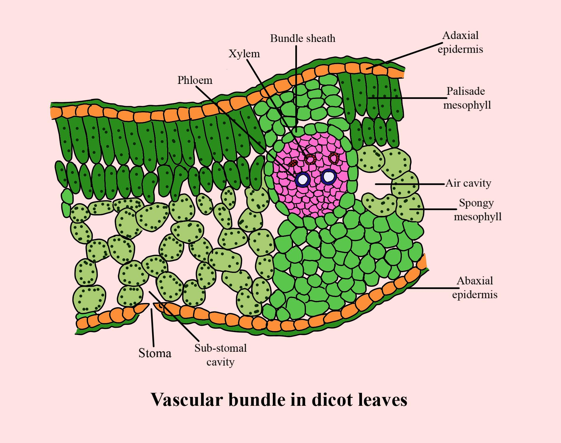
Dicot leaf diagram refers to a detailed, labelled illustration showing the internal cross-sectional structure of the leaf of dicotyledonous plants. This concept is important in areas like plant anatomy, practical biology, and board exam preparation. Diagrams help students recognize tissues such as mesophyll, vascular bundles (veins and midrib), epidermis, and stomata, supporting topics like photosynthesis and tissue differentiation.
Structure of Dicot Leaf Diagram
A typical dicot leaf diagram displays three main regions:
- Upper and Lower Epidermis: Thin layers covering the leaf's upper (adaxial) and lower (abaxial) surfaces, sometimes with a waxy cuticle for protection against water loss and injury.
- Mesophyll: Layer between the epidermises, divided into two parts: palisade parenchyma (elongated cells rich in chloroplasts, just below the upper epidermis) and spongy parenchyma (loosely arranged cells with air cavities, near the lower epidermis).
- Vascular Bundles: Comprised of xylem and phloem for water/mineral and food transport; bundled together in the veins and midrib, surrounded by bundle sheath cells for protection.
The lower surface typically has more stomata, important for gas exchange and transpiration.
Key Parts of a Dicot Leaf (Label and Function)
| Part | Location | Function |
|---|---|---|
| Upper Epidermis | Outer upper layer | Protection, sometimes covered with cuticle |
| Palisade Parenchyma | Below upper epidermis | Photosynthesis (rich in chloroplasts) |
| Spongy Parenchyma | Below palisade, above lower epidermis | Air exchange, minor photosynthesis |
| Vascular Bundles | Veins/Midrib | Transport of water, minerals, food |
| Lower Epidermis | Outer lower layer | Has more stomata for gas exchange |
| Stomata | Mainly in lower epidermis | Controls water loss and gas exchange |
Identification Tips and Monocot vs Dicot Leaf
- Mesophyll differentiation: Only dicot leaves have both palisade and spongy mesophyll. Monocots have undifferentiated mesophyll.
- Number of stomata: Dicots have more stomata on lower surface; monocots have equal numbers on both sides.
- Venation: Dicots: reticulate (net-like) venation. Monocots: parallel venation.
- Bundle sheath cells: Well-developed in dicots.
For a detailed difference, visit Difference Between Monocot and Dicot Leaf.
Worked Example – Observing Dicot Leaf Under Microscope
1. Take a section of the leaf using a blade.
2. Place it on a glass slide with water and cover-slip.
3. Observe the arrangement: upper epidermis, palisade cells, spongy mesophyll, vascular bundles, and lower epidermis.
4. Draw and neatly label all visible tissues as shown above.
Tip: Always note the side with more stomata as the lower surface.
Common Mistakes to Avoid
- Confusing dicot leaf diagram with monocot (look at mesophyll and venation).
- Mixing label positions for upper/lower epidermis or forgetting to show bundle sheath.
Real-World Applications
The concept of dicot leaf diagram is used in fields like botany, agriculture, and environmental biology. It is vital for understanding photosynthesis and plant nutrition. In practical labs and exams, drawing and identifying the correct tissues can help score well. Vedantu brings such concepts alive with high-quality visuals and stepwise guidance.
Practice Questions
- Draw and label a neat diagram of the cross-section of a dicot leaf.
- Describe the roles of palisade and spongy parenchyma in the dicot leaf.
- List three anatomical differences between dicot and monocot leaves.
- Why are more stomata present on the lower surface of a dicot leaf?
In this article, we explored dicot leaf diagram, its key parts, structure, practical significance, and tips for identifying it. Practicing labelled diagrams and understanding the roles of tissues builds exam confidence. To learn more, keep practicing with Vedantu.
Related Concepts & Useful Vedantu Links
- Difference Between Monocot and Dicot Leaf
- Plant Cell
- Photosynthesis Process
- Difference Between Monocot and Dicot Root
- Lamina of a Plant Leaf
- Stomata
- Cell Structure and Function
- Nutrition in Plants
- Plant Tissues
- Basic Internal Anatomy of Leaf
- Difference Between Monocot and Dicot Stem


FAQs on Dicot Leaf Structure with Detailed Diagram and Explanation
1. What is a dicot leaf diagram?
A dicot leaf diagram is a labeled drawing that shows the internal and external structure of a leaf from a dicotyledonous plant. It typically represents:
- The epidermis (upper and lower layers)
- The mesophyll differentiated into palisade parenchyma and spongy parenchyma
- The vascular bundle containing xylem and phloem
- Features like stomata and cuticle
2. What are the main parts labeled in a dicot leaf diagram?
The main parts labeled in a dicot leaf diagram include the epidermis, mesophyll, and vascular tissues. These parts are:
- Upper epidermis – protective outer layer
- Lower epidermis – contains many stomata
- Palisade mesophyll – elongated cells rich in chloroplasts
- Spongy mesophyll – loosely arranged cells with air spaces
- Vascular bundle – includes xylem (water transport) and phloem (food transport)
3. What is the function of palisade mesophyll in a dicot leaf?
The palisade mesophyll is responsible for most of the photosynthesis in a dicot leaf. It consists of tightly packed, elongated cells located below the upper epidermis and contains a large number of chloroplasts. This arrangement:
- Maximizes light absorption
- Increases efficiency of photosynthesis
- Helps in glucose production
4. How does a dicot leaf differ from a monocot leaf?
A dicot leaf differs from a monocot leaf mainly in venation pattern and internal structure. Key differences include:
- Reticulate venation in dicots vs parallel venation in monocots
- Mesophyll differentiated into palisade and spongy layers in dicots
- Usually fewer vascular bundles compared to monocots
5. What is the role of stomata in a dicot leaf?
The stomata in a dicot leaf regulate gas exchange and transpiration. These small pores, mostly found in the lower epidermis, allow:
- Entry of carbon dioxide for photosynthesis
- Release of oxygen
- Loss of water vapor through transpiration
6. What is reticulate venation in a dicot leaf?
Reticulate venation is a vein pattern in which veins form a network-like structure in a dicot leaf. In this pattern:
- A main vein called the midrib is present
- Smaller veins branch out and form a net-like arrangement
- It provides support and efficient transport of water and nutrients
7. What is the function of the vascular bundle in a dicot leaf?
The vascular bundle in a dicot leaf transports water, minerals, and food. It contains:
- Xylem – transports water and minerals from roots to leaves
- Phloem – transports prepared food from leaves to other parts of the plant
8. Why is the lower epidermis important in a dicot leaf?
The lower epidermis is important because it contains most of the stomata in a dicot leaf. Its functions include:
- Protection of internal tissues
- Regulation of gas exchange
- Control of transpiration
9. What type of mesophyll is found in a dicot leaf?
A dicot leaf contains two types of mesophyll: palisade mesophyll and spongy mesophyll. These are:
- Palisade mesophyll – tightly packed cells for photosynthesis
- Spongy mesophyll – loosely arranged cells with large air spaces for gas exchange
10. How do you draw and label a dicot leaf diagram step by step?
To draw and label a dicot leaf diagram, follow clear structural steps showing all major tissues. Steps include:
- Draw an outline of the leaf cross-section
- Add the upper epidermis with a thin cuticle
- Sketch elongated palisade mesophyll cells below it
- Draw loosely arranged spongy mesophyll with air spaces
- Insert a vascular bundle with xylem (upper side) and phloem (lower side)
- Complete with the lower epidermis showing stomata


