

What Is the Function of Each Part of a Compound Microscope
The concept of mention the function of each microscope part is essential in biology and helps students understand laboratory work, cell observation, and practical exam questions effectively.
Understanding the Function of Each Microscope Part
Mention the function of each microscope part means to state what each component of a microscope does. Knowing this is important in topics like cell structure, biology experiments, and understanding the anatomy of scientific instruments. Whether for practical classes, viva, or board exams, listing major microscope parts and their uses is a high-frequency biology question.
List of Microscope Parts and Their Functions
Each microscope part serves a key function. For example, the eyepiece magnifies the image, the objective lens increases detail, the stage holds the specimen, and the focus knobs sharpen the view. Learn all major microscope parts and their uses below.
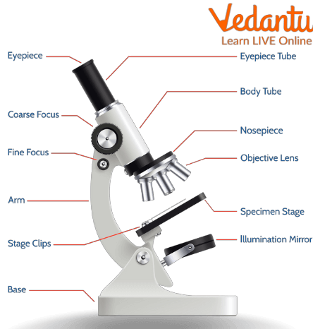
Microscope Parts and Functions Table
| Microscope Part | Function |
|---|---|
| Eyepiece (Ocular Lens) | The lens you look through; magnifies the image, usually 10x or 15x. |
| Body Tube | Connects the eyepiece to the objective lenses; maintains the correct distance. |
| Revolving Nosepiece (Turret) | Holds objective lenses and rotates to switch between different magnifications. |
| Objective Lenses | Main magnifying lenses (usually 4x, 10x, 40x, 100x) to view the specimen at different levels of detail. |
| Arm | Supports the tube and connects it to the base; also used for carrying the microscope. |
| Stage | Flat platform where you place the slides; holds the specimen in position. |
| Stage Clips | Hold the slide securely on the stage. |
| Coarse Adjustment Knob | Moves the stage up and down for rough focusing of the specimen. |
| Fine Adjustment Knob | Moves the stage slightly to sharpen the image for precise focusing. |
| Base | Bottom part; supports and stabilizes the entire microscope. |
| Mirror/Illuminator | Provides the light source to illuminate the specimen; may be adjustable. |
| Condenser | Focuses light onto the specimen for clear viewing. |
| Diaphragm (Iris) | Adjusts the intensity and size of the cone of light reaching the specimen. |
| Rack Stop | Prevents the stage from moving too close to the objective lenses, protecting slides and lenses. |
How the Microscope Parts Work Together
Here’s the step-by-step use of a microscope and how each part functions during specimen observation:
- Place the slide on the stage, securing it with stage clips.
- Switch the objective lens using the nosepiece to select desired magnification.
- Look through the eyepiece to view the image.
- Adjust the mirror/illuminator to provide sufficient light.
- Use the coarse adjustment knob to bring specimen roughly into focus.
- Use the fine adjustment knob for sharp, clear focus.
- The diaphragm and condenser help optimize light for clarity.
Sample Exam Answers: State the Function of Each Part
Q: State the function of the objective lenses.
A: Objective lenses magnify the specimen at different strengths for detailed observation.
Q: What is the use of the diaphragm?
A: The diaphragm controls the amount of light passing through the specimen.
Q: What is the function of the fine adjustment knob?
A: The fine adjustment brings the image into sharp focus, especially under high-power objectives.
Tips and Tricks to Remember Microscope Functions
- Link “Eyepiece is for Eye”, “Objective is for the Object (specimen)”.
- Remember “Stage is like a platform for the slide”.
- Use the acronym “F.O.C.U.S.”: Fine/Coarse knobs for Focusing, Objective lens for Observation, Condenser/Diaphragm for Controlling light, Use Stage/Clips for Slide positioning.
- Draw and label the diagram while revising — visual memory helps recall functions quickly during exams.
Practice Questions
- Mention the function of each microscope part in your own words.
- Draw and label a compound microscope showing all parts and their functions.
- Explain how the condenser and diaphragm affect image clarity.
- Compare the function of the eyepiece and objective lenses.
Common Mistakes to Avoid
- Mixing up coarse and fine adjustment knobs.
- Forgetting to mention the light source in answers.
- Drawing incorrect or incomplete microscope diagrams in exams.
Real-World Applications
Understanding the functions of microscope parts helps students while observing plant and animal cells (Cell Structure and Function), studying tissues (Tissues), or discovering important biological facts (Discovery of Cells). At Vedantu, such concepts are explained with visuals and stepwise clarity to help you succeed in exams and labs.
In this article, we explored how to mention the function of each microscope part, included a detailed table for revision, given sample answers, and shared tips for remembering each part’s use. Keep practicing with Vedantu for lab confidence and excellent board results!
Explore Related Biology Topics & Experiments
- Compound Microscope Parts – Get more details and diagrams.
- Human Body and Its Movements – Explore practical lab applications.
- Cell Structure and Function – Connect microscope use with cell biology.
- Labeled Diagram of Human Ear – Sharpen your diagram labeling skills.
- Nutrition in Plants – See how microscopes are used in plant studies.
- Amoeba – Classic observation: single-celled organisms.
- Discovery of Cells – See why microscopes revolutionized biology.
- Tissues – Deepen your knowledge of tissue examination with a microscope.
- Cell – Understand why every part of the microscope is essential for studying cells.
- Brain Facts – Practice labeled diagrams and observation skills.
- Biotic and Abiotic – Understand the microscopic and macroscopic world connections.
- Neurons and Nerves – See nerve cells under the microscope in biology visuals.


FAQs on Functions of Microscope Parts in Detail
1. What is the function of the eyepiece in a microscope?
The eyepiece (ocular lens) magnifies the image formed by the objective lens so it can be viewed clearly by the eye. It is the top lens you look through in a compound microscope.
- Usually provides 10× magnification (sometimes 15×).
- Works with the objective lens to give total magnification.
- May contain a pointer in student microscopes for identifying structures.
2. What is the function of the objective lenses in a microscope?
The objective lenses provide the primary magnification and form the initial enlarged image of the specimen. They are located on the revolving nosepiece.
- Common magnifications: 4× (scanning), 10× (low power), 40× (high power), and 100× (oil immersion).
- Higher magnification gives greater detail but a smaller field of view.
- Oil immersion lens increases resolution using immersion oil.
3. What is the function of the stage in a microscope?
The stage supports and holds the slide containing the specimen in place during observation. It is the flat platform below the objective lenses.
- Has a central opening to allow light to pass through the specimen.
- Often includes stage clips or a mechanical stage to secure the slide.
- Allows movement of the slide left, right, forward, and backward.
4. What is the function of the coarse adjustment knob?
The coarse adjustment knob moves the stage or body tube up and down rapidly for rough focusing. It is mainly used under low power.
- Helps bring the specimen into general focus.
- Should not be used under high power to avoid damaging the slide.
- Works by changing the distance between the objective lens and specimen.
5. What is the function of the fine adjustment knob?
The fine adjustment knob sharpens the image after coarse focusing by making small, precise movements. It is essential for clear viewing under high magnification.
- Used after the coarse adjustment knob.
- Improves image clarity and detail.
- Important when using 40× or 100× objective lenses.
6. What is the function of the diaphragm in a microscope?
The diaphragm controls the amount of light that passes through the specimen. It is located below the stage and above the light source.
- Adjusts brightness and contrast.
- Helps improve image clarity.
- Often part of the iris diaphragm system.
7. What is the function of the light source or mirror in a microscope?
The light source or mirror provides illumination so the specimen can be seen clearly. It directs light upward through the stage opening.
- Modern microscopes use a built-in electric bulb.
- Older models use a mirror to reflect external light.
- Essential for observing transparent biological specimens.
8. What is the function of the condenser in a microscope?
The condenser concentrates and focuses light onto the specimen for clearer and brighter images. It is located beneath the stage.
- Improves image resolution.
- Works together with the diaphragm.
- Especially important at high magnification.
9. What is the function of the revolving nosepiece?
The revolving nosepiece holds the objective lenses and allows the user to switch between different magnifications. It rotates to align a selected objective lens with the specimen.
- Provides quick change of magnification.
- Clicks into position for accurate alignment.
- Supports multiple objective lenses.
10. What is the function of the arm and base of a microscope?
The arm and base provide structural support and stability to the microscope. They hold all other parts in position.
- Arm: Connects the body to the base and is used for carrying the microscope.
- Base: Supports the entire microscope and often houses the light source.
- Ensure balance and reduce vibrations during observation.


