

Detailed Anatomy of Lungs Including Lobes Bronchi and Alveoli
The concept of structure of lungs is essential in biology and helps explain real-world biological processes and exam-level questions effectively.
Understanding Structure of Lungs
Structure of lungs refers to the detailed anatomy and organization of the human lungs, including the arrangement of airways (bronchi and bronchioles), air sacs (alveoli), and protective membranes. This concept is important in areas like the human respiratory system, gas exchange, and respiratory functions. Learning the structure of lungs is crucial for board exam diagrams, understanding how we breathe, and identifying lung diseases.
Main Parts of Lung Structure
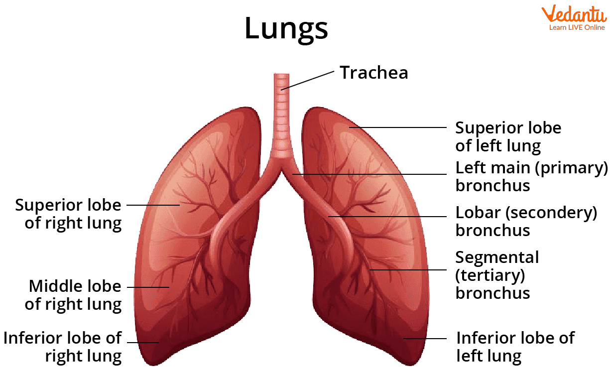
The human lungs are paired, cone-shaped organs located in the thoracic cavity, protected by the rib cage. Their structure can be broken down as follows:
- Bronchi: Two main branches (left and right) arising from the trachea, entering each lung. Bronchi further divide into smaller tubes.
- Bronchioles: Smaller subdivisions of bronchi, spreading throughout the lungs and ending at the alveoli.
- Alveoli: Millions of tiny, thin-walled air sacs where gaseous exchange occurs. They maximize the surface area for oxygen and carbon dioxide transfer.
- Lobes: The right lung has three lobes (superior, middle, inferior), while the left lung has two lobes (superior, inferior) and a cardiac notch for the heart.
- Pleura: A double-layered membrane (visceral and parietal pleura) surrounds each lung. The space between contains pleural fluid to prevent friction during breathing.
- Hilum: The region on the lung’s medial surface where bronchi, blood vessels, and nerves enter and exit.
Functions of Lung Parts
Each part of the lung has a special function that helps us breathe efficiently:
- Bronchi & Bronchioles: Passage for air to flow from trachea to alveoli.
- Alveoli: Site of oxygen and carbon dioxide exchange with blood (gas exchange).
- Pleura & Pleural fluid: Reduces friction and protects lungs during inhalation and exhalation.
- Lobes: Allow division of work within the lungs and greater surface area.
- Blood Vessels: Carry deoxygenated blood to the lungs and return oxygenated blood to the heart.
Here’s a helpful table to understand structure of lungs better:
Structure of Lungs Table
| Part | Structure | Main Function |
|---|---|---|
| Bronchi | Large, main air tubes from trachea | Carry air into each lung |
| Bronchioles | Smallest air branches | Carry air to alveoli |
| Alveoli | Tiny, round air sacs | Gas exchange (O2/CO2) |
| Pleura | Double serous membrane | Protect & reduce friction |
| Lobes | Right: 3, Left: 2 | Divide lung for function |
Common Mistakes to Avoid
- Confusing alveoli with bronchioles or placing them at the wrong end of the lung branches.
- Forgetting the difference in number of lobes: Right lung (3 lobes), Left lung (2 lobes).
- Missing the pleura membrane in labelled diagrams.
- Ignoring the cardiac notch in the left lung during labelling.
Worked Example – Biological Process
Let’s understand the path of air during breathing:
1. Air enters through the nose/mouth and travels down the trachea.
2. The trachea divides into two bronchi, entering each lung.
3. Bronchi branch into many bronchioles, which further lead to alveoli.
4. In alveoli, oxygen passes into the blood, and carbon dioxide is released from blood into air sacs for exhalation.
Final Understanding: This efficient branching and huge alveolar surface enable quick gas exchange, keeping us alive and active.
Practice Questions
- Draw and label a neat diagram of the human lung showing major structural parts.
- Explain the functions of alveoli.
- Why is the left lung smaller than the right lung?
- What is the role of the pleural membrane in breathing?
- How does lung structure help in gas exchange?
Real-World Applications
The concept of structure of lungs is used in medicine to diagnose and treat lung diseases, in sports science for understanding endurance, and in biotechnology for developing artificial lungs. Vedantu helps students relate such topics to practical examples and inspires careers in healthcare and research.
Page Summary
In this article, we explored the structure of lungs, the function of key parts, and how it all connects to our breathing and exam success. For more diagrams, detailed explanations, and practice, keep learning with Vedantu’s Biology resources.
Related Concepts & Further Reading
- Human Respiratory System
- Respiratory and Lung Volumes
- Pulmonary Alveolus
- Function of Lungs
- Bronchi
- Difference Between Right and Left Lung
- Mechanism of Breathing
- Lung Diseases
- Human Body and Its Movements
- Respiration in Fish


FAQs on Structure Of Lungs and Their Anatomical Organization
1. What is the structure of lungs?
The structure of lungs consists of a pair of spongy, elastic organs divided into lobes and made up of branching airways that end in tiny air sacs called alveoli.
- The right lung has three lobes, while the left lung has two lobes.
- Each lung is enclosed by a double-layered membrane called the pleura.
- Inside, the airways branch from the bronchi into bronchioles and finally into alveoli.
- The lungs are supported by connective tissue and rich blood vessels for gas exchange.
2. What are the main parts of the lungs?
The main parts of the lungs include the bronchi, bronchioles, alveoli, and the pleural membranes.
- Bronchi: Two primary branches from the trachea entering each lung.
- Bronchioles: Smaller branching tubes inside the lungs.
- Alveoli: Microscopic air sacs where gas exchange occurs.
- Pleura: Protective double membrane covering the lungs.
3. How many lobes are present in the human lungs?
Human lungs have five lobes in total, with three in the right lung and two in the left lung.
- Right lung: Superior, middle, and inferior lobes.
- Left lung: Superior and inferior lobes.
- The left lung has fewer lobes to accommodate the position of the heart.
4. What is the structure and function of alveoli?
The alveoli are tiny, thin-walled air sacs that serve as the main site of gas exchange in the lungs.
- They have a single-cell-thick wall for rapid diffusion.
- Surrounded by a network of capillaries.
- Contain surfactant to prevent collapse.
- Allow exchange of oxygen and carbon dioxide by diffusion.
5. How do bronchi and bronchioles differ in structure?
The bronchi are larger air passages with cartilage support, while bronchioles are smaller branches without cartilage.
- Bronchi: Contain cartilage rings and ciliated epithelium.
- Bronchioles: Lack cartilage and have smooth muscle walls.
- Bronchioles regulate airflow by constricting or dilating.
6. What is the role of the pleura in lung structure?
The pleura is a double-layered membrane that protects the lungs and reduces friction during breathing.
- Visceral pleura: Covers the lung surface.
- Parietal pleura: Lines the chest cavity.
- Pleural fluid: Between the layers to allow smooth movement.
7. How are the lungs adapted for efficient gas exchange?
The lungs are adapted for efficient gas exchange by having a large surface area, thin walls, and a rich blood supply.
- Millions of alveoli increase surface area.
- Alveolar walls are one cell thick for rapid diffusion.
- Dense capillary networks maintain concentration gradients.
- Moist surface aids in dissolving gases.
8. What type of tissue makes up the lungs?
The lungs are primarily made of epithelial tissue, connective tissue, and smooth muscle tissue.
- Simple squamous epithelium lines the alveoli.
- Ciliated columnar epithelium lines the bronchi.
- Elastic connective tissue allows expansion and recoil.
- Smooth muscle controls bronchiole diameter.
9. Why is the left lung smaller than the right lung?
The left lung is smaller than the right lung because it accommodates space for the heart.
- The indentation is called the cardiac notch.
- The left lung has two lobes instead of three.
- This structural difference ensures proper placement of thoracic organs.
10. How does air travel through the lungs step by step?
Air travels through the lungs in a branching pathway from the trachea to the alveoli for gas exchange.
- Step 1: Air enters through the trachea.
- Step 2: It divides into two primary bronchi.
- Step 3: Bronchi branch into smaller bronchioles.
- Step 4: Bronchioles end in clusters of alveoli.
- Step 5: Oxygen diffuses into blood and carbon dioxide diffuses out.


