

Labeled Diagram of Human Lungs with Parts and Functions
The human respiratory system enables breathing by drawing in oxygen and removing carbon dioxide. It consists of multiple parts, including the nose, pharynx, larynx, trachea, bronchi, bronchioles, and alveoli. All these parts work together to keep our cells well supplied with oxygen and to expel waste gases.
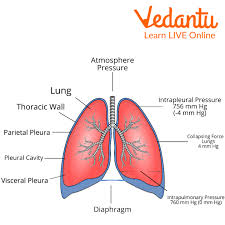
Location and Gross Structure of the Lungs
Our lungs occupy most of the space in the chest (thoracic) cavity. They are covered by a thin membrane called the pleura and are situated on either side of the heart.
Right Lung: Divided into three lobes (superior, middle, and inferior). It is slightly larger than the left lung to accommodate the heart’s position.
Left Lung: Divided into two lobes (superior and inferior), separated by an oblique fissure.
Each lung has a pointed top region, known as the apex, that extends just above the first rib, while the broad base rests on the diaphragm.
Respiratory Pathway and Air Flow
Air enters the body primarily through the nose, where it is warmed, filtered, and moistened. It then moves through the pharynx and the larynx (voice box) into the trachea.
Nose/Mouth → Pharynx → Larynx → Trachea → Bronchi → Bronchioles → Alveoli
In the pharynx, air from the nose and mouth converges, then passes to the larynx.
The larynx leads air into the trachea (windpipe), which is supported by rings of cartilage to prevent collapsing.
The trachea branches into two main bronchi (one for each lung), which further subdivide into smaller bronchi and then into bronchioles.
Eventually, air reaches tiny air sacs called alveoli, where oxygen is transferred into the blood, and carbon dioxide is released from the blood into the alveolar air to be exhaled.
Functions of the Lungs
Gas Exchange: The primary function is to add oxygen to the bloodstream and remove carbon dioxide.
Regulation of Blood pH: By controlling carbon dioxide levels, the lungs help regulate acidity in the blood.
Protection: Fine hairs, mucus, and immune cells in the airways trap and eliminate foreign particles.
Pulmonary Segments and Bronchioles
Each lung is further divided into smaller regions called pulmonary segments. The right lung has 10 pulmonary segments, while the left lung typically has 8 to 10 segments.
Bronchi: The main bronchi branch repeatedly, gradually becoming narrower to form bronchioles.
Bronchioles: These are small tubes without cartilage in their walls. They eventually lead to clusters of alveoli.
Alveoli: The Site of Gaseous Exchange
Each bronchiole ends in tiny, spongy sacs called alveoli. A single air sac is called an alveolus. Millions of alveoli in each lung create a large surface area for gas exchange. Oxygen from inhaled air diffuses into the surrounding capillaries, while carbon dioxide diffuses from the blood into the alveoli to be exhaled.
Role of the Diaphragm in Breathing
The diaphragm is a sheet of muscle located beneath the lungs, separating the chest cavity from the abdominal cavity. During inhalation, the diaphragm contracts and moves downward, increasing space in the chest cavity and pulling air into the lungs. During exhalation, it relaxes, moving upwards and forcing air out of the lungs. One full cycle of inhalation and exhalation is one complete breath.
Diagram of Lungs with Labelling
A simple lung diagram typically shows the trachea branching into the bronchi, which then divide into smaller bronchioles and end in alveolar sacs. A human lungs diagram with parts highlighted helps learners understand each component’s placement. Make sure to note important features like the lobes, fissures, and the structure of the lungs diagram to see how the air passageway connects to the alveoli.
Additional Points about the Lungs
Protective Mucus: The lining of the airways produces mucus, which traps dust and microorganisms.
Cilia: Tiny hair-like structures (cilia) push mucus upwards towards the throat, where it can be swallowed or coughed out.
Number of Alveoli: A newborn baby’s lungs have 20–50 million alveoli, while adults have around 300 million alveoli in each lung.
Short Quiz (With Answers)
1. Which lung is larger in humans?
Answer: The right lung is larger because of the heart’s position slightly to the left.
2. What is the function of the alveoli?
Answer: Alveoli are tiny air sacs where oxygen and carbon dioxide are exchanged between the air and blood.
3. Where does air travel immediately after it passes through the nose?
Answer: The air moves into the pharynx.
4. Which structure prevents food from entering the windpipe?
Answer: The epiglottis in the larynx helps prevent food from going into the windpipe.
5. What is the role of the diaphragm during inhalation?
Answer: It contracts and moves downwards, creating more space in the chest cavity and pulling air into the lungs.
Related Topics


FAQs on Human Lungs Diagram and Structure Explained
1. What is the human lungs diagram?
A human lungs diagram is a labeled illustration that shows the structure and main parts of the lungs and related respiratory organs. It typically includes:
- The right lung and left lung
- The trachea (windpipe)
- The bronchi and bronchioles
- The alveoli (air sacs)
- The diaphragm
2. What are the main parts labeled in a human lungs diagram?
The main parts labeled in a human lungs diagram are the trachea, bronchi, bronchioles, alveoli, and diaphragm. These parts include:
- Trachea – tube that carries air to the lungs
- Bronchi – two branches from the trachea entering each lung
- Bronchioles – smaller branches inside the lungs
- Alveoli – tiny air sacs where gas exchange occurs
- Diaphragm – muscle that helps in breathing
3. What is the function of the lungs in the human body?
The primary function of the lungs is to perform gas exchange by supplying oxygen to the blood and removing carbon dioxide. This process involves:
- Inhaling oxygen-rich air into the lungs
- Oxygen diffusing into blood in the alveoli
- Carbon dioxide diffusing from blood into the alveoli
- Exhaling carbon dioxide out of the body
4. How do the alveoli work in the lungs?
The alveoli work by allowing oxygen and carbon dioxide to diffuse between air and blood through thin walls. Each alveolus:
- Has a very thin, moist wall (one cell thick)
- Is surrounded by a network of capillaries
- Provides a large surface area for efficient gas exchange
5. What is the difference between the right lung and left lung?
The main difference between the right lung and left lung is that the right lung has three lobes, while the left lung has two lobes. Specifically:
- Right lung – three lobes (superior, middle, inferior)
- Left lung – two lobes (superior, inferior)
- The left lung has a cardiac notch to accommodate the heart
6. How does air travel through the lungs step by step?
Air travels through the respiratory tract in a specific pathway from the nose to the alveoli. The steps are:
- 1. Air enters through the nose or mouth
- 2. Passes through the trachea
- 3. Moves into the bronchi
- 4. Travels through smaller bronchioles
- 5. Reaches the alveoli for gas exchange
7. What is the role of the diaphragm in breathing?
The diaphragm is a dome-shaped muscle that controls breathing by changing the volume of the chest cavity. During:
- Inhalation – the diaphragm contracts and flattens, increasing chest volume
- Exhalation – the diaphragm relaxes and moves upward, decreasing chest volume
8. Why are the lungs spongy in structure?
The lungs are spongy because they contain millions of tiny air-filled sacs called alveoli. This spongy structure:
- Provides a very large surface area for gas exchange
- Allows the lungs to expand and contract easily
- Makes breathing efficient and flexible
9. What is gas exchange in the human lungs?
Gas exchange in the human lungs is the process by which oxygen enters the blood and carbon dioxide leaves it. This occurs in the alveoli through diffusion due to concentration differences:
- Oxygen diffuses from alveolar air into capillary blood
- Carbon dioxide diffuses from blood into the alveoli
10. How many lobes are there in human lungs?
There are five lobes in total in the human lungs, with three in the right lung and two in the left lung. Specifically:
- Right lung – 3 lobes
- Left lung – 2 lobes


