

Structure and Development of the Embryo Sac in Angiosperms
The embryo sac is a vital structure in the reproductive process of flowering plants, serving as the site where fertilisation and seed development begin. Understanding its formation, structure, and functional roles is essential for Biology students, especially at the Class 12 level. This topic page will guide you through the embryo sac’s definition, development, significance, and real-world applications in agriculture and plant sciences.
What is an Embryo Sac? (Embryo Sac Definition)
The embryo sac is the female gametophyte found within the ovule of angiosperms (flowering plants). It functions as the central unit for female reproduction and fertilisation. The typical embryo sac consists of seven cells with eight nuclei and plays a pivotal role in double fertilisation, which leads to the formation of both the plant embryo and endosperm. This structure is essential for sexual reproduction in plants.
Key Stages of Embryo Sac Development
Embryo sac development starts with a process called megasporogenesis, which takes place inside the megasporangium of the ovule. Understanding these steps builds a solid foundation for plant reproduction studies and is often highlighted in embryo sac notes and MCQs.
- Formation of Megaspore Mother Cell: A hypodermal cell in the ovule's nucellus enlarges to become the megaspore mother cell (MMC).
- Meiosis in MMC: The MMC undergoes meiosis, resulting in four haploid megaspores arranged linearly.
- Degeneration: Three megaspores degenerate, leaving one functional megaspore at the chalazal end (monosporic development, often the Polygonum type).
- Mitosis: The surviving megaspore undergoes three mitotic divisions, creating an embryo sac with eight nuclei.
- Cellular Organisation: The eight nuclei arrange into seven cells—three at each end and two polar nuclei in the center—which finally fuse to form a diploid secondary nucleus.
During this sequence, only the functional megaspore matures into the complete embryo sac, which is crucial for egg apparatus formation and successful fertilisation in plants.
Structure of Embryo Sac
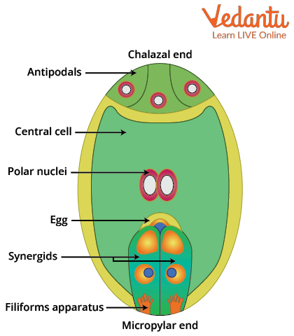
A mature embryo sac in angiosperms displays a unique organisation. It is often referred to as a 7-celled, 8-nucleate structure, which is commonly asked in embryo sac MCQs and questions. This arrangement ensures the efficient process of double fertilisation, a major difference between angiosperms and other plants.
- Egg Apparatus: Located at the micropylar end, it consists of one egg cell and two synergids. The synergids help direct the growing pollen tube during fertilisation.
- Central Cell: The largest cell in the centre contains two polar nuclei, which later fuse to form a diploid secondary nucleus (endosperm mother cell).
- Antipodal Cells: Three small cells at the chalazal end, which typically degenerate after fertilisation.
All these cells are surrounded by the embryo sac’s thin wall and play distinct roles during seed formation. The central cell ensures the development of nutritious endosperm for the growing embryo. For a comparison, check out the dicot embryo page to see how the embryo sac connects to further stages of development.
Role of Embryo Sac in Plant Reproduction
The embryo sac performs the key functions of female reproductive success in flowering plants. The process is a classic topic in embryo sac class 12 short notes and MCQs, as it highlights double fertilisation and its significance.
- Double Fertilisation: One male gamete fuses with the egg cell, forming the zygote (future embryo). The other male gamete fuses with the diploid central cell, forming the endosperm, which nourishes the embryo.
- Synergids: These regulate the pollen tube’s entry and discharge, ensuring the male gametes reach their correct targets.
- Antipodal Cells: Although their role is less clear, they may help in nutrient transfer or degeneration post-fertilisation.
The embryo sac’s ability to manage multiple fertilisation events distinguishes angiosperms. This process is not found in animals or gymnosperms. For deeper insight into plant reproductive systems, explore sexual reproduction in flowering plants on Vedantu.
Types of Embryo Sac Development
There are different types of embryo sac development, but the Polygonum type is the most common. In this monosporic development, only one megaspore out of four forms the complete embryo sac. The terminology and distinctions here make for excellent embryo sac MCQs and explain its widespread presence in plant reproduction.
- Monosporic (Polygonum type): Originates from a single megaspore (most common).
- Bisporic: Involves two nuclei from two different megaspores.
- Tetrasporic: Involves all four nuclei combining without walls separating them.
Understanding these types is important in advanced topics like megasporogenesis, which is the process by which the embryo sac forms inside the ovule. For more background, browse megasporogenesis and related reproductive processes on Vedantu.
Real-World Significance & Examples
The embryo sac is crucial in agriculture, plant breeding, and food production. By manipulating embryo sac development, scientists can increase seed yield in crops like rice, wheat, and maize. In environmental science, understanding the embryo sac’s function can help address climate-related challenges that impact seed viability. For more on plant science applications, see food science and life science.
- Example 1: Improving hybrid seed production in crops depends on precise embryo sac management.
- Example 2: Conservation efforts for endangered plants often involve studying the embryo sac to ensure successful reproduction.
- Example 3: Biotechnological advances allow for detection and correction of seed defects at the embryo sac stage.
In summary, the embryo sac’s study enhances food security, supports biodiversity, and is foundational in plant genetics and breeding.
Difference Between Ovule and Embryo Sac
| Feature | Ovule | Embryo Sac |
|---|---|---|
| Definition | Structure inside ovary that develops into a seed | Female gametophyte inside the ovule |
| Components | Integuments, nucellus, embryo sac | Egg cell, synergids, antipodals, central cell |
| Role | Protects and nourishes female gametophyte | Site of fertilisation and endosperm formation |
While the ovule encloses and supports the embryo sac, the embryo sac itself is responsible for all the essential reproductive events leading to seed formation in flowering plants.
Quick Embryo Sac Short Notes
If you need rapid revision, these embryo sac short notes will help:
- Embryo sac = female gametophyte of angiosperms, usually 7-celled, 8-nucleate.
- Develops from a single functional megaspore after meiosis.
- Composed of egg apparatus (egg + two synergids), three antipodals, and a central cell with two polar nuclei.
- Central for double fertilisation—unique to angiosperms.
- Key topic for plant reproduction, genetics, and MCQs.
Downloadable Embryo Sac Resources
To master the topic, students can refer to Vedantu’s concise embryo sac notes, detailed diagrams, and topic-wise explanations relevant for Class 12 and entrance exams. For visual learners, try creating your own embryo sac diagram or organize an embryo sac ppt to summarize the steps and the key differences from other plant reproductive structures.
Practice Embryo Sac MCQs & Sample Questions
Boost your exam preparation by practising multiple choice questions (MCQs) and short-answer samples based on real-world and theoretical concepts about the embryo sac. Exploring these questions will improve your understanding of fertilisation, development, and structure-function relationships. For more MCQs and Class 12 biology resources, visit Vedantu’s biology sections and related reproductive biology pages.
In summary, the embryo sac is central to plant reproductive biology and food production, linking cell division, fertilisation, and seed formation. Mastery of this topic prepares students for higher studies and agricultural applications, as well as a deeper appreciation of plant life cycles and biodiversity. Vedantu’s expert, student-friendly content supports learning with easy-to-digest explanations and interactive revision materials.


FAQs on Embryo Sac in Flowering Plants
1. What is an embryo sac in plants?
An embryo sac is the female gametophyte of flowering plants that develops inside the ovule and contains the egg cell. It is formed from a functional megaspore through the process of megasporogenesis and megagametogenesis.
- Located inside the ovule of the ovary
- Typically 7-celled and 8-nucleate in most angiosperms
- Contains the egg cell, synergids, antipodals, and polar nuclei
2. Where is the embryo sac located?
The embryo sac is located inside the ovule, which is present within the ovary of a flower. It develops in the nucellus region of the ovule.
- Ovule is attached to the placenta inside the ovary
- Embryo sac lies embedded in the nucellar tissue
- Protected by one or two integuments
3. What is the structure of a typical embryo sac?
A typical angiosperm embryo sac is 7-celled and 8-nucleate in structure. It is organized into distinct regions and cell types.
- Micropylar end: 1 egg cell + 2 synergids
- Central cell: 2 polar nuclei
- Chalazal end: 3 antipodal cells
4. How is the embryo sac formed?
The embryo sac is formed from a diploid megaspore mother cell through meiosis followed by mitotic divisions. The process occurs in two main stages:
- Megasporogenesis: The megaspore mother cell undergoes meiosis to produce four haploid megaspores; usually only one remains functional.
- Megagametogenesis: The functional megaspore undergoes three mitotic divisions to form eight nuclei, which organize into the mature embryo sac.
5. What is the function of the embryo sac?
The main function of the embryo sac is to enable fertilization and seed formation in flowering plants. It performs multiple roles:
- Houses the egg cell for fertilization
- Facilitates entry of the pollen tube through synergids
- Forms the endosperm after fusion of a male gamete with the polar nuclei
6. What are synergids and antipodals in the embryo sac?
Synergids and antipodals are accessory cells present in the embryo sac that support fertilization and embryo sac function. They differ in position and role:
- Synergids: Two cells at the micropylar end that guide the pollen tube using a specialized structure called the filiform apparatus.
- Antipodals: Three cells at the chalazal end that may help in nourishment and usually degenerate after fertilization.
7. What is double fertilization in relation to the embryo sac?
Double fertilization is a unique process in angiosperms where two male gametes fuse with two different nuclei inside the embryo sac. It involves:
- One male gamete fusing with the egg cell to form the zygote (syngamy).
- The second male gamete fusing with the two polar nuclei to form the triploid endosperm (triple fusion).
8. What is the Polygonum type embryo sac?
The Polygonum type embryo sac is the most common type of embryo sac found in angiosperms, characterized by a 7-celled, 8-nucleate structure. It develops from a single functional megaspore.
- Three mitotic divisions produce eight nuclei
- Nuclei arrange into egg apparatus, central cell, and antipodals
- Seen in plants like Polygonum and most flowering species
9. What is the difference between ovule and embryo sac?
The ovule is the entire structure inside the ovary that develops into a seed, while the embryo sac is the female gametophyte located inside the ovule. Key differences include:
- Ovule: Contains nucellus, integuments, and embryo sac; diploid structure.
- Embryo sac: Located within the nucellus; haploid female gametophyte.
- Ovule becomes a seed after fertilization, whereas the embryo sac forms the embryo and endosperm.
10. How many nuclei are present in a mature embryo sac?
A mature angiosperm embryo sac typically contains eight nuclei arranged within seven cells. These nuclei are distributed as follows:
- 1 nucleus in the egg cell
- 2 nuclei in two synergids
- 3 nuclei in three antipodal cells
- 2 polar nuclei in the central cell


