

What is the difference between voluntary and involuntary muscles in structure and control
The muscular system is responsible for body movements, posture, and the circulation of substances like blood and food within the body. Humans have more than 700 muscles, accounting for nearly 40% of total body weight. These muscles are attached to bones, blood vessels, and various internal organs. They work by contracting and relaxing, enabling our bodies to move and perform essential functions.
Muscles in our body can be grouped into three main types:
Skeletal Muscles: Usually attached to bones and responsible for voluntary movements.
Smooth Muscles: Found inside organs such as blood vessels and intestines, controlling involuntary actions.
Cardiac Muscles: Located only in the heart, ensuring continuous pumping of blood.
Before understanding the difference between voluntary and involuntary muscles, let us see how they are broadly classified and how they function.
Key Differences Between Voluntary and Involuntary Muscles
When you explore the difference between voluntary and involuntary muscles with examples, you will notice that voluntary muscles primarily enable movements you control consciously, while involuntary muscles maintain critical functions inside the body without your active decision.
Classification of Muscles in the Human Body
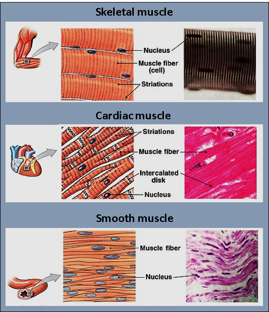
Skeletal Muscles
They were found attached to the skeleton by tendons.
Under voluntary control (you can decide when to move your arm or leg).
Appear striated (striped) under a microscope.
Contains multiple nuclei in each cell.
Smooth Muscles
Present in the walls of internal organs like the intestines, stomach, and blood vessels.
Involuntary in nature (work automatically without conscious control).
Spindle-shaped cells with a single nucleus.
Not striated under a microscope.
Cardiac Muscles
Found only in the heart.
Involuntary in action, as the heart beats continuously without conscious effort.
Appear striated and branched.
Usually, they have one nucleus per cell but may occasionally have more.
Voluntary Muscles
Voluntary muscles are controlled by the somatic nervous system. You can move them consciously—like raising your arm or walking. These muscles:
It has a cylindrical shape and can be quite long.
Possess multiple nuclei per fibre.
Display striations (light and dark bands).
Requires a high amount of energy, which is why they can get tired after prolonged activity.
Voluntary and involuntary muscles examples for the voluntary category include:
Muscles of the arms and legs (e.g. biceps, triceps).
Muscles in the abdominal wall.
The tongue.
The diaphragm (though it can also function involuntarily for breathing).
Involuntary Muscles
Involuntary muscles work under the autonomic nervous system. They handle essential internal processes like digestion and blood flow. You cannot consciously control them. Involuntary muscles are of two main types:
Smooth Muscles
Found in the walls of organs, including the stomach, intestines, uterus, and blood vessels.
These muscles help move substances like food and blood through the body.
They have spindle-shaped cells with a single nucleus each.
Cardiac Muscles
Found only in the heart.
Have striations and branched fibres.
Work continuously to pump blood around the body without fatigue.
Some voluntary and involuntary muscles examples for the involuntary category are:
Smooth Muscles: In the walls of the alimentary canal, blood vessels, respiratory tract, and ducts of glands.
Cardiac Muscles: In the heart, ensuring continuous heartbeat.
Additional Points about Voluntary and Involuntary Motor Muscles
Voluntary muscles (skeletal muscles) can be consciously controlled, so they are also referred to as voluntary motor muscles.
Involuntary muscles (smooth and cardiac) act independently of conscious thought, which is why they are sometimes called involuntary motor muscles.
A unique example is the diaphragm. While it primarily functions involuntarily for breathing, you can also control it voluntarily to an extent (holding your breath, for instance).
Quick Quiz (with Answers)
Test your understanding with this short quiz:
Which type of muscle is found in the walls of the stomach?
A. Skeletal
B. Smooth
C. Cardiac
D. None of the above
Answer: B. Smooth
Which muscle type never gets fatigued?
A. Skeletal
B. Cardiac
C. Smooth
D. Voluntary muscles only
Answer: B. Cardiac
Which nervous system primarily controls voluntary muscles?
A. Somatic nervous system
B. Autonomic nervous system
C. Peripheral nervous system
D. Central nervous system
Answer: A. Somatic nervous system
What is the primary function of cardiac muscles?
A. Move limbs
B. Aid in digestion
C. Pump blood
D. Support posture
Answer: C. Pump blood
Related Topics


FAQs on Voluntary and Involuntary Muscles Key Differences and Functions
1. What is the difference between voluntary and involuntary muscles?
The main difference between voluntary muscles and involuntary muscles is that voluntary muscles are controlled consciously, while involuntary muscles function automatically without conscious control.
- Voluntary muscles are attached to bones and help in body movements like walking and writing.
- Involuntary muscles are found in internal organs and control processes like digestion and heartbeat.
- Voluntary muscles are also called skeletal muscles, whereas involuntary muscles include smooth muscles and cardiac muscle.
2. What are voluntary muscles?
Voluntary muscles are muscles that work under conscious control and are responsible for body movements.
- They are also known as skeletal muscles.
- They are attached to bones by tendons.
- They help in actions like running, lifting objects, and speaking.
- They appear striated (striped) under a microscope.
3. What are involuntary muscles?
Involuntary muscles are muscles that function automatically without conscious effort.
- They are controlled by the autonomic nervous system.
- They are found in internal organs such as the stomach, intestines, and blood vessels.
- Types include smooth muscles and cardiac muscle.
- They regulate vital processes like digestion, breathing, and blood circulation.
4. Where are voluntary and involuntary muscles found in the body?
Voluntary muscles are attached to bones, while involuntary muscles are located in internal organs.
- Voluntary muscles: Arms, legs, face, neck, and trunk.
- Smooth muscles (involuntary): Stomach, intestines, urinary bladder, blood vessels.
- Cardiac muscle (involuntary): Found only in the heart.
5. How are voluntary muscles controlled?
Voluntary muscles are controlled by signals from the brain through the somatic nervous system.
- The cerebrum initiates movement.
- Motor neurons transmit impulses to skeletal muscles.
- The muscle fibers contract in response to nerve stimulation.
6. How are involuntary muscles controlled?
Involuntary muscles are controlled automatically by the autonomic nervous system without conscious effort.
- The sympathetic and parasympathetic divisions regulate their activity.
- They respond to internal stimuli such as hormones and stretch.
- They maintain essential life functions like heartbeat and digestion.
7. What are examples of voluntary and involuntary muscles?
Examples of voluntary muscles include biceps and quadriceps, while examples of involuntary muscles include stomach muscles and heart muscle.
- Voluntary muscles: Biceps, triceps, thigh muscles.
- Smooth muscles: Walls of the intestine and blood vessels.
- Cardiac muscle: Muscle of the heart.
8. Are cardiac muscles voluntary or involuntary?
Cardiac muscles are involuntary muscles that contract automatically to pump blood throughout the body.
- They are found only in the heart.
- They are striated but not under conscious control.
- Their rhythm is regulated by the sinoatrial (SA) node.
9. Do voluntary and involuntary muscles differ in structure?
Yes, voluntary and involuntary muscles differ in structure, appearance, and cell shape.
- Skeletal muscles: Long, cylindrical, multinucleated, and striated fibers.
- Smooth muscles: Spindle-shaped, single nucleus, non-striated fibers.
- Cardiac muscle: Branched, striated fibers with intercalated discs.
10. Why are involuntary muscles important for survival?
Involuntary muscles are essential for survival because they control vital automatic functions of the body.
- They maintain heartbeat and blood circulation.
- They enable digestion through peristalsis.
- They regulate breathing by controlling airway diameter.


