


What is the Role of Centrioles in NEET Biology Syllabus?
Centriole is a key cell structure that often appears in NEET Biology questions, especially in cell biology. Understanding centrioles helps students grasp crucial processes like cell division and organization of the cytoskeleton. This concept is foundational in Biology, making it an essential topic for NEET aspirants aiming for clear conceptual understanding and scoring accuracy in the exam.
What is a Centriole?
A centriole is a small, cylindrical organelle found in most animal cells. It typically exists in pairs and plays a crucial role in organizing microtubules during cell division. Centrioles help form the spindle fibers that separate chromosomes during mitosis and meiosis. In a simple sense, centrioles are like tiny organizers in the cell, ensuring that the genetic material is distributed accurately when a cell divides.
Core Ideas and Fundamentals of Centriole
Basic Structure of Centrioles
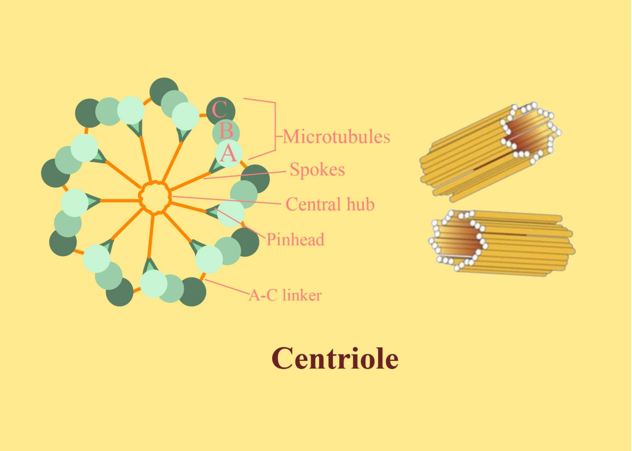
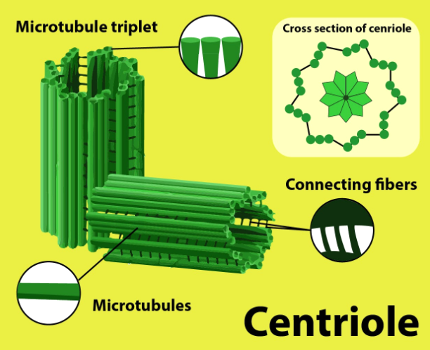
Centrioles are shaped like short cylinders made up of microtubules arranged in a specific pattern. Typically, each centriole consists of nine triplet microtubules arranged in a ring. These structures are usually found near the nucleus in the cell’s centrosome region, where they exist as a pair oriented at right angles to each other.
Position and Duplication
In animal cells, centrioles are usually located just outside the nucleus in the centrosome. Before cell division begins, each centriole duplicates so that each daughter cell receives a pair.
Functions of Centrioles
- Organizing microtubules to form spindle fibers during cell division
- Helping in the formation of cilia and flagella in eukaryotic cells
- Maintaining the structure of the cytoskeleton
Important Sub-Concepts Related to Centrioles
Centrosome
The centrosome is the region near the cell nucleus where a pair of centrioles is found. It acts as the main microtubule-organizing center (MTOC) and is essential in directing the cell cycle progression and division.
Mitotic Spindle Formation
During mitosis, centrioles help in forming spindle fibers. These fibers attach to chromosomes and ensure their even distribution between the two daughter cells. An error in spindle formation can lead to improper chromosome separation, resulting in genetic disorders.
Cilia and Flagella Formation
Centrioles form basal bodies, which are the starting points for the formation of cilia and flagella. These structures are vital for cell movement and the movement of substances across cell surfaces.
Principles and Relationships Involving Centrioles
There are no complex formulas for centrioles, but students should understand their relationships and roles. The arrangement of microtubules is described as a ‘9+0’ pattern in a centriole (nine triplets with no central pair), and the duplication of centrioles happens once per cell cycle, ensuring that each daughter cell inherits one pair.
Key Features of Centrioles
- Cylindrical, microtubule-based structure
- Found in most animal cells but absent in plant cells
- Each centriole is made of nine microtubule triplets organized in a ring
- Centriole pairs are always present at right angles to each other
Importance of Centrioles for NEET
Centrioles are often tested in NEET through conceptual MCQs on cell organelles, cell division, and cytoskeleton. Understanding centrioles is important for answering questions about mitosis, meiosis, and differences between plant and animal cells. A clear understanding of this concept forms the base for learning about genetic inheritance, chromosome behavior, and overall cell function. It is closely connected with the broader topics of cell biology, molecular biology, and genetics that are highly relevant in NEET.
How to Study Centrioles Effectively for NEET
- Draw and label centriole structure diagrams to aid memory.
- Focus on understanding the role of centrioles in cell division and how errors can affect the outcome.
- Practice distinguishing features between centrioles, cilia, flagella, and centrosomes.
- Solve NEET-level MCQs involving cell organelles and their functions.
- Create mind maps linking centrioles with related cell structures and processes.
- Regularly revise definitions and diagrams for quick recollection during exams.
Common Mistakes Students Make with Centrioles
- Confusing centrioles with centrosomes or basal bodies
- Assuming centrioles are present in plant cells (they are absent)
- Forgetting the 9+0 arrangement of microtubules in centrioles
- Mixing up the roles of centrioles in cell division versus cilia/flagella formation
- Neglecting diagram practice, which can cause confusion in labelling questions
Quick Revision Points for Centrioles
- Centriole: cylindrical organelle - 9 microtubule triplets, no central microtubule (9+0 pattern)
- Only found in animal cells, not in plant cells
- Located in the centrosome near the nucleus, always as a pair at right angles
- Duplicate once per cell cycle, ensuring proper cell division
- Organize spindle fiber formation; also form basal bodies for cilia and flagella
- Mistakes in centriole function can disrupt chromosome segregation during division
FAQs on Centriole in NEET Biology: Structure and Function Explained
1. What is a centriole in biology?
Centriole is a cylindrical cell structure found mainly in animal cells and some lower plants, essential for cell division and forming cilia and flagella.
Key points about centriole in NEET:
- Comprised of nine triplets of microtubules arranged in a ring
- Helps organize the mitotic spindle during cell division (mitosis and meiosis)
- Plays a role in the formation of cilia and flagella
- Usually located within the centrosome
2. What is the function of centriole?
Centriole plays a crucial role in cell division and the organization of microtubules.
Functions of centriole in NEET-relevant syllabus:
- Helps form and organize the spindle fibers during mitosis and meiosis
- Assists in the formation of cilia and flagella
- Maintains proper positioning of cell organelles by organizing the microtubule network
3. What is the structure of centriole?
Centriole is a barrel-shaped structure made up of microtubules arranged in a specific pattern.
Key structural details:
- Composed of nine triplet microtubules arranged in a cylindrical shape
- The triplets are held together by proteins
- Lacks a membrane, making it a non-membranous cell organelle
4. Is centriole present in plant cells?
In most cases, centrioles are absent in higher plant cells but are present in animal cells and some lower plants.
NEET students should remember:
- Animal cells (except some fungi) contain centrioles
- Higher plant cells usually lack centrioles but can perform cell division without them
- Some lower plants and algae may have centrioles
5. What happens if centrioles are absent from a cell?
If centrioles are absent, animal cells face difficulty in organizing spindle fibers during mitosis, but plant cells adapt using other mechanisms.
Effects of centriole absence:
- Possible abnormal cell division in animal cells
- Formation of improper spindle apparatus
- Plant cells utilize other microtubule organizing centers for division
6. Which cell organelle is formed by centrioles?
Cilia and flagella are cell organelles formed with the help of centrioles.
Centrioles also contribute to:
- Spindle fiber formation during cell division
- Basal body structure for cilia and flagella development
7. Do bacteria have centrioles?
Bacteria lack centrioles, as they are prokaryotic cells.
- Centrioles are only found in eukaryotic cells (mainly animal cells and some lower plants)
- Bacteria divide using different mechanisms (binary fission)
8. What is the difference between a centriole and a centrosome?
Centriole is a single cylindrical structure, while centrosome consists of two centrioles arranged at right angles.
Key differences:
- Centriole: One barrel-shaped microtubule structure
- Centrosome: Contains a pair of centrioles plus pericentriolar material
- Centriole forms the core of the centrosome, which acts as the main microtubule organizing center (MTOC)
9. Are centrioles membrane-bound organelles?
No, centrioles are non-membranous organelles made up of microtubules.
- Unlike organelles such as mitochondria or ER, centrioles lack a surrounding membrane
- They play important roles in cell division and cilia formation
10. Why are centrioles important for NEET exam preparation?
Centrioles are a frequently tested topic in NEET due to their essential roles in cell division and organelle formation.
For NEET exam:
- Understand structure, location, and function of centrioles
- Differentiate between centrioles and centrosomes
- Know which cells contain centrioles
- Be able to answer MCQs on their involvement in mitosis, cilia, and flagella
11. What is the role of centrioles during cell division?
Centrioles help organize spindle fibers, ensuring accurate separation of chromosomes during cell division.
In NEET terms:
- Centrioles duplicate before mitosis/meiosis
- Move to opposite poles of the cell
- Assist in spindle apparatus formation
- Guide chromosome movement
12. How many centrioles are present in a typical animal cell?
A typical animal cell contains a pair of centrioles, oriented at right angles to each other.
Quick facts:
- 2 centrioles per centrosome
- Paired arrangement: one mother and one daughter centriole