

Detailed Meiosis Diagram with Phases and Chromosomal Events
Meiosis is a vital type of cell division in biology responsible for producing gametes—such as sperm and egg cells—that carry half the genetic material of a parent cell. This reduction is essential for maintaining chromosome number and enabling sexual reproduction in animals and plants. Meiosis differs from other forms of cell division because it produces four genetically unique haploid cells from a single diploid cell, introducing genetic diversity across generations.
The meiosis process involves two consecutive divisions, known as Meiosis I and Meiosis II. Each division contains specific phases that aid in chromosome number reduction and genetic shuffling. Understanding these phases and their visual representation in diagrams helps students recognize chromosome behavior and anticipate outcomes of sexual reproduction.
Overview of Meiosis: Steps and Significance
Meiosis ensures the correct chromosome number in sexually reproducing organisms and results in genetic variation. The process can be summarized as:
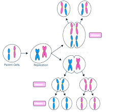
- A diploid cell (2n) undergoes one round of DNA replication.
- Two divisions—Meiosis I and Meiosis II—create four haploid cells (n).
- Each haploid cell has a unique combination of genetic material.
Stages of Meiosis
Meiosis is divided into two main phases, each with distinct stages. This breakdown is essential for board and competitive exam preparation.
| Division | Phase | Event |
|---|---|---|
| Meiosis I | Prophase I | Chromosomes condense, homologous chromosomes pair and crossing over occurs. |
| Metaphase I | Homologous pairs align at the cell's equator. | |
| Anaphase I | Homologous chromosomes separate, sister chromatids stay together. | |
| Telophase I | Two haploid cells form; chromosomes may decondense slightly. | |
| Meiosis II | Prophase II | Chromosomes condense again in each haploid cell. |
| Metaphase II | Chromosomes align singly at the cell equator. | |
| Anaphase II | Sister chromatids separate to opposite poles. | |
| Telophase II | Four unique haploid cells formed after cell division. |
Key Definitions in Meiosis
| Term | Explanation |
|---|---|
| Homologous Chromosomes | Chromosome pairs from each parent, similar in structure and genes. |
| Crossing Over | Exchange of genetic material between homologous chromosomes during Prophase I. |
| Ploidy | The number of chromosome sets (2n = diploid, n = haploid). |
| Gametes | Sex cells (sperm or egg) produced after meiosis. |
| Genetic Variation | Diversity in genetic material produced through meiosis. |
Step-by-Step Example: Chromosome Number Change
Consider a diploid cell with 2n = 4 chromosomes:
- Before meiosis: 4 chromosomes (diploid)
- After Meiosis I: 2 chromosomes per cell (haploid, but with sister chromatids)
- After Meiosis II: 4 cells with 2 single chromosomes each (haploid)
Comparison: Mitosis vs. Meiosis
| Feature | Mitosis | Meiosis |
|---|---|---|
| Number of Divisions | 1 | 2 |
| Number of Cells Produced | 2 | 4 |
| Ploidy of Cells | Diploid (2n) | Haploid (n) |
| Genetic Variation | No | Yes |
| Role | Growth and repair | Gamete formation |
Why is Meiosis Important?
Meiosis is crucial for maintaining a stable chromosome number and providing genetic variety in populations. Crossing over and the random assortment of chromosomes lead to new gene combinations, supporting evolution and adaptation.
Practice: Meiosis Questions for Students
- Draw the stages of meiosis in a cell with 2n = 4 chromosomes and label each step.
- List two key differences between mitosis and meiosis.
- Describe the event of crossing over and its biological consequence.
- How does meiosis contribute to genetic diversity?
Continue Learning About Meiosis
- Meiosis Definition
- Meiosis Phases
- Meiosis I Stages and Process
- Meiosis II Cell Division
- Stages of Meiosis
- Meiosis Cell Division
- MCQs on Meiosis
Meiosis is a foundation of genetics and sexual reproduction, helping future generations inherit a balanced set of chromosomes and diverse traits. A strong grasp of meiosis diagrams and concepts helps learners prepare confidently for their Biology studies and exams.


FAQs on Meiosis Diagram and Stages of Cell Division
1. What is meiosis?
Meiosis is a type of cell division that reduces the chromosome number by half to produce four genetically different haploid cells. It occurs in reproductive cells to form gametes such as sperm and eggs. Key features include:
- Two successive divisions: Meiosis I and Meiosis II
- Reduction of chromosome number from diploid (2n) to haploid (n)
- Generation of genetic variation through crossing over and independent assortment
2. What are the stages shown in a meiosis diagram?
A meiosis diagram shows two main divisions—Meiosis I and Meiosis II—each with four stages. The stages are:
- Prophase I, Metaphase I, Anaphase I, Telophase I
- Prophase II, Metaphase II, Anaphase II, Telophase II
These stages illustrate chromosome pairing, separation, and the formation of four haploid daughter cells.
3. What happens in Prophase I of meiosis?
In Prophase I, homologous chromosomes pair up and exchange genetic material through crossing over. This stage is crucial for genetic variation. Key events include:
- Synapsis: pairing of homologous chromosomes
- Formation of bivalents or tetrads
- Exchange of DNA at chiasmata
- Breakdown of the nuclear membrane
4. What is the difference between meiosis I and meiosis II?
The main difference between Meiosis I and Meiosis II is that homologous chromosomes separate in Meiosis I, while sister chromatids separate in Meiosis II. Specifically:
- Meiosis I: Reduction division; chromosome number is halved
- Meiosis II: Similar to mitosis; sister chromatids are pulled apart
- Result: Four genetically distinct haploid cells
5. Why is meiosis important?
Meiosis is important because it produces haploid gametes and increases genetic variation in sexually reproducing organisms. Its significance includes:
- Maintaining constant chromosome number across generations
- Creating variation through crossing over and independent assortment
- Supporting evolution and adaptation
6. What does a meiosis diagram show about chromosome number?
A meiosis diagram shows the reduction of chromosome number from diploid (2n) to haploid (n). It illustrates:
- Starting cell with two sets of chromosomes
- Separation of homologous chromosomes in Meiosis I
- Final formation of four haploid cells, each with one set of chromosomes
This reduction is essential for sexual reproduction.
7. How does crossing over appear in a meiosis diagram?
In a meiosis diagram, crossing over appears as an exchange of segments between homologous chromatids during Prophase I. It is shown by:
- Overlapping chromatids forming chiasmata
- Swapping of corresponding DNA segments
- Recombinant chromosomes with mixed parental traits
This process increases genetic diversity in gametes.
8. What is the end result of meiosis?
The end result of meiosis is four genetically different haploid daughter cells. These cells:
- Contain half the original chromosome number
- Are genetically unique due to recombination
- Develop into gametes such as sperm or ova in animals
9. How is meiosis different from mitosis?
Meiosis differs from mitosis because it produces four haploid cells with genetic variation, while mitosis produces two identical diploid cells. Key differences include:
- Meiosis has two divisions; mitosis has one
- Crossing over occurs only in meiosis
- Meiosis is for gamete formation; mitosis is for growth and repair
10. What are homologous chromosomes in a meiosis diagram?
Homologous chromosomes are pairs of chromosomes that carry the same genes but may have different alleles. In a meiosis diagram, they:
- Pair during Prophase I
- Form bivalents or tetrads
- Separate during Anaphase I
Each pair consists of one maternal and one paternal chromosome.


