

What is IgG antibody structure function types and significance
The concept of IgG antibody is essential in biology and helps explain real-world biological processes and exam-level questions effectively.
Understanding IgG Antibody
IgG antibody, also known as Immunoglobulin G, is the most abundant type of antibody found in human blood and extracellular fluids. IgG plays a vital role in the body's immune response by neutralizing bacteria, viruses, and toxins, and is involved in immunity, infection diagnosis, and medical testing. This concept is important in areas like immunity, humoral immunity, and medical diagnostics.
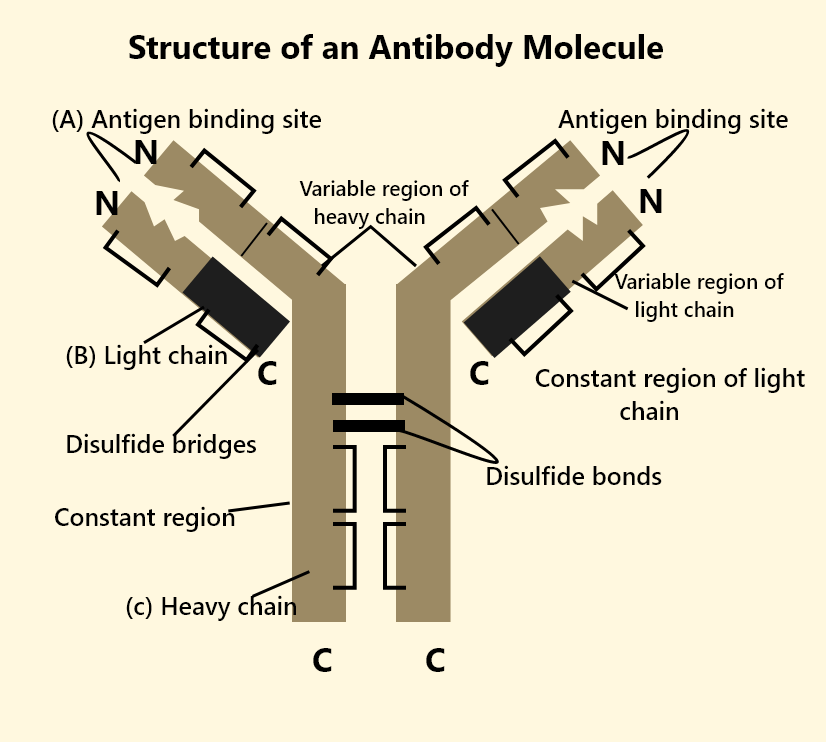
Structure of IgG Antibody
IgG antibody has a distinct Y-shaped structure. Each molecule consists of four peptide chains: two identical heavy (gamma) chains and two identical light chains, held together by disulfide bonds. The arms (Fab fragments) are responsible for binding antigens, while the stem (Fc region) determines the biological function of the antibody. The variable regions of the Fab arms contribute to the antibody's ability to recognize a wide array of pathogens. This structure makes IgG flexible and efficient in targeting invaders.
Functions of IgG Antibody
The IgG antibody offers protection through several key roles in the human immune system:
- Neutralization: Binds to toxins and pathogens, preventing them from harming cells.
- Opsonization: Tags microbes for easier recognition and destruction by phagocytes.
- Complement Activation: Triggers a cascade of immune proteins to remove pathogens.
- Antibody-dependent cell-mediated cytotoxicity (ADCC): Helps recruit natural killer cells to attack infected or abnormal cells.
- Crosses Placenta: Only antibody class providing passive immunity to newborns by transferring from mother to fetus.
Subclasses of IgG Antibodies
There are four subclasses of IgG in humans: IgG1, IgG2, IgG3, and IgG4. Each has slightly different roles and properties. For example, IgG1 and IgG3 are especially effective at activating the complement system and responding to protein antigens, while IgG2 is more involved with responses to polysaccharide antigens.
IgG Antibody in Testing and Diagnosis
The IgG antibody test measures the level of IgG in blood and is widely used to diagnose previous infection or exposure to pathogens such as measles, rubella, and herpes simplex virus. A result of "IgG antibody positive" means the person has encountered the pathogen before, either through infection or vaccination, and has developed a memory immune response.
Interpretation of IgG levels helps doctors assess immunity, determine vaccination needs, and detect certain autoimmune conditions or immunodeficiencies. Remember, high or low IgG levels should always be interpreted by a healthcare professional as results can vary due to different medical reasons.
Here’s a helpful table to understand IgG antibody subclasses better:
IgG Antibody Subclass Table
| Subclass | Main Role | Serum % | Placental Transfer |
|---|---|---|---|
| IgG1 | Response to proteins, strong complement activation | ~60% | Yes |
| IgG2 | Response to polysaccharides, weak complement activation | ~32% | Limited |
| IgG3 | Strong complement activation, antiviral/antibacterial | ~4% | Yes |
| IgG4 | Chronic antigen exposure, minimal complement activation | ~4% | Yes |
Difference Between IgG and IgM
IgG and IgM antibodies are often compared in medical diagnostics. Understanding their differences helps in interpreting results from infection tests:
| Feature | IgG | IgM |
|---|---|---|
| Appearance in Infection | Later, indicates past infection or immunity | First, indicates current or recent infection |
| Structure | Monomer (Y-shaped) | Pentamer (5 Y-shaped units) |
| Placental Transfer | Yes | No |
| Test Example | IgG positive = past exposure/immunity | IgM positive = current/recent infection |
Common Mistakes to Avoid
- Confusing IgG antibody with IgM or IgA antibodies in test results.
- Assuming a positive IgG means you are currently infected (it usually means past infection or immunity).
- Not considering all subclasses and their unique properties.
Real-World Applications
The concept of IgG antibody is used in medicine for diagnosing diseases, checking vaccine effectiveness, and monitoring immune status in conditions like autoimmune disorders. Vedantu helps students relate these concepts to practical scenarios, exam cases, and health awareness. IgG testing is also vital in blood banks, pregnancy health, and epidemiological studies.
In this article, we explored IgG antibody, its structure, subclasses, key functions, clinical uses, and how to avoid common mistakes. To learn more and build confidence, keep practicing with Vedantu.
Explore More Related Topics
- Immunity
- Humoral Immunity
- Immunoglobulin Structure
- Antibodies
- Antigen-Antibody Reaction Types
- Immunology
- Virus
- Measles
- Microbiology
- NEET Biology MCQs


FAQs on IgG Antibody Structure and Role in Immunity
1. What is IgG antibody?
The IgG antibody is the most abundant type of immunoglobulin in human blood and plays a major role in long-term immune protection. It is a type of immunoglobulin (Ig) produced by plasma cells in response to infections.
Key features of IgG include:
- Accounts for about 70–75% of total antibodies in serum
- Provides long-term immunity after infection or vaccination
- Can cross the placenta to protect the fetus
- Involved in neutralization and opsonization of pathogens
2. What is the structure of IgG antibody?
The IgG antibody has a Y-shaped structure composed of four polypeptide chains. It consists of:
- Two identical heavy chains (γ chains)
- Two identical light chains
- Variable regions that bind to specific antigens
- A constant region (Fc region) that interacts with immune cells
The antigen-binding sites are located at the tips of the Y-shaped arms, allowing IgG to specifically recognize and bind foreign antigens.
3. What is the function of IgG antibody?
The main function of IgG antibodies is to protect the body by recognizing and eliminating pathogens. IgG performs several immune functions:
- Neutralization – blocks toxins and viruses from entering cells
- Opsonization – tags pathogens for phagocytosis
- Complement activation – triggers the classical complement pathway
- Antibody-dependent cellular cytotoxicity (ADCC) – helps immune cells kill infected cells
These roles make IgG essential for adaptive immunity and long-term protection.
4. How does IgG provide long-term immunity?
IgG provides long-term immunity by persisting in the bloodstream and rapidly responding to previously encountered antigens. After first exposure to a pathogen:
- Memory B cells are formed
- On re-exposure, memory B cells quickly produce large amounts of IgG antibodies
- This leads to a faster and stronger secondary immune response
Because IgG has a long half-life (about 21 days), it remains in circulation to maintain lasting immune protection.
5. Can IgG cross the placenta?
Yes, IgG is the only class of antibody that can cross the placenta to provide passive immunity to the fetus. This transfer occurs through:
- Binding of IgG to Fc receptors (FcRn) in the placenta
- Transport into the fetal circulation
Maternal IgG protects newborns against infections during the first few months of life until their own immune system becomes fully functional.
6. What is the difference between IgG and IgM?
The main difference between IgG and IgM is that IgM is produced first during an infection, while IgG provides long-term immunity. Key differences include:
- IgM: First antibody in primary response, pentamer structure, short-lived
- IgG: Dominant in secondary response, monomer structure, long-lasting
- IgG can cross the placenta; IgM cannot
Clinically, high IgM suggests recent infection, while IgG indicates past exposure or immunity.
7. What are the subclasses of IgG?
IgG has four subclasses in humans: IgG1, IgG2, IgG3, and IgG4. These subclasses differ in structure and immune function:
- IgG1: Most abundant, effective against protein antigens
- IgG2: Important for response to polysaccharide antigens
- IgG3: Strong complement activator
- IgG4: Involved in chronic antigen exposure and allergic responses
Each subclass has slightly different roles in immune defense and disease.
8. How is IgG involved in complement activation?
IgG activates the complement system by binding to the C1 complex in the classical pathway. When IgG binds to an antigen:
- The Fc region becomes exposed
- C1 binds to the Fc portion of two adjacent IgG molecules
- This triggers the classical complement pathway
Complement activation leads to pathogen lysis, inflammation, and enhanced phagocytosis.
9. What does a high IgG level indicate?
A high IgG level usually indicates past infection, chronic infection, or an active immune response. Elevated IgG may be seen in:
- Previous exposure to a pathogen
- Chronic infections
- Autoimmune diseases
- After vaccination
In diagnostic tests, the presence of specific IgG antibodies suggests prior immunity rather than a recent infection.
10. Where is IgG found in the body?
IgG is primarily found in the blood plasma and extracellular fluid, where it circulates to detect and neutralize pathogens. It is present in:
- Serum
- Lymph
- Tissue fluids
- Placental circulation (during pregnancy)
Because IgG can diffuse easily into tissues, it plays a key role in systemic immune defense throughout the body.


