

What Is the Procedure to Prepare a Temporary Mount of Leaf Peel to Show Stomata
Stomata are the elliptical openings on leaves, guarded by two cells, which when swollen with water allow stomata to open and when flaccid allow stomata to close. The guard cells of the stomata possess large vacuoles, chloroplast, and nucleus. Stomata help the plant to take in carbon dioxide needed for photosynthesis. They look like tiny mouths opening and closing as they help in transpiration.
The bigger/wider the leaf, the greater the rate of transpiration as more surface means more stomata will be present. Plants possessing smaller stomata have lower levels of evaporation and survive harsh conditions better. All green plants are termed as producers as they produce their own food from solar energy, and carry out the vital processes of photosynthesis and respiration where gaseous exchange between the tissue of plants and the atmosphere is an essential component of the whole ecosystem. This is carried out through the tiny openings known as stomata.
Types of Stomata
A plant survives better with faster growth if it possesses a higher number of stomata and a wet climate. The lower the number of stomata, the lower the rate of photosynthesis, and a drier climate is not ideal for plant growth.
There are four types of stomata:
Moss Type: These are found in certain mosses.
Gymnospermous: These are naked seed plants deeply sunken to reduce water loss during transpiration.
Coniferous: Sunken stomata and the guard cells are elliptical.
Gramineous: These are found in grade families, two guard cells and two subsidiary cells are present too.
On the basis of arrangement, the types of stomata are:
Anomocytic Stomata - This type of stomata is embedded in the epidermal cells having fixed shape and size where there is no fixed number of cells surrounding the stomata.
Anisocytic Stomata - In this arrangement, three subsidiary unequal cells are surrounding the stomata.
Paracytic Stomata - Here the stomatal pore and guard cells surround the stomata.
Diacytic Stomata - It is surrounded by subsidiary cells lying perpendicular to the guard cells.
Gramineous Stomata - Stomata has two dumbbell shaped guard cells and subsidiary cells which lie parallel to guard cells.
Coniferous Stomata - It is found on the surface of the leaves of gymnosperm plants and is found below the leaf surface.
Experimental Setup
Materials Required:
To prepare a temporary mount of a leaf peel to show stomata, we need to follow the correct method of arrangement. We need some essential equipment such as needles, forceps, watch glass, dropper, slides, coverslip, blotting paper, safranin, glycerine, and a compound microscope.
Procedure:
First and foremost, fold a leaf to pull apart and take the peel off the leaf from the lower surface. Peeling the leaf with a blade requires patience and dexterity. We must immediately dip the transparent peel off the leaf in water kept in watch glass to avoid shrinkage and crumpling. Let it remain in the water for a while and in the watch glass, add a few drops of glycerine so that the peel remains hydrated.
Let it rest, then add a few drops of safranin which are red in colour through a dropper. Now we take out the peel with the help of forceps and put it gently on the glass slide. We blot away the excess glycerin and safranin with blotting paper. On examination, under a compound microscope, we clearly observe the epidermal cells containing stomata on the lower surface of the leaf.
Experimental Observations
The epidermal cells are seen in an irregular manner with no intracellular space between them. Stomata and guard cells both are observed clearly on the surface. Guard cells possess a nucleus and chloroplasts too. They possess a thin outer and thick inner cover. The number of stomata is less compared to those found on the lower surface of the leaf. The reason behind this is to prevent excessive loss of water through evaporation which happens because of exposure to direct sunlight.
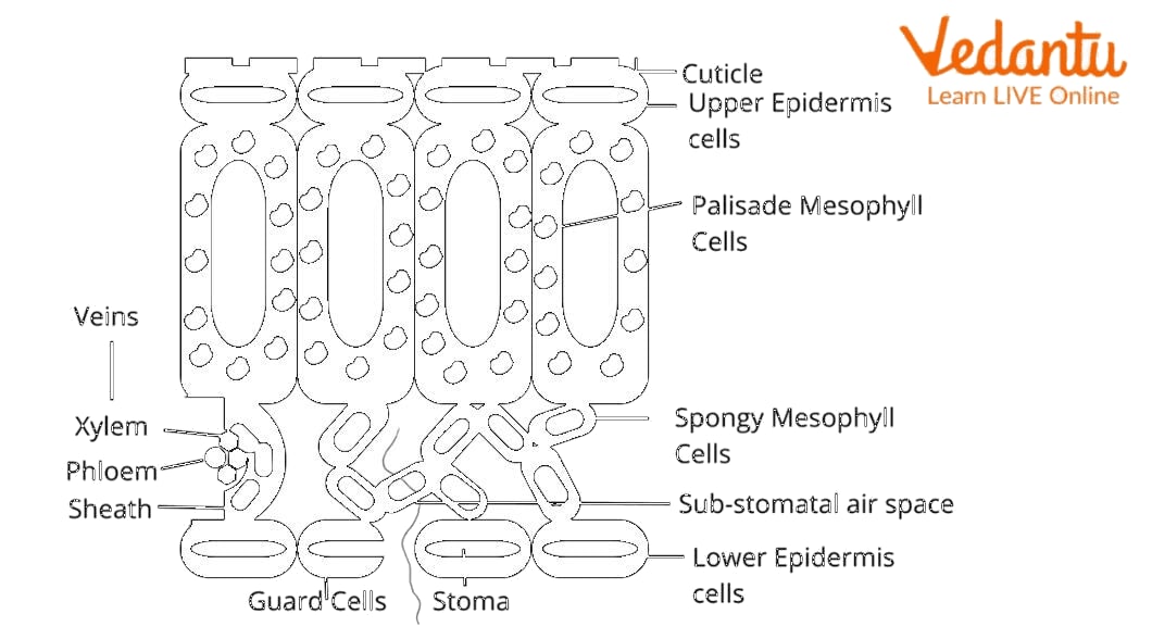
Leaf Observed Under Microscope
Interesting Fact
An interesting fact is that the stomata are studied during research if a plant has undergone any kind of stress due to excessive heat, drought, or any kind of harsh conditions.
Important Questions
1. What are light induced stomatal responses?
Ans: Light induced stomatal responses were first reported by Darwin (1989). Stomata open up in response to light that includes blue and red light. Red light makes it possible for stomata to open by photosynthesis in the guard cell chloroplasts. Blue light brings about stomatal opening. Phototropins in the guard cell act as receptors for blue light and opening of stomata.
2. Which hormone is responsible for stomatal colour?
Ans: The hormone which is responsible for the colour of stomata is abscisic acid. It is a plant hormone largely involved in the growth and development of the plant.
Key Features of Preparing a Temporary Mount of a Leaf Peel to Show Stomata
The peel should be cut to a proper size and kept hydrated with glycerine
A few drops of safranin help in magnifying the stomata and guard cells with ease.
A coverslip should be placed in such a manner as to avoid air bubbles.
Stomata are better seen on the lower surface of a dicot leaf


FAQs on Preparing a Temporary Mount of Leaf Peel to Observe Stomata Under Microscope
1. What is the aim of preparing a temporary mount of leaf peel to show stomata?
The aim of preparing a temporary mount of leaf peel is to observe stomata under a microscope. This experiment helps to:
- Study the structure of stomatal apparatus
- Identify guard cells and the stomatal pore
- Understand the role of stomata in transpiration and gaseous exchange
2. What are stomata in plants?
Stomata are tiny pores present on the epidermis of leaves that regulate gas exchange and water loss. Each stoma consists of:
- Two guard cells
- A central stomatal pore
3. What materials are required to prepare a temporary mount of leaf peel?
The materials required for preparing a temporary mount of leaf peel include basic laboratory equipment and reagents. These are:
- Fresh leaf (commonly Tradescantia or Rhoeo)
- Slide and coverslip
- Forceps and needle
- Safranin stain
- Glycerine
- Brush and dropper
- Compound microscope
4. How do you prepare a temporary mount of leaf peel to show stomata step by step?
A temporary mount of leaf peel is prepared by peeling, staining, and mounting the epidermis on a slide for microscopic observation. The steps are:
- Peel a thin layer of the lower epidermis using forceps.
- Place the peel in water in a watch glass.
- Stain it with safranin for 1–2 minutes.
- Wash off excess stain with water.
- Transfer the peel to a slide and add a drop of glycerine.
- Place a coverslip gently to avoid air bubbles.
- Observe under a compound microscope.
5. Why is safranin used in the temporary mount of leaf peel?
Safranin is used to stain the leaf peel so that the guard cells and epidermal cells become clearly visible under the microscope. It:
- Provides contrast to transparent cells
- Highlights the stomatal apparatus
- Makes microscopic observation easier
6. Why is the lower epidermis of the leaf usually taken to show stomata?
The lower epidermis is usually taken because it contains a higher number of stomata in most dicot leaves. This helps in:
- Reducing excessive water loss
- Protecting stomata from direct sunlight
- Making observation easier due to greater stomatal density
7. What is the structure of the stomatal apparatus?
The stomatal apparatus consists of two guard cells surrounding a central stomatal pore. Its structure includes:
- Kidney-shaped guard cells (in dicots)
- Thick inner walls and thin outer walls
- Chloroplasts present inside guard cells
8. What precautions should be taken while preparing a temporary mount of leaf peel?
Proper precautions ensure a clear and undamaged temporary mount of leaf peel. Important precautions include:
- Take a very thin and transparent epidermal peel
- Avoid folding or tearing the peel
- Do not over-stain with safranin
- Place the coverslip gently to prevent air bubbles
- Use clean slides and coverslips
9. What will you observe under the microscope in a leaf peel mount?
Under the microscope, you will observe numerous stomata surrounded by epidermal cells. The main observations include:
- Kidney-shaped guard cells
- A central stomatal pore
- Irregularly shaped epidermal cells
10. What is the function of stomata observed in the leaf peel experiment?
The primary function of stomata is to regulate gaseous exchange and transpiration in plants. Specifically, they:
- Allow carbon dioxide to enter for photosynthesis
- Release oxygen into the atmosphere
- Control water vapor loss by opening and closing


