

Gastrointestinal Tract Structure and Function in Human Digestion
The gastrointestinal tract is a one-of-a-kind system. Despite the fact that people use it on a daily basis, most people only have a basic understanding of what it is and how it works. Food enters the mouth, is digested, and used for energy and nutrients; what cannot be used is expelled from the body. The system is much more complex.
Here in this article we will study different parts of this alimentary canal and will look into their functions as well that you may not be aware of. The GI tract is a complex and fascinating part of your daily life that most people ignore until something goes wrong with it. Make sure that you take good care of it.
Alimentary Canal
Alimentary canal is a long muscular tube that extends from the mouth to the stomach and is part of the digestive system. It extends approximately 9 metres in length.
It includes different organs such as the stomach and intestine. It aids in the digestion of meals and the absorption of nutrients from food.
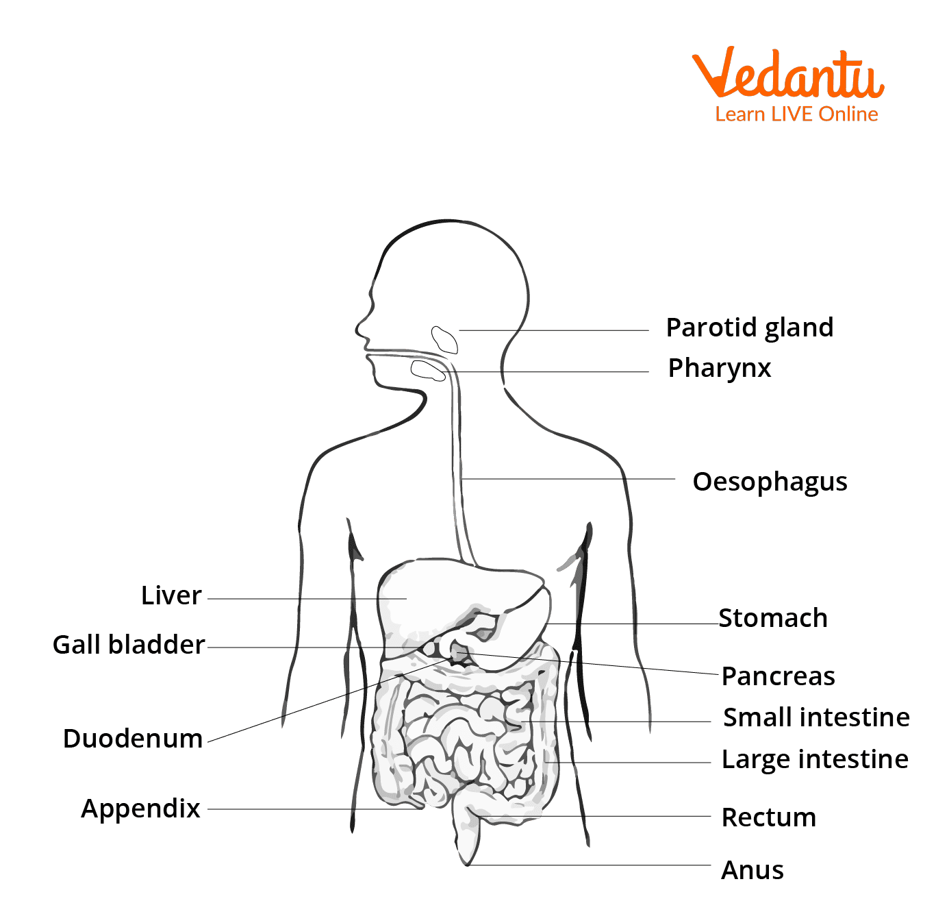
It contains glands that are related to it. The glands are located outside of the gastrointestinal system and are linked to it through ducts like salivary glands, liver and pancreas.
The layers present in the gastrointestinal system are mucous membrane (mucosa), submucosa, muscularis externa, and serosa or adventitia. The mucosa and submucosa projections enhance the surface area available for absorption. The lamina propria and submucosa possess glands.
Alimentary Canal Parts
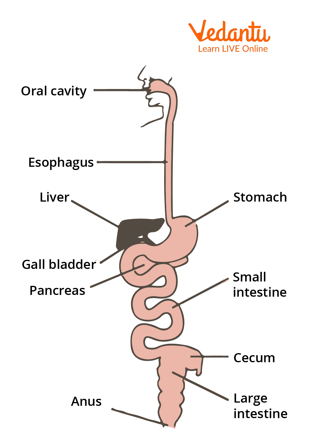
A tube that travels through the body in one direction is called the alimentary canal. From the mouth, it extends to the anus. About 9 metres make up the alimentary canal. The mouth, oesophagus, stomach, small intestine and large intestine are examples of continuous muscular tubes that run throughout the body (digestive tract). They are capable of digesting and absorbing nutrients.
Mouth
The mouth is the initial part of the alimentary canal. It includes teeth and salivary glands. The teeth help in the mechanical breakdown of food while the salivary glands release saliva that mixes with food to make it soft.
Pharynx
The pharynx is a tube that connects the respiratory and digestive systems. It is linked directly with the larynx and the oesophagus. It's a muscular tube that runs between the tongue and the soft palate. Nasopharynx, oropharynx and laryngeal pharynx are the three types of the pharynx.
Oesophagus
The oesophagus is a lengthy, thin muscular tube that delivers food from the pharynx to the stomach. It contains a lot of longitudinal folds that facilitate the transfer of food.
Stomach
The stomach is a storehouse as well as a digestive organ with little absorption. The cardia, fundus, body and pylorus are the four primary areas. Mucosa, submucosa, muscularis externa and serosa are the four layers of the stomach.
These layers produce mucus, acid and pepsin, which are mixed with food. The stomach wall has chief cells and parietal cells. Pepsinogen and lipase are found in chief cells. Pepsinogen is a proenzyme that is transformed into pepsin by the stomach's acid pH. Parietal cells help in the formation of gastric acid and release some factors for digestion.
Small Intestine
Duodenum, jejunum and ileum are small Intestine structures. Absorption is one of the functions of the small intestine. The villi help with these activities by increasing the surface area of the gut, allowing for greater absorption. Thus, it is a nutrition absorber. Hormones and chemicals are released into the small intestine by the exocrine liver and pancreas.
Large Intestine
It includes the colon (ascending, transverse and descending colon). The purpose of the large intestine is to accept the soupy digestion, remove the water and convert it to faeces. It is located next to the small intestine.
Rectum
The rectum is the intestine's swollen end section. After food digestion, it stores the waste.
Anus
The anus is the hole and end point of the alimentary canal. The external aperture is known as the anus. Defecation is controlled by layers of the muscle. It has sphincters that help in the excretion of the waste outside the body.
Structure of Alimentary Canal
Alimentary Canal Structure
Digestive System Diagram
The digestive system involves the alimentary canal and some other accessory organs that help in digestion. The alimentary canal is a long hollow tube that helps in food absorption and digestion. The digestive organs are accessory organs which assist the main digestive tract. Teeth, tongue, salivary glands and liver are examples. Food is never allowed to enter through them.
Digestive System Diagram
Flow Chart of Digestive System

Flow Chart of Digestive System
Digestive Process Flow Chart
The mouth, throat and salivary glands make up the oral cavity, which is where early digestion takes place.
Ingestion → Digestion → Absorption → Excretion.
Ingestion involves the uptake of food. It involves the mouth pharynx. Digestion helps in food breakdown in small particles. Absorption occurs in the small intestine. The digested food is absorbed and passed into blood circulation. Excretion helps in the removal of waste material from the body after the complete digestion of the food.
Functions of Alimentary Canal
The alimentary canal has the main function of food digestion and absorption. It has different organs and parts that help in food digestion. Teeth help in the mechanical breakdown of food and the mouth helps in food intake. The oesophagus helps in carrying the food particles to the stomach. The stomach helps in storing and maintaining the pH of the food. The small intestine absorbs food particles and passes them to blood. The large Intestine helps in extra water absorption from undigested food.
Conclusion
Our mouth not only chews food but also warms or cools it to a temperature suitable for digestion. The food travels through the oesophagus from our mouth to our stomach in about 7 seconds. Over 500 different types of bacteria live in our digestive tract. While many of them are beneficial, they can be extremely dangerous if they spread to other parts of the body.
Food is digested and absorbed in the body with the help of the gastrointestinal tract. The organs and part of this tract help in the digestion of food. The stomach intestines are important parts and the main digestion and absorption occur here. The above articles help us to understand the structure and function of parts of the alimentary canal. It will be useful to clear concepts about food digestion in the body.


FAQs on Gastrointestinal Tract Anatomy and Digestive Functions
1. What is the gastrointestinal tract?
The gastrointestinal tract (GI tract) is a continuous muscular tube that runs from the mouth to the anus and is responsible for digestion and absorption of nutrients. It is a major part of the digestive system and performs both mechanical and chemical digestion.
- Begins at the mouth and ends at the anus
- Includes organs such as the esophagus, stomach, small intestine, and large intestine
- Functions in ingestion, digestion, absorption, and elimination
2. What are the main parts of the gastrointestinal tract in order?
The main parts of the gastrointestinal tract in order are mouth, pharynx, esophagus, stomach, small intestine, and large intestine. These organs form a continuous pathway for food movement.
- Mouth – ingestion and initial digestion
- Pharynx – passageway for swallowed food
- Esophagus – transports food to the stomach
- Stomach – mechanical and chemical digestion
- Small intestine – major site of digestion and absorption
- Large intestine – water absorption and feces formation
3. What is the function of the gastrointestinal tract?
The primary function of the gastrointestinal tract is to digest food, absorb nutrients, and eliminate waste. It converts complex food into usable molecules for the body.
- Ingestion – taking in food
- Digestion – mechanical and chemical breakdown
- Absorption – uptake of nutrients into blood and lymph
- Elimination – removal of indigestible waste
4. How does digestion occur in the gastrointestinal tract?
Digestion in the gastrointestinal tract occurs through coordinated mechanical and chemical processes that break food into absorbable molecules. This process happens step by step along the tract.
- Mouth – chewing and action of salivary amylase
- Stomach – churning and protein digestion by pepsin
- Small intestine – enzymatic digestion by pancreatic enzymes and bile
5. What is the difference between the small intestine and large intestine?
The small intestine mainly digests and absorbs nutrients, while the large intestine primarily absorbs water and forms feces. They differ in structure and function.
- Small intestine: long (~6 m), has villi and microvilli, major site of nutrient absorption
- Large intestine: shorter (~1.5 m), lacks villi, absorbs water and electrolytes
6. What is peristalsis in the gastrointestinal tract?
Peristalsis is the wave-like muscular contraction that moves food through the gastrointestinal tract. It ensures unidirectional movement from the esophagus to the anus.
- Caused by coordinated contraction of circular and longitudinal muscles
- Occurs in the esophagus, stomach, and intestines
- Prevents backflow of food under normal conditions
7. What is the role of the stomach in the gastrointestinal tract?
The stomach stores food and begins protein digestion through mechanical churning and gastric secretions. It acts as a temporary reservoir between the esophagus and small intestine.
- Secretes hydrochloric acid (HCl) to create an acidic environment
- Releases pepsin to digest proteins
- Forms semi-liquid chyme for entry into the duodenum
8. Where does most nutrient absorption occur in the gastrointestinal tract?
Most nutrient absorption occurs in the small intestine, particularly in the jejunum and ileum. Its structure maximizes surface area for efficient uptake.
- Presence of villi and microvilli
- Rich supply of blood capillaries and lacteals
- Absorption of carbohydrates, proteins, lipids, vitamins, and minerals
9. What are the layers of the gastrointestinal tract wall?
The wall of the gastrointestinal tract has four main layers: mucosa, submucosa, muscularis externa, and serosa. These layers support digestion and movement.
- Mucosa – innermost layer for secretion and absorption
- Submucosa – contains blood vessels, nerves, and glands
- Muscularis externa – circular and longitudinal muscles for peristalsis
- Serosa – protective outer covering
10. Why is the gastrointestinal tract important for overall health?
The gastrointestinal tract is essential for overall health because it provides nutrients, supports immunity, and maintains fluid balance. Proper GI function is vital for survival.
- Supplies energy through nutrient absorption
- Contains gut microbiota that aid digestion and immunity
- Absorbs water and electrolytes to maintain homeostasis


