

What are the 12 cranial nerves and their functions with diagram
Cranial nerves are special nerves that arise directly from the brain and the brainstem rather than from the spinal cord. They are found on both sides of the body and serve as important communication lines carrying information to and from the brain. These nerves primarily govern the movements and sensations of the head and neck region.
Unlike spinal nerves, which emerge from segments of the spinal cord, cranial nerves originate in the cranium (skull) and exit through openings known as foramina. There are 12 cranial nerves in order, each with specific roles in controlling muscle movements, collecting sensory information, and regulating vital functions. If you have ever wondered, “What are the 12 cranial nerves and their function?” you will find a detailed explanation in this guide.
12 Cranial Nerves in Order: Names and Overview
The 12 cranial nerve names are conventionally labelled using Roman numerals (I–XII) based on their order from the front of the brain to the back. Among them, Cranial Nerves I (Olfactory) and II (Optic) are considered parts of the central nervous system because of their direct connection to the brain without a peripheral nerve structure.
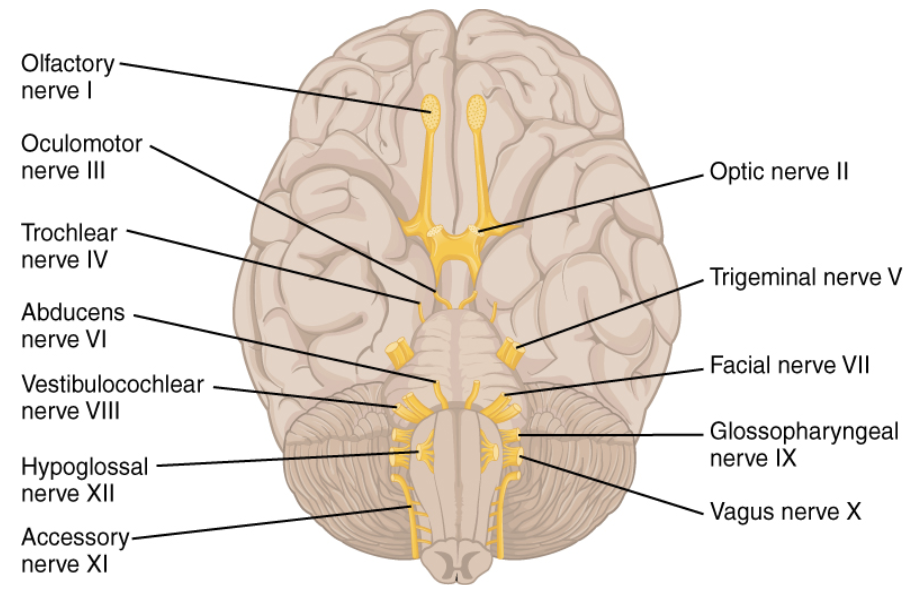
Below is the standard cranial nerves list in order:
Olfactory (I)
Optic (II)
Oculomotor (III)
Trochlear (IV)
Trigeminal (V)
Abducens (VI)
Facial (VII)
Vestibulocochlear (VIII)
Glossopharyngeal (IX)
Vagus (X)
Accessory (XI)
Hypoglossal (XII)
Detailed Cranial Nerves List with Functions
Key Points:
Olfactory (I) and Optic (II) nerves are purely sensory and are central nervous system structures.
Oculomotor (III), Trochlear (IV), and Abducens (VI) focus on eye movement.
Trigeminal (V) has both sensory (face sensation) and motor (chewing) functions.
The facial (VII) nerve handles facial expressions and tastes on the front part of the tongue.
Vestibulocochlear (VIII) supports hearing and balance.
Glossopharyngeal (IX) and Vagus (X) assist in taste, swallowing, and parasympathetic regulation of internal organs.
The accessory (XI) nerve mainly controls head and shoulder movement.
The Hypoglossal (XII) nerve controls the tongue’s movements.
Cranial Nerves and Their Functions in Detail
Olfactory Nerve (I):
Detects and transmits smell-related signals.
Damage may affect the sense of smell and taste.
Optic Nerve (II):
Carries visual information from the retina to the brain.
Damage may result in visual impairment or blindness in affected areas.
Oculomotor Nerve (III):
Controls most of the eye muscles for movement, eyelid elevation, and pupil constriction.
Damage can lead to double vision, drooping eyelids, and misalignment of the eyes.
Trochlear Nerve (IV):
Innervates the superior oblique muscle for downward and inward eye movement.
Damage often presents as difficulty in downward gaze, especially when reading.
Trigeminal Nerve (V):
The largest cranial nerve has three major divisions (ophthalmic, maxillary, and mandibular).
Conveys facial sensations (touch, pain, temperature) and controls jaw muscles for chewing.
Abducens Nerve (VI):
Innervates the lateral rectus muscle to move the eyeball laterally.
Damage results in the inability to move the eye outward, causing double vision.
Facial Nerve (VII):
Manages facial expressions, tear and saliva production, and taste (front 2/3 of the tongue).
Injury might cause facial droop or paralysis on one or both sides.
Vestibulocochlear (VIII):
Divided into vestibular (balance) and cochlear (hearing) components.
Dysfunction can lead to hearing loss, vertigo, and balance issues.
Glossopharyngeal (IX):
Aids in swallowing, saliva secretion, and taste on the back 1/3 of the tongue.
Injury disrupts taste and gag reflexes and can cause swallowing difficulties.
Vagus (X):
The “wandering” nerve has widespread parasympathetic control over most thoracic and abdominal organs.
Involved in heart rate, blood pressure, digestion, and throat sensation.
Accessory (XI):
Controls sternocleidomastoid and trapezius muscles, aiding in head and shoulder movement.
Damage leads to weakness in head rotation and shoulder elevation.
Hypoglossal (XII):
Regulates tongue movement, which is crucial for speech and swallowing.
Injury results in tongue deviation towards the damaged side.
How to Memorise 12 Cranial Nerves (12 Cranial Nerves Mnemonic)
Students often ask, “How to memorise 12 cranial nerves?” or look for a “12 cranial nerves mnemonic” to help them remember the 12 cranial nerve names. One popular mnemonic is:
“On Occasion, Our Trusty Truck Acts Funny — Very Good Vehicle Any How”
Where each first letter stands for a cranial nerve in order:
O – Olfactory
O – Optic
O – Oculomotor
T – Trochlear
T – Trigeminal
A – Abducens
F – Facial
V – Vestibulocochlear
G – Glossopharyngeal
V – Vagus
A – Accessory
H – Hypoglossal
Feel free to create your mnemonic or tune a familiar phrase to suit your memory. Repetition and flashcards are also extremely helpful when mastering cranial nerves and their functions.
Cranial Nerves Vs. Spinal Nerves
Cranial Nerves:
Originates directly from the brain and brainstem.
Responsible for sensory and motor control of the head, neck, and certain internal organs.
There are 12 cranial nerves in order.
Spinal Nerves:
Arise from the spinal cord segments.
Primarily relay information between the spinal cord and the rest of the body (trunk and limbs).
There are 31 pairs of spinal nerves in humans.
Both cranial nerves and spinal nerves form part of the peripheral nervous system except for the Olfactory (I) and Optic (II) nerves, which are structurally connected to the central nervous system.
Unique Facts about the Cranial Nerves
Longest pathway: The Vagus (X) is the longest cranial nerve, wandering through multiple organs.
Most complex: The Trigeminal (V) has three divisions, handling both facial sensation and motor functions like chewing.
The first two CNS, Olfactory (I) and Optic (II), are often classified as part of the central nervous system because they are structurally similar to tracts within the brain.
Quick Quiz on Cranial Nerves
Which cranial nerve is responsible for hearing and balance?
A) Facial (VII)
B) Vestibulocochlear (VIII)
C) Glossopharyngeal (IX)
Name the cranial nerve mainly involved in shoulder elevation and head-turning.
A) Accessory (XI)
B) Trigeminal (V)
C) Vagus (X)
Which cranial nerve deals with the sense of smell?
A) Optic (II)
B) Oculomotor (III)
C) Olfactory (I)
What is the primary function of the Hypoglossal (XII) nerve?
A) Moving the tongue
B) Eye movement
C) Facial sensation
Quiz Answers:
B) Vestibulocochlear (VIII)
A) Accessory (XI)
C) Olfactory (I)
A) Moving the tongue
Related Topics


FAQs on Cranial Nerves Structure Types and Functions in Humans
1. What are cranial nerves?
The cranial nerves are 12 pairs of nerves that arise directly from the brain and primarily supply the head and neck. Unlike spinal nerves, they originate from the cerebrum and brainstem and are numbered I to XII using Roman numerals. They may be sensory, motor, or mixed in function and control activities such as smell, vision, facial movement, hearing, and swallowing.
- Total of 12 pairs
- Originate from the brain or brainstem
- Can be sensory, motor, or mixed
2. What are the names of the 12 cranial nerves in order?
The 12 cranial nerves in order are Olfactory, Optic, Oculomotor, Trochlear, Trigeminal, Abducens, Facial, Vestibulocochlear, Glossopharyngeal, Vagus, Accessory, and Hypoglossal. They are numbered I to XII from anterior to posterior in the brain.
- I – Olfactory
- II – Optic
- III – Oculomotor
- IV – Trochlear
- V – Trigeminal
- VI – Abducens
- VII – Facial
- VIII – Vestibulocochlear
- IX – Glossopharyngeal
- X – Vagus
- XI – Accessory
- XII – Hypoglossal
3. What is the function of cranial nerves?
The main function of the cranial nerves is to control sensory and motor activities of the head and neck. They transmit impulses between the brain and structures such as the eyes, ears, nose, tongue, and facial muscles.
- Sensory functions: smell, vision, hearing, taste
- Motor functions: eye movement, facial expression, tongue movement
- Mixed functions: swallowing and salivation
4. Which cranial nerves are sensory, motor, and mixed?
Cranial nerves are classified as sensory, motor, or mixed based on the type of fibers they contain. This classification helps in understanding their functional roles.
- Sensory nerves: I (Olfactory), II (Optic), VIII (Vestibulocochlear)
- Motor nerves: III (Oculomotor), IV (Trochlear), VI (Abducens), XI (Accessory), XII (Hypoglossal)
- Mixed nerves: V (Trigeminal), VII (Facial), IX (Glossopharyngeal), X (Vagus)
5. What is the difference between cranial nerves and spinal nerves?
The key difference between cranial nerves and spinal nerves is their origin and area of distribution. Cranial nerves arise from the brain, while spinal nerves arise from the spinal cord.
- Cranial nerves: 12 pairs, mainly supply head and neck
- Spinal nerves: 31 pairs, supply trunk and limbs
- Cranial nerves may be sensory, motor, or mixed, whereas spinal nerves are typically mixed
6. What is the role of the vagus nerve?
The vagus nerve (cranial nerve X) controls parasympathetic functions of the heart, lungs, and digestive tract. It is the longest cranial nerve and extends from the brainstem to the abdomen.
- Regulates heart rate
- Controls digestion and gut motility
- Contributes to swallowing and voice production
7. What is the function of the trigeminal nerve?
The trigeminal nerve (cranial nerve V) provides sensation to the face and controls muscles involved in chewing. It is the largest cranial nerve and has three main branches.
- Ophthalmic branch – sensation from forehead and eyes
- Maxillary branch – sensation from upper jaw
- Mandibular branch – sensation from lower jaw and motor control of mastication
8. Where do cranial nerves originate in the brain?
Most cranial nerves originate from the brainstem, except the first two which arise from the forebrain. Their nuclei are located in specific regions of the brain.
- I (Olfactory) – Cerebrum
- II (Optic) – Diencephalon
- III–XII – Mainly from the midbrain, pons, and medulla oblongata
9. What happens if a cranial nerve is damaged?
Damage to a cranial nerve causes loss of the specific sensory or motor function it controls. The symptoms depend on which nerve is affected.
- Damage to Optic nerve (II) → vision loss
- Damage to Facial nerve (VII) → facial paralysis (Bell’s palsy)
- Damage to Vestibulocochlear nerve (VIII) → hearing or balance problems
10. How do cranial nerves control eye movements?
Eye movements are controlled by three cranial nerves: Oculomotor (III), Trochlear (IV), and Abducens (VI). These nerves innervate the extraocular muscles that move the eyeball.
- Oculomotor nerve – controls most eye muscles and pupil constriction
- Trochlear nerve – controls the superior oblique muscle
- Abducens nerve – controls lateral rectus muscle for outward movement


