

What is the structure and function of the spinal cord
The spinal cord is an integral part of the CNS, serving as the main communication pathway between the brain and the rest of the body. It is a long, tubular structure made up of nerve fibres that runs through the vertebral column and controls various motor and sensory functions.
Understanding the spinal cord structure and function is essential for comprehending how signals travel between the brain and body, enabling movements, reflexes, and autonomic functions.
Also Read: Nervous System
Spinal Cord Anatomy
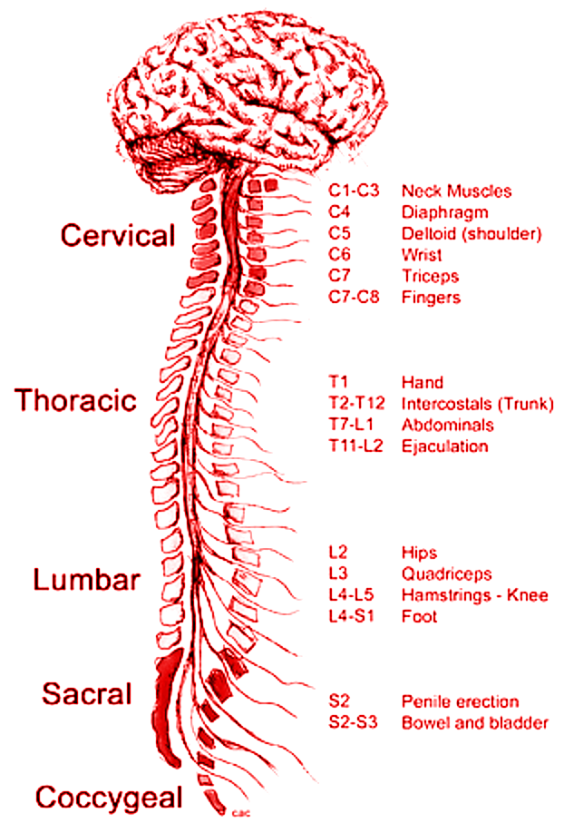
The human spinal cord is typically 40 cm long and 2 cm wide in adults, extending from the medulla oblongata to the lumbar region of the vertebral column. It is divided into five main regions:
Cervical cord – Located in the neck region.
Thoracic cord – Found in the upper and mid-back.
Lumbar cord – Situated in the lower back.
Sacral cord – At the base of the spine.
Coccygeal – The tail end of the spinal cord.
Each region contains spinal nerves that connect to different parts of the body. There are 31 pairs of spinal nerves, classified as follows:
8 cervical nerves (C1-C8)
12 thoracic nerves (T1-T12)
5 lumbar nerves (L1-L5)
5 sacral nerves (S1-S5)
1 coccygeal nerve (Co1)
These spinal nerves transmit signals between the brain and body, controlling reflexes, muscle movements, and sensory functions.
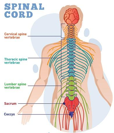
Spinal Cord Diagram – Labelled and Easy
A labelled spinal cord diagram helps in better visualising its structure. Below is an easy-to-understand spinal cord diagram that shows its different parts.
Key Points:
The spinal cord has different segments, each with specific nerve connections.
It is housed within the vertebral column, which protects it from injuries.
Grey and white matter play distinct roles in sensory and motor functions.
Structure of the Spinal Cord
The spinal cord runs through a hollow cavity inside the vertebral column, protected by three layers of meninges:
Dura mater – The outermost layer.
Arachnoid mater – The middle protective layer.
Pia mater – The innermost layer, closely attached to the spinal cord.
Cross-section of the Spinal Cord
A cross-section of the spinal cord reveals grey and white matter:
Grey matter – Located in the centre, shaped like a butterfly. It contains nerve cells and the central canal filled with cerebrospinal fluid (CSF).
White matter – Surrounds the grey matter, consisting of axons that transmit nerve signals.
The spinal cord structure and function are crucial for relaying sensory (afferent) signals to the brain and motor (efferent) signals from the brain to the body.
Spinal Cord Function
The spinal cord serves as a vital link between the brain and body, performing multiple functions:
Relays messages between the brain and the body.
Controls reflex actions without brain involvement.
Provides structural support to maintain body posture.
Coordinates movement and muscle control.
Regulates autonomic functions, such as heart rate and digestion.
Facilitates pain and temperature sensation transmission.
The spinal cord function is crucial in coordinating reflexes and ensuring smooth bodily movements.
Spinal Cord Injuries
Damage to the spinal cord can cause severe health issues due to the loss of communication between the brain and body. Injuries can be classified as:
1. Complete Injury
Total loss of motor and sensory function below the injury site.
Results in paralysis of affected body parts.
2. Incomplete Injury
Partial loss of function, with some movement or sensation still present.
Types of Paralysis Due to Spinal Cord Injury
Tetraplegia (Quadriplegia) – Affects all four limbs and torso.
Paraplegia – Affects the lower limbs and lower body.
Symptoms include:
Loss of voluntary movements
Numbness or loss of sensation
Reflex abnormalities
Breathing difficulties in severe cases
Early diagnosis and treatment can help in managing spinal cord injuries effectively.
Spinal Cord Nerves & Types
The 31 pairs of spinal nerves are divided into different categories based on their location:
Cervical Nerves (C1-C8) – Control neck, arms, and diaphragm movements.
Thoracic Nerves (T1-T12) – Control chest and upper abdominal muscles.
Lumbar Nerves (L1-L5) – Control lower back, hips, and leg functions.
Sacral Nerves (S1-S5) – Control pelvic organs, lower limbs, and feet.
Coccygeal Nerve (Co1) – Controls the tailbone region.
These spinal cord nerves play a significant role in maintaining motor and sensory functions throughout the body.
Unique Information
1. Role of the Spinal Cord in Reflex Actions: The spinal cord is responsible for reflexes, which are automatic responses to stimuli. The reflex arc involves sensory neurons, interneurons, and motor neurons to execute quick responses.
2. How the Spinal Cord Adapts to Injuries: In some cases, the spinal cord can retrain the brain to use alternative pathways for nerve signal transmission, helping people recover partial movements after injuries.
3. The Spinal Cord and Neuroplasticity: Neuroplasticity enables the spinal cord to form new neural connections even after minor injuries, improving recovery chances.
Related Links:


FAQs on Spinal Cord Anatomy and Role in the Nervous System
1. What is the spinal cord?
The spinal cord is a long, cylindrical part of the central nervous system (CNS) that connects the brain to the rest of the body. It lies inside the vertebral column and acts as a major pathway for nerve signals.
- Transmits sensory information from the body to the brain
- Sends motor commands from the brain to muscles and glands
- Controls many rapid reflex actions independently of the brain
2. What is the function of the spinal cord?
The main function of the spinal cord is to transmit nerve impulses and control reflex activities. It performs three key roles:
- Sensory function: Carries sensory impulses from receptors to the brain
- Motor function: Sends motor impulses from the brain to skeletal muscles
- Reflex center: Acts as the center for simple reflexes like withdrawal reflex
3. Where is the spinal cord located?
The spinal cord is located inside the vertebral canal of the vertebral column. It extends from the base of the brain at the medulla oblongata to about the level of the first or second lumbar vertebra (L1–L2) in adults.
- Protected by vertebrae
- Surrounded by meninges
- Cushioned by cerebrospinal fluid (CSF)
4. What are the main parts of the spinal cord?
The main parts of the spinal cord include gray matter, white matter, and spinal nerves. Structurally, it consists of:
- Gray matter: H-shaped inner region containing neuron cell bodies
- White matter: Outer region containing myelinated nerve fibers
- Dorsal and ventral roots: Form the spinal nerves
- Central canal: Contains cerebrospinal fluid
5. How does the spinal cord control reflex actions?
The spinal cord controls reflex actions through a rapid pathway called a reflex arc. A reflex arc works in the following steps:
- 1. A receptor detects a stimulus (e.g., heat).
- 2. A sensory neuron carries the impulse to the spinal cord.
- 3. An interneuron processes the signal in the gray matter.
- 4. A motor neuron sends a response to a muscle or gland.
6. What is the difference between the brain and the spinal cord?
The brain is the main control center of the nervous system, while the spinal cord mainly transmits signals and controls reflexes. Key differences include:
- Location: Brain is inside the skull; spinal cord is inside the vertebral column
- Function: Brain handles thinking, memory, and complex actions; spinal cord handles signal transmission and reflexes
- Structure: Brain has highly folded cortex; spinal cord is cylindrical with central gray matter
7. How many spinal nerves are there in humans?
Humans have 31 pairs of spinal nerves arising from the spinal cord. These are classified into:
- 8 cervical nerves
- 12 thoracic nerves
- 5 lumbar nerves
- 5 sacral nerves
- 1 coccygeal nerve
8. What is the role of gray matter and white matter in the spinal cord?
In the spinal cord, gray matter processes information while white matter conducts nerve impulses. Their roles are:
- Gray matter: Contains neuron cell bodies and is involved in reflex integration
- White matter: Contains ascending and descending tracts that transmit impulses to and from the brain
9. What happens if the spinal cord is damaged?
Damage to the spinal cord can cause loss of movement, sensation, or both below the level of injury. The effects depend on the location and severity of damage:
- Paralysis: Loss of voluntary muscle control
- Loss of sensation: Inability to feel touch, pain, or temperature
- Impaired reflexes: Reflex actions may be reduced or exaggerated
10. Why is the spinal cord important for the body?
The spinal cord is important because it serves as the main communication pathway between the brain and the body. Its importance includes:
- Enabling voluntary movements like walking and writing
- Allowing sensory perception such as pain and temperature
- Coordinating protective reflexes
- Maintaining basic motor coordination


