

Detailed Anatomy of Kidney Including Nephron Structure and Functions
The kidneys are a vital part of the human excretory system, responsible for filtering blood, removing waste products, and regulating fluid and electrolyte balance. Each kidney is a bean-shaped organ located just below and behind the liver in the peritoneal cavity. On top of each kidney sits an adrenal (suprarenal) gland. The kidneys continuously filter all of the body's blood throughout the day, helping to purify it and maintain homeostasis.
Structure of the Human Kidney
The human kidney is externally protected by three main layers. The outermost layer is the renal fascia, a tough connective tissue that provides support. Inside the fascia lies the perirenal fat capsule, which anchors the kidneys and offers cushioning. The innermost covering is the renal capsule, closely attached to the kidney's surface.
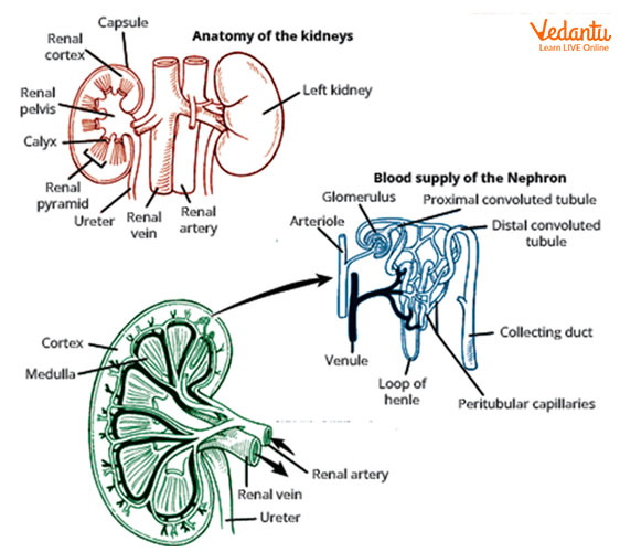
Internally, the kidney consists of three main regions:
-
Renal Cortex: The outermost region, granular in appearance due to millions of nephrons.
-
Renal Medulla: Located beneath the cortex, composed of several cone-shaped renal pyramids. The tips (renal papillae) point inward toward the renal pelvis.
-
Renal Pelvis: A funnel-shaped cavity in the center, which collects urine and leads to the ureter.
Renal pyramids and their adjoining cortex form units called kidney lobes. Between the pyramids are renal columns, which provide space for blood vessels and nerves. The renal pelvis branches into major calyces, and each major calyx divides into minor calyces, which collect urine from different pyramids.
| Part | Description | Function |
|---|---|---|
| Renal Cortex | Outer, granular layer | Contains most of the nephrons |
| Renal Medulla | Middle, striped region | Formed by renal pyramids; concentration of urine |
| Renal Pelvis | Central funnel-shaped cavity | Collects urine, channels to ureter |
| Major & Minor Calyces | Cup-like extensions of the pelvis | Collect urine from the pyramids |
| Renal Columns | Regions between pyramids | Pathway for blood vessels and nerves |
Blood Supply and Filtration
An extensive network of blood vessels enters the kidney at the hilum. The renal artery branches to segmental, interlobar, arcuate, and cortical radiate arteries, supplying blood to the nephrons. The renal vein carries filtered blood away. The arrangement of these vessels supports efficient filtration of blood and removal of waste.
The Nephron: Structural and Functional Unit
Each kidney contains over one million nephrons, the microscopic units that filter blood and form urine. Nephrons are found mainly in the cortex, but extend into the medulla. There are two nephron types: cortical (deep in cortex, about 85%) and juxtamedullary (near medulla, about 15%).
The main parts of a nephron are:
-
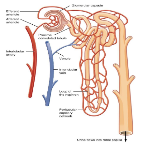
Renal Corpuscle: Located in the cortex, formed by the glomerulus (capillary network) and Bowman’s (glomerular) capsule.
-
Renal Tubule: Emerges from the corpuscle, divided into:
-
Proximal Convoluted Tubule (PCT): Closest to glomerulus, remains in cortex.
-
Loop of Henle: Forms a U-shaped loop descending into the medulla, then returns to the cortex.
-
Distal Convoluted Tubule (DCT): Located in cortex, connects to collecting duct.
-
-
Collecting Duct: Receives filtrate from multiple nephrons, descends through medullary pyramids, empties into the renal pelvis.
| Nephron Part | Main Function |
|---|---|
| Glomerulus | Filtration of blood plasma |
| Bowman's Capsule | Collects the filtrate |
| Proximal Convoluted Tubule | Reabsorption of water, glucose, and ions |
| Loop of Henle | Concentrates urine; reabsorbs water and salts |
| Distal Convoluted Tubule | Selective secretion and absorption; pH regulation |
| Collecting Duct | Final concentration of urine |
Capillary Networks Associated with the Nephron
Blood enters each nephron via an afferent arteriole, forms the glomerular capillary bed, and exits via the efferent arteriole. The efferent arteriole then creates a network called the peritubular capillaries (in cortical nephrons) or the vasa recta (around the loop of Henle in juxtamedullary nephrons). These vessels play a key role in reabsorption and secretion during urine formation.
Significance and Functions of the Kidney
The kidneys maintain body fluid balance, filter metabolic wastes, regulate electrolytes, and contribute to acid-base balance. They also support blood pressure control through specialized hormones and help in producing active vitamin D. Efficient kidney function is indispensable for overall health and well-being.
Practice Questions and Deeper Learning
-
Draw and label a simple kidney diagram and explain the function of each region.
-
Describe the stepwise path of blood flow through the kidney.
-
Explain the difference between cortical and juxtamedullary nephrons.
If you wish to learn about kidney disorders such as renal calculi. Explore related topics including nephron function, for further learning.


FAQs on Structure of Kidney and Its Detailed Anatomy
1. What is the structure of the kidney?
The structure of the kidney consists of an outer renal cortex, an inner renal medulla, and a central renal pelvis that collects urine. Each kidney is bean-shaped and covered by a tough renal capsule.
- Renal cortex: Outer region containing glomeruli and convoluted tubules.
- Renal medulla: Inner region made of renal pyramids and loops of Henle.
- Renal pelvis: Funnel-shaped cavity that drains urine into the ureter.
2. What are the main parts of a human kidney?
The main parts of a human kidney are the renal cortex, renal medulla, and renal pelvis. These regions work together in filtration and excretion.
- Cortex: Contains renal corpuscles and proximal and distal convoluted tubules.
- Medulla: Contains renal pyramids, loops of Henle, and collecting ducts.
- Pelvis: Collects urine before it enters the ureter.
3. What is a nephron and what is its structure?
A nephron is the structural and functional unit of the kidney responsible for urine formation. Each kidney contains about one million nephrons.
- Renal corpuscle: Composed of the glomerulus and Bowman’s capsule.
- Renal tubule: Includes proximal convoluted tubule (PCT), loop of Henle, and distal convoluted tubule (DCT).
- Collecting duct: Receives urine from multiple nephrons.
4. What is the difference between renal cortex and renal medulla?
The renal cortex is the outer region of the kidney containing glomeruli, while the renal medulla is the inner region containing renal pyramids and loops of Henle.
- Cortex: Site of blood filtration in glomeruli.
- Medulla: Concentrates urine through long loops of Henle and collecting ducts.
5. What is the structure of Bowman’s capsule?
The Bowman’s capsule is a double-walled cup-shaped structure that surrounds the glomerulus in the nephron. It forms part of the renal corpuscle.
- Parietal layer: Outer simple squamous epithelium.
- Visceral layer: Inner layer made of specialized cells called podocytes.
- Capsular space: Collects the glomerular filtrate.
6. What are renal pyramids in the kidney?
The renal pyramids are cone-shaped structures in the renal medulla that contain loops of Henle and collecting ducts. They play a key role in urine concentration.
- Base faces the cortex.
- Apex forms the renal papilla, which drains urine into minor calyces.
- Contain tubules that transport urine toward the pelvis.
7. How is blood supplied to the kidney?
The kidney receives blood through the renal artery, which branches into smaller arteries for filtration. Proper blood supply is essential for kidney function.
- Renal artery → segmental arteries → interlobar arteries.
- Interlobar arteries → arcuate arteries → interlobular arteries.
- Interlobular arteries form afferent arterioles leading to glomeruli.
8. What is the hilum of the kidney?
The hilum of the kidney is a concave indentation on the medial side where blood vessels, nerves, and the ureter enter or leave. It acts as the entry and exit point of the kidney.
- Renal artery enters.
- Renal vein exits.
- Ureter emerges from the renal pelvis.
9. What is the function of the renal pelvis?
The renal pelvis is a funnel-shaped cavity that collects urine from the calyces and channels it into the ureter. It acts as a temporary storage area for urine.
- Receives urine from minor and major calyces.
- Narrows to form the ureter.
- Ensures smooth flow of urine to the urinary bladder.
10. How many nephrons are present in each kidney?
Each human kidney contains approximately 1 million nephrons, which are responsible for filtration and urine formation. The exact number may vary slightly among individuals.
- Nephrons are located in both cortex and medulla.
- They perform filtration, reabsorption, and secretion.
- Loss of nephrons reduces kidney efficiency.


