


Understanding the Human Heart: Basics to Advance
The human heart is a strong muscular organ and the main organ of the circulatory system. Its job is to pump blood throughout the body in a continuous and well-coordinated manner. By doing this, the heart ensures that oxygen and nutrients reach every cell, while carbon dioxide and other waste materials are carried away for removal.
The heart is often described as roughly fist-sized and slightly cone-shaped or pyramid-like with rounded borders. Its muscular walls contract rhythmically in response to electrical impulses, causing the heart to beat in a regular pattern. This rhythmic contraction and relaxation maintain blood flow and blood pressure.
Where is the Heart Located in the Human Body?
The heart is located in the chest cavity, between the two lungs. It lies slightly behind and a little to the left of the sternum or breastbone. Although many students think the heart is completely on the left side, that is not fully correct. The heart is more centrally placed but tilts slightly toward the left. The left lung is slightly smaller than the right lung to make room for the heart. The rib cage surrounds and protects it.
This location is very important because it helps us understand why the apex beat of the heart is usually felt more on the left side of the chest.
in the thoracic cavity
between the lungs
behind the sternum
slightly toward the left side
Human Heart Shape, Size and Position
The human heart shape is often compared to an upside-down pyramid with rounded edges. It is not perfectly round and not exactly like the heart symbol people draw. In reality, it is a hollow, muscular, cone-like organ. Large blood vessels enter and leave the heart from its upper side, while the lower pointed part is called the apex.
In terms of size, the heart is usually about the size of a person's fist. In adults, it weighs about 10 ounces on average, though this can vary by body size and sex.
Important points for NEET:
The heart is fist-sized
The apex points downward, forward, and to the left
Its base is broader and directed upward
It lies obliquely in the thoracic cavity
Structure of the Human Heart
The structure of the human heart is specialised for continuous pumping. It includes:
heart walls
four chambers
four valves
major blood vessels
coronary blood supply
electrical conduction system
protective covering called the pericardium
All these parts work together in a coordinated manner. The heart is not just a muscle mass. It is a highly organised pumping organ with different structural divisions, each with a specific function.
The heart is divided into right and left sides by a muscular partition called the septum. This prevents mixing of oxygenated and deoxygenated blood. This separation is one of the major evolutionary advantages in mammals and birds because it allows efficient double circulation.
Layers of the Human Heart Wall
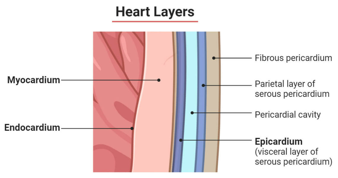
The wall of the heart has three major layers:
1. Endocardium
This is the innermost layer of the heart wall. It lines the heart's chambers from the inside and provides a smooth surface for blood flow.
2. Myocardium
This is the middle muscular layer and the thickest part of the heart wall. The myocardium is responsible for contraction. Since the heart must pump blood continuously, this layer is made of specialised cardiac muscle.
3. Epicardium
This is the outer protective layer of the heart wall. It forms one layer of the pericardium.
Pericardium
Outside the heart wall is the pericardium, a protective sac surrounding the heart. It contains fluid that reduces friction during heartbeats and prevents rubbing against nearby organs.
For NEET, remember:
endocardium = inner layer
myocardium = muscular middle layer
epicardium = outer layer
pericardium = covering sac
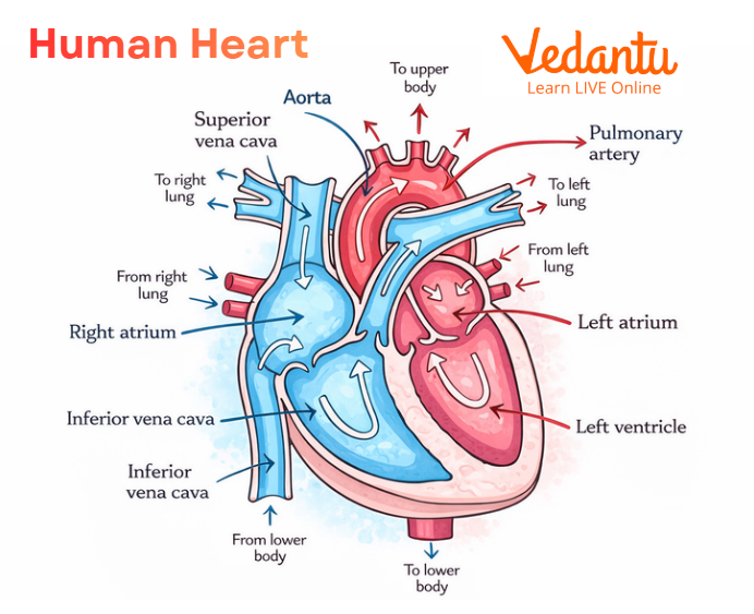
How Many Chambers are There in the Human Heart?
There are four chambers in the human heart.
These are:
right atrium
right ventricle
left atrium
left ventricle
The upper two chambers are called atria, and the lower two chambers are called ventricles. Each side of the heart has one atrium and one ventricle.
Right Atrium
The right atrium receives oxygen-poor blood from the body through two large veins:
superior vena cava
inferior vena cava
It then passes this blood to the right ventricle.
Right Ventricle
The right ventricle pumps oxygen-poor blood to the lungs through the pulmonary artery, where it becomes oxygenated.
Left Atrium
The left atrium receives oxygen-rich blood from the lungs through the pulmonary veins and then sends it to the left ventricle.
Left Ventricle
The left ventricle is the thickest and strongest chamber. It pumps oxygen-rich blood to the rest of the body through the aorta. Its wall is thicker than that of the right ventricle because it has to pump blood to the entire body under higher pressure.
Human Heart Valves
There are four main heart valves:
Atrioventricular Valves
These are located between atria and ventricles.
Tricuspid Valve
Present between the right atrium and right ventricle.
Mitral Valve
Present between the left atrium and left ventricle. It is also called the bicuspid valve.
Semilunar Valves
These are present where blood leaves the ventricles.
Pulmonary Valve
Present between the right ventricle and the pulmonary artery.
Aortic Valve
Present between the left ventricle and the aorta.
Valves are clinically important because faulty valves can lead to heart murmurs, regurgitation, or stenosis.
Blood Vessels Connected to the Human Heart
The heart is connected to major blood vessels that bring blood to and from it. These include arteries, veins, and capillaries.
Arteries
Arteries usually carry oxygen-rich blood away from the heart to tissues. The major exception is the pulmonary artery, which carries oxygen-poor blood from the right ventricle to the lungs.
Veins
Veins usually carry oxygen-poor blood back to the heart. The major exception is the pulmonary veins, which carry oxygen-rich blood from the lungs to the left atrium.
Major Vessels of the Heart
superior vena cava
inferior vena cava
pulmonary artery
pulmonary veins
aorta
These vessels are routinely labelled in a human heart diagram.
Coronary Circulation
The heart muscle itself also needs oxygen and nutrients. These are supplied by the coronary arteries, which run on the surface of the heart.
Left Coronary Artery
This divides into two major branches:
circumflex artery
left anterior descending artery (LAD)
The circumflex artery supplies the left atrium and the side and back of the left ventricle. The LAD supplies the front and lower portion of the left ventricle and the front of the septum.
Right Coronary Artery
This supplies the right atrium, right ventricle, lower part of the left ventricle, and the back of the septum.
The coronary circulation is clinically important because blockage of these arteries can cause a heart attack.
Electrical Conduction System of the Heart
The heart does not beat randomly. A specialised electrical conduction system regulates its contractions. This system controls the rhythm and pace of the heartbeat.
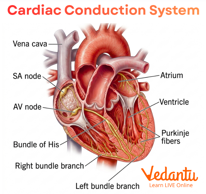
The main components are:
Sinoatrial Node (SA Node)
This is the natural pacemaker of the heart. It generates electrical impulses that initiate each heartbeat.
Atrioventricular Node (AV Node)
This receives impulses from the atria and relays them to the ventricles.
Bundle of His
This carries impulses from the AV node downward.
Right and Left Bundle Branches
These conduct impulses to the respective ventricles.
Purkinje Fibres
These spread the impulse through the ventricles and cause ventricular contraction.
This system is essential for the synchronised contraction of atria and ventricles.
Function of the Human Heart
The Function of the human heart is much more than simply pumping blood. It performs several related roles.
1. Pumps Blood Throughout the Body
This is the primary function. Blood delivers oxygen and nutrients to tissues and removes carbon dioxide and waste materials.
2. Maintains Blood Pressure
The pumping action of the heart helps maintain blood pressure, which is needed for efficient blood flow.
3. Controls Rhythm and Speed of Heartbeat
The heart’s conduction system and its interaction with the nervous and endocrine systems regulate heart rate.
4. Supports Gas Exchange Indirectly
By sending deoxygenated blood to the lungs and oxygenated blood to tissues, the heart supports respiration and gas transport.
5. Works with Other Systems
The nervous system sends signals that can slow the heart during rest or increase its rate during stress. The endocrine system also affects the heart through hormones, including thyroid hormones, which can increase or decrease heart rate.
Human Heart Rate
The human heart rate is the number of times the heart beats per minute. It is controlled by the SA node and regulated by the nervous and endocrine systems. When the body is at rest, the heart beats more slowly. During exercise, stress, fear, or excitement, the heart beats faster to meet the increased oxygen demand.
For NEET, the most important concept is that heart rate is:
controlled by pacemaker activity
influenced by the autonomic nervous system
influenced by hormones
adjusted according to body needs
A faster heart rate does not always mean disease. It may be a normal response to exercise or stress. However, extremely fast, slow, or irregular heart rates may indicate cardiac problems.
Pathway of Blood Through the Heart
The sequence is:
Body tissues send oxygen-poor blood through the superior and inferior vena cava
Blood enters the right atrium
It passes through the tricuspid valve into the right ventricle
The right ventricle pumps it through the pulmonary valve into the pulmonary artery
Blood reaches the lungs and becomes oxygenated
Oxygen-rich blood returns through pulmonary veins
It enters the left atrium
It passes through the mitral valve into the left ventricle
The left ventricle pumps it through the aortic valve into the aorta
Blood is distributed to the whole body
This complete double circulation is one of the most important physiological concepts for NEET.
Common Heart Conditions
The heart can be affected by many disorders. Common examples include:
arrhythmia
cardiomyopathy
congestive heart failure
coronary artery disease
heart attack
valve disease
high blood pressure
high cholesterol
pericarditis
diabetes-related cardiovascular complications
These conditions can interfere with the heart's normal pumping ability or the flow of blood through the coronary vessels.
Symptoms of Heart Problems
Some common signs of heart problems include:
chest pain
palpitations
dizziness
shortness of breath
fatigue
swelling in the lower body
These symptoms are clinically important and also relevant to applied biological understanding.
Tests Used to Check Heart Health
Doctors may use several tests to assess heart function, such as:
blood pressure measurement
electrocardiogram (EKG)
echocardiogram
chest X-ray
blood tests
cardiac catheterisation
CT scan
heart MRI
stress test
These are important to know in general biology and health education contexts.
How to Keep the Heart Healthy?
Keeping the heart healthy is important because cardiovascular disease is common. Good habits include:
maintaining a healthy body weight
eating fruits, vegetables, and whole grains
staying physically active
limiting sodium intake
avoiding smoking and tobacco
managing stress in healthy ways
Taking medicines properly if prescribed
This is not only useful in health science but also helps with real-life awareness.
Also Read:
FAQs on Human Heart: Structure, Function, Diagram, Chambers, Location and Heart Rate | NEET Biology
1. What are the 7 parts of the human heart?
The 7 main parts of the human heart are usually explained as:
Right atrium
Right ventricle
Left atrium
Left ventricle
Tricuspid valve
Pulmonary valve
Mitral valve
Aortic valve
When grouped simply, people often count the four chambers and the main valves as the heart's key parts.
2. What is the human heart in full detail?
The human heart is a muscular organ about the size of a fist. It is located in the chest, slightly to the left, between the lungs. Its main job is to pump blood throughout the body.
The heart has:
4 chambers – right atrium, right ventricle, left atrium, and left ventricle
4 valves – tricuspid, pulmonary, mitral, and aortic
3 layers – endocardium, myocardium, and epicardium
A covering called the pericardium
The right side of the heart receives oxygen-poor blood and sends it to the lungs. The left side receives oxygen-rich blood from the lungs and pumps it to the whole body. The heart also has an electrical system that controls the heartbeat.
3. What is the 10 heart function?
A heart working at 10% function means it is pumping very poorly. This usually refers to a very low ejection fraction, which shows how much blood the heart pumps out with each beat.
At 10% function:
The heart cannot supply enough blood to the body
The person may feel very weak or breathless
There is a high risk of collapse, organ damage, or death
It is a serious medical emergency
So, 10% heart function means the heart is severely weakened.
4. What is the hardest-working muscle in the body?
The heart is the hardest-working muscle in the body because it works continuously without rest. It beats all day and night to pump blood, oxygen, and nutrients throughout the body.
5. What are the 4 types of the heart?
If this question means the 4 main chambers of the heart, they are:
Right atrium
Right ventricle
Left atrium
Left ventricle
These four chambers help receive and pump blood.
6. What are 5 signs your heart is in danger?
Five common warning signs that your heart may be in danger are:
Chest pain or pressure
Shortness of breath
Dizziness or lightheadedness
Pain spreading to the arm, neck, jaw, or back
Unusual tiredness or fatigue
These symptoms should not be ignored.
7. What is heart failure?
Heart failure is a condition in which the heart becomes too weak or too stiff to pump blood properly. It does not mean the heart has stopped, but it means the heart is not working as efficiently as it should.
8. How powerful is the heart?
The heart is a very powerful muscle. It beats around 100,000 times a day and pumps thousands of litres of blood through the body daily. This constant pumping keeps all organs alive and working.
9. What damages the heart the most?
The things that damage the heart the most include:
Smoking
Unhealthy diet
Lack of exercise
Long-term stress
High blood pressure
High cholesterol
Diabetes
Family history can also increase the risk of heart disease.
10. What are the top 3 most common heart diseases?
The top 3 most common heart diseases are:
Coronary artery disease
Atrial fibrillation
Heart failure
These conditions affect blood flow, heart rhythm, or the heart's pumping ability.