

What Is an Electrocardiograph and How Does It Work
An Electrocardiograph (commonly referred to as ECG) is a medical marvel that lets us record and interpret the electrical signals produced by our heart. These signals enable the heart muscles to contract and relax, creating the rhythmic beat that pumps blood throughout the body. Learning about the ECG full form and knowing how an electrocardiograph machine works can be transformative in understanding heart health and diagnosing potential cardiac issues early.
In this guide, we’ll delve into the ECG diagram, break down its key components (P wave, QRS complex, T wave), explore what information ecg gives about a person, and compare electrocardiogram vs electrocardiograph to clear any lingering doubts. We’ll also look at ECG test results, common medical uses, and what are 3 reasons a person would get an EKG. By the end, you’ll see why an ECG is a vital tool for students, doctors, and patients alike.
ECG Full Form in Medical Science
ECG full form: Electrocardiogram.
ECG full form in medical usage is the same, but often you might also hear it called an EKG (from the German term “Elektrokardiogramm”).
Although many use ECG and EKG interchangeably, these abbreviations point to the same test. When we discuss electrocardiogram vs electrocardiograph, remember that an electrocardiogram is the actual recording or printout of the heart’s activity, while an electrocardiograph machine is the device used to capture these signals.
How Does an Electrocardiograph Machine Work?
An electrocardiograph machine detects the heart’s electrical pulses via electrodes placed on the skin. These electrical signals spread through the body, and the machine translates them into tracings (spikes and dips) on paper or a digital screen. Typically:
Electrodes Placement
Sticky electrodes are placed on the wrists, ankles, and various spots on the chest.
Standard tests often use three leads (one on each wrist and one on the left ankle), but a comprehensive 12-lead test includes additional chest electrodes for a more detailed view.
Signal Detection
The machine picks up minute voltage changes resulting from each heartbeat.
These signals are converted into wave patterns that form the familiar ecg diagram (P wave, QRS complex, T wave).
Recording
The final readout is the electrocardiogram (ECG or EKG), illustrating each phase of heart muscle depolarisation and repolarisation.
Breaking Down the ECG Diagram
A standard ecg diagram displays three main wave components:
P Wave
Represents atrial depolarisation (the electrical activity causing the atria to contract).
If the P wave is abnormal, it can suggest issues like atrial enlargement.
QRS Complex
Illustrates ventricular depolarisation. This is when the ventricles contract, marking the start of systole.
Variations in the shape or timing can indicate conduction blocks or underlying heart diseases.
T Wave
Shows the repolarisation of the ventricles (returning to a resting state). This marks the end of systole.
Abnormal T waves may point to electrolyte imbalances or ischaemia.
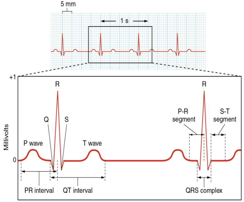
By counting the QRS complexes, healthcare professionals derive the heart rate from your ecg test results. Any noticeable irregularities in wave patterns can reveal potential arrhythmias, conduction problems, or structural anomalies in the heart.
Electrocardiogram vs Electrocardiograph
Electrocardiogram (ECG): The result or output – the paper or digital record of your heart’s electrical activity.
Electrocardiograph: The machine or device used to perform the recording.
So, when you hear about reading an ECG, you’re talking about interpreting the chart produced by the electrocardiograph machine.
Explore, the Electrocardiogram
Types of ECG Tests
Medical practitioners use different ECG tests depending on the clinical scenario:
Resting ECG
Conducted when you’re lying down or sitting quietly.
Good for routine checks or when investigating common symptoms like palpitations.
Exercise ECG (Stress Test)
Monitors the heart while you walk or run on a treadmill (or pedal a stationary bike).
Helps evaluate heart performance under physical stress, often used to investigate chest pain or exercise intolerance.
24-hour ECG (Holter Monitor)
Uses a portable device worn for a day (or longer) to capture heart activity during daily routines.
Beneficial for detecting intermittent arrhythmias or abnormalities that may not show up in a short, resting exam.
What are 3 Reasons a Person Would Get an EKG?
If you’re wondering what are 3 reasons a person would get an ekg, here are the most common:
Suspected Heart Attack
If a patient experiences chest pain, an immediate ECG can reveal signs of myocardial infarction.
Unexplained Fainting or Dizziness
Episodes of fainting (syncope) can be caused by underlying heart rhythm issues detectable on ecg test results.
Check-Up for Heart-Related Symptoms
Palpitations, shortness of breath, or irregular heartbeat often prompt an ECG to find potential arrhythmias.
What Information Does ECG Give About a Person?
You might ask, what information does ecg give about a person. An ECG provides a window into:
Heart Rate and Rhythm
Tells if it’s regular, slow (bradycardia), or fast (tachycardia).
Conduction Pathway
Detects blocks or delays (e.g., bundle branch blocks) in electrical signal travel.
Chamber Size
Abnormal wave patterns can hint at enlargement of ventricles or atria.
Past or Current Damage
Signs of old heart attacks or ongoing ischaemia.
Medication Effects
Some drugs alter the ECG pattern, so regular checks confirm proper dosing and safety.
Medical Uses and Significance
Apart from diagnosing common arrhythmias, an ECG can also help in identifying or managing:
Seizures and Fainting
Some neurological symptoms can mimic cardiac issues, so an ECG helps rule out heart causes.
Pulmonary Embolism
Certain ECG changes can be associated with acute embolism in the lungs.
Deep Vein Thrombosis (DVT)
While not a direct test for DVT, an ECG may detect heart strain if there’s a significant pulmonary complication.
Structural Heart Diseases
Ventricular hypertrophy or hypertrophic cardiomyopathy can alter ecg test results.
Monitoring Medication or Devices
For patients on heart-specific medication or with implanted devices like pacemakers, ECG checks if everything works smoothly.
Leads and Advanced Monitoring
3-lead vs 12-lead ECG
A simple 3-lead set-up is often used for basic monitoring (in ambulances or quick checks).
A 12-lead ECG offers a comprehensive view of the heart from multiple angles, vital for detailed diagnosis.
Wearable Technology
Modern portable devices can record short ECG segments, beneficial in alerting users to irregular rhythms in real time.
ECG in Sports Medicine
Screening athletes for hidden cardiac anomalies, such as hypertrophic cardiomyopathy, helps prevent sudden cardiac events.
Process of Getting an ECG: Step-by-Step
Preparation
You might be asked to lie still and relax. Jewellery or electronic devices near the chest area might be removed.
Electrode Placement
Small sticky patches (electrodes) are placed on your arms, legs, and chest.
Recording
The electrocardiograph machine detects and records electrical impulses.
Interpretation
Healthcare professionals analyse the resulting ecg diagram for any abnormalities.
ECG Test Results
Often available immediately; a doctor can provide a quick assessment of your heart’s rhythm and rate.
Why is an ECG Done?
Doctors recommend ECGs to:
Check for heart disease in patients with risk factors (e.g., high blood pressure, diabetes, high cholesterol).
Evaluate the thickness of heart walls (hypertrophy).
Monitor potential side effects of medications.
Check if a mechanical device (pacemaker) is functioning properly.
Assess heart health pre- and post-surgery or during follow-up visits.
Also, read Systolic and Diastolic Blood Pressure
Interactive Quiz: Test Your Knowledge
1. Which part of the ecg diagram indicates ventricular depolarisation?
a) P wave
b) QRS complex
c) T wave
2. What are 3 reasons a person would get an ekg? Choose the correct set:
a) Stomach pain, earache, vision test
b) Suspected heart attack, dizziness, heart murmur
c) Knee pain, headache, cough
3. The ecg full form in medical terms is:
a) Electrocardiogram
b) Electroencephalogram
c) Electromyogram
4. Which device gives the printed record, electrocardiogram vs electrocardiograph?
a) The electrocardiogram is the machine
b) The electrocardiograph is the recorded paper
c) The electrocardiograph is the machine
5. What information does ecg give about a person?
a) Heart rate, chamber size, conduction pathway
b) Lung capacity, muscle mass, height
c) Brain waves and reflexes
Check Your Answers
b) QRS complex
b) Suspected heart attack, dizziness, heart murmur (ECG can also be for other heart-related symptoms, but these three are classic reasons)
a) Electrocardiogram
c) The electrocardiograph is the machine; the electrocardiogram is the record
a) Heart rate, chamber size, conduction pathway


FAQs on Electrocardiograph and Its Role in Recording Heart Activity
1. What is an electrocardiograph?
An electrocardiograph is a medical device that records the electrical activity of the heart over time. It detects tiny electrical impulses generated by the cardiac muscle during each heartbeat and converts them into a graphical tracing called an electrocardiogram (ECG or EKG). This recording helps assess heart rhythm, rate, and overall cardiac function.
2. How does an electrocardiograph work?
An electrocardiograph works by detecting and amplifying the heart’s electrical signals through skin electrodes. The process includes:
- Placement of electrodes on the chest, arms, and legs
- Detection of electrical impulses generated by depolarization and repolarization of cardiac cells
- Amplification of these signals by the machine
- Recording of the signals as waves (P, QRS, T) on paper or a digital screen
These waves represent different phases of the cardiac cycle.
3. What is the difference between electrocardiograph and electrocardiogram?
An electrocardiograph is the device used to record heart activity, while an electrocardiogram (ECG) is the actual tracing or record produced. In simple terms:
- Electrocardiograph = the machine
- Electrocardiogram = the graph or output
Both terms are related but refer to different components of cardiac electrical monitoring.
4. What do the P, QRS, and T waves represent in an ECG?
The P, QRS, and T waves in an ECG represent specific electrical events in the heart. Specifically:
- P wave: Atrial depolarization (contraction of the atria)
- QRS complex: Ventricular depolarization (contraction of the ventricles)
- T wave: Ventricular repolarization (recovery phase of ventricles)
These waves together reflect the sequence of electrical conduction during one cardiac cycle.
5. Why is an electrocardiograph important?
An electrocardiograph is important because it helps detect abnormalities in heart rhythm, rate, and electrical conduction. It is commonly used to identify:
- Arrhythmias (irregular heartbeat)
- Myocardial infarction (heart attack)
- Conduction blocks
- Electrolyte imbalances affecting cardiac function
It is a non-invasive and quick method to assess cardiac health.
6. How are electrodes placed in a standard ECG?
In a standard 12-lead ECG, electrodes are placed on the limbs and chest to record electrical activity from different angles. The placement includes:
- Four limb electrodes on both arms and legs
- Six precordial (chest) leads placed at specific positions on the thorax
This arrangement provides a comprehensive view of the heart’s electrical activity in multiple planes.
7. What is meant by depolarization and repolarization in ECG?
Depolarization is the electrical activation of cardiac muscle cells leading to contraction, while repolarization is the return of these cells to their resting state. In an ECG:
- Depolarization produces the P wave and QRS complex
- Repolarization produces the T wave
These processes are driven by the movement of ions such as sodium and potassium across cardiac cell membranes.
8. What is a normal heart rate on an electrocardiogram?
A normal resting heart rate on an electrocardiogram ranges from 60 to 100 beats per minute in adults. This rate is typically regulated by the sinoatrial (SA) node, the natural pacemaker of the heart. Rates below 60 bpm are called bradycardia, while rates above 100 bpm are called tachycardia.
9. What are the types of electrocardiograph tests?
The main types of electrocardiograph tests vary based on monitoring duration and purpose. These include:
- Resting ECG: Recorded while the patient is at rest
- Stress ECG (exercise ECG): Recorded during physical activity
- Holter monitoring: Continuous 24–48 hour recording
Each type helps evaluate heart function under different physiological conditions.
10. Can an electrocardiograph detect a heart attack?
Yes, an electrocardiograph can detect a heart attack by identifying characteristic changes in the ECG tracing. During a myocardial infarction, the ECG may show:
- ST-segment elevation or depression
- Abnormal Q waves
- T wave inversion
These changes reflect damage to the cardiac muscle due to reduced blood supply.


