

Well Labeled Diagram of Human Heart with Parts and Blood Flow Explained
The human heart is a vital organ that keeps us alive by pumping blood throughout the body. It works tirelessly to deliver oxygen and nutrients to every cell while carrying away waste products. In this article, we will explore the diagram of the heart, its structure, the heart diagram with labelling, the section of the human heart diagram, and a simple flow chart of heart function. This content is presented in a way that will help students of all levels understand the topic easily.
Structure of the Human Heart
The heart has four chambers:
Upper Chambers (Atria): These include the right atrium and left atrium (also called auricles). They receive blood returning to the heart.
Lower Chambers (Ventricles): These include the right ventricle and left ventricle. They pump blood out of the heart to the lungs or the rest of the body.
Major Blood Vessels
Arteries: Transport oxygenated blood from the heart to various body areas. However, the pulmonary artery is unique; it carries oxygen-poor blood to the lungs.
Veins: Transport blood low in oxygen from the body back to the heart. The pulmonary vein is an exception, carrying oxygen-rich blood from the lungs to the heart.
Layers of the Heart Wall
The heart wall has three layers:
Epicardium (outer layer)
Myocardium (middle layer): This muscular layer is responsible for the heart’s contractions.
Endocardium (inner layer)
Valves of the Heart
The heart contains four valves to prevent backflow of blood:
Aortic Valve: Stops blood from flowing back into the left ventricle from the aorta.
Mitral Valve: Prevents blood returning to the left atrium when the left ventricle contracts.
Pulmonary Valve: Stops blood from flowing back into the right ventricle from the pulmonary artery.
Tricuspid Valve: Prevents blood returning to the right atrium when the right ventricle contracts.
Blood Flow Chart of the Heart
Below is a simple flow chart of heart circulation:
Blood lacking oxygen enters the right atrium from the body through the vena cava.
It then moves into the right ventricle through the tricuspid valve.
The right ventricle pushes this blood to the lungs via the pulmonary artery, where it gets oxygen.
Oxygenated blood travels back to the left atrium from the lungs through the pulmonary vein.
It then passes through the mitral valve into the left ventricle.
The left ventricle pumps this oxygen-rich blood into the aorta, distributing it throughout the body.
Human Heart Diagram (Labelled)
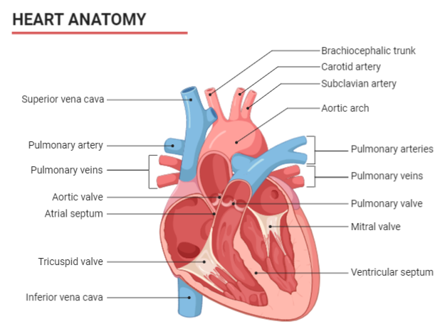
When looking at a human heart diagram, you will notice clear divisions of the atria and ventricles, along with the valves positioned at the boundaries of these chambers. Each component works together to ensure continuous blood circulation.
Quick Quiz
1. Which blood vessel carries oxygenated blood from the lungs to the heart?
Answer: Pulmonary vein
2. Name the valve that prevents backflow of blood into the left ventricle.
Answer: Aortic valve
3. What is the middle layer of the heart wall called?
Answer: Myocardium
4. How many chambers are there in the human heart?
Answer: Four (two atria and two ventricles)
5. Which side of the heart handles deoxygenated blood?
Answer: Right side (right atrium and right ventricle)
Heart Attack Warning Signs
Common warning signs include:
Pain in the chest or discomfort
Shortness of breath
Nausea or vomiting
Excessive sweating
Paying attention to these signs and seeking medical help early can save lives.
Related Topics


FAQs on Diagram of Heart and Its Detailed Structure
1. What is a diagram of the heart?
A diagram of the heart is a labeled visual representation that shows the structure and main parts of the human heart. It typically includes:
- The four chambers: right atrium, right ventricle, left atrium, and left ventricle
- Major blood vessels such as the aorta, vena cava, pulmonary artery, and pulmonary veins
- The four valves: tricuspid, bicuspid (mitral), pulmonary, and aortic valves
2. What are the main parts shown in a heart diagram?
The main parts shown in a heart diagram are the chambers, valves, septum, and major blood vessels. These include:
- Four chambers – two atria (upper) and two ventricles (lower)
- Septum – muscular wall dividing right and left sides
- Heart valves – prevent backflow of blood
- Major vessels – aorta, pulmonary artery, pulmonary veins, superior and inferior vena cava
3. How does blood flow through the heart step by step?
Blood flows through the heart in a specific sequence known as double circulation. The steps are:
- Deoxygenated blood enters the right atrium through the vena cava.
- It moves to the right ventricle through the tricuspid valve.
- Blood is pumped to the lungs via the pulmonary artery.
- Oxygenated blood returns to the left atrium through pulmonary veins.
- It passes into the left ventricle via the mitral valve.
- The left ventricle pumps blood to the body through the aorta.
4. What is the function of the four chambers in the heart?
The four chambers of the heart work together to receive and pump blood efficiently. Their functions are:
- Right atrium – receives deoxygenated blood from the body
- Right ventricle – pumps blood to the lungs
- Left atrium – receives oxygenated blood from the lungs
- Left ventricle – pumps oxygenated blood to the entire body
5. What is the role of valves in the heart diagram?
The heart valves ensure one-way flow of blood and prevent backflow between chambers. The four valves are:
- Tricuspid valve – between right atrium and right ventricle
- Mitral (bicuspid) valve – between left atrium and left ventricle
- Pulmonary valve – between right ventricle and pulmonary artery
- Aortic valve – between left ventricle and aorta
6. Why is the left ventricle thicker than the right ventricle?
The left ventricle has a thicker muscular wall because it pumps blood to the entire body at high pressure. In contrast:
- The right ventricle only pumps blood to the nearby lungs.
- Systemic circulation requires greater force than pulmonary circulation.
7. What is the septum in the heart diagram?
The septum is the muscular wall that separates the right and left sides of the heart. It consists of:
- Interatrial septum – divides the two atria
- Interventricular septum – divides the two ventricles
8. What is double circulation in relation to the heart diagram?
Double circulation means blood passes through the heart twice in one complete cycle. It includes:
- Pulmonary circulation – heart → lungs → heart
- Systemic circulation – heart → body → heart
9. What are the major blood vessels connected to the heart?
The major blood vessels connected to the heart carry blood to and from the lungs and body. These include:
- Aorta – carries oxygenated blood to the body
- Pulmonary artery – carries deoxygenated blood to the lungs
- Pulmonary veins – bring oxygenated blood from lungs
- Superior and inferior vena cava – bring deoxygenated blood from the body
10. How do you label a simple diagram of the human heart for exams?
To label a simple diagram of the human heart for exams, include the most important anatomical parts clearly and accurately. Follow these steps:
- Draw and label the four chambers
- Mark the major blood vessels
- Label the four valves
- Indicate the septum
- Use arrows to show direction of blood flow


