


Alimentary Canal Structure and Functions Explained for NEET Students
Alimentary Canal Anatomy is a fundamental topic in NEET Biology, forming the core of understanding the human digestive system. It refers to the structure and organization of the tube-like pathway through which food passes, digests, and gets absorbed in the body. Mastering this concept is essential for NEET aspirants because questions related to digestive tract parts, their functions, and related processes are frequently asked in the exam. A clear grasp of the alimentary canal's anatomy also helps build a solid foundation for related chapters in physiology and human health.
What is Alimentary Canal Anatomy?
The alimentary canal anatomy deals with the structure, parts, and arrangement of the continuous muscular tube called the alimentary canal or gastrointestinal tract. This canal starts at the mouth and ends at the anus. It is responsible for the entire process of ingestion, digestion, absorption, and egestion of food. In simple terms, it maps out the 'roadway' food takes as it is broken down and absorbed into the body, highlighting the features, length, and functions of each segment.
Core Ideas and Fundamentals of Alimentary Canal Anatomy
General Overview of the Human Alimentary Canal
The human alimentary canal is a long, muscular, coiled tube about 6-9 meters in length. It is designed to transport food while allowing various digestive processes to occur at specific regions. Each part has structural adaptations suited to its specialized function.
Main Components of the Alimentary Canal
- Mouth (Oral Cavity): Entry point for food, contains teeth, tongue, and salivary glands for mechanical and chemical breakdown.
- Pharynx: Common passage for food and air; leads food to the esophagus while preventing entry into the windpipe.
- Esophagus: Muscular tube passing food to the stomach via peristalsis.
- Stomach: Muscular, J-shaped organ where food is churned and mixed with gastric juices for protein digestion.
- Small Intestine: Longest portion (duodenum, jejunum, ileum); main site of enzyme-based digestion and nutrient absorption.
- Large Intestine: Absorbs water and salts, forms feces; includes caecum, colon, and rectum.
- Rectum and Anus: End region for storage and elimination of undigested waste.
Wall Structure of the Alimentary Canal
The wall of the alimentary canal consists of four basic layers in most parts:
- Serosa: Outermost protective layer (except in the mouth and pharynx).
- Muscularis: Responsible for peristaltic movements; contains circular and longitudinal muscle layers.
- Submucosa: Connective tissue layer containing blood vessels, nerves, and glands.
- Mucosa: Innermost layer; produces mucus and contains cells for absorption and enzyme secretion.
Important Sub-Concepts Related to Alimentary Canal Anatomy
Accessory Digestive Organs
While not part of the canal itself, organs like the liver, pancreas, and gall bladder play vital roles by supplying digestive juices and enzymes directly into the canal, especially into the small intestine.
Regional Specialization
Each segment of the alimentary canal has adaptations (such as villi in the small intestine, rugae in the stomach) that optimize its function. Understanding these specializations is essential for NEET questions on structure-function relationship.
Peristalsis
Peristalsis is the wave-like muscular contraction that propels food along the alimentary canal. Recognizing how these contractions occur and their regulation is important for understanding digestive physiology.
Key Features and Structural Relationships in Alimentary Canal Anatomy
Some relevant facts and relationships regarding the anatomy of the alimentary canal include:
- The total length of the alimentary canal is about 6-9 meters in an adult human.
- The small intestine (about 6 meters) is the longest section, specially adapted for absorption.
- Layers of the wall (mucosa, submucosa, muscularis, serosa) remain consistent but show regional modifications for specialized functions.
- Specialized structures like sphincters (gastroesophageal, pyloric, ileocecal, anal) regulate transit between compartments.
Why is Alimentary Canal Anatomy Important for NEET?
A thorough understanding of alimentary canal anatomy is crucial for NEET due to its central role in the digestive system and overall human physiology. Many NEET Biology questions test knowledge of structural details, functional relationships, and clinical correlations related to the digestive tract. Questions can involve identifying parts, understanding layer functions, specializations, comparison of regions, or the impact of digestive disorders. Additionally, this topic supports foundational understanding for physiology, nutrition, and related medical subjects.
How to Study Alimentary Canal Anatomy Effectively for NEET
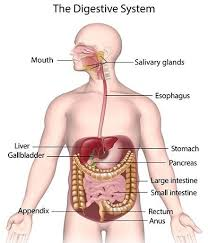
- Start by visually familiarizing yourself with labelled diagrams (like the Human Digestive System image above), and practice drawing and labelling key parts.
- Break the canal into its main parts and review the structure, function, and adaptations of each.
- Use mnemonics or memory aids to recall the sequence of regions.
- Revise the four basic layers of the canal and understand modifications seen in different regions.
- Solve NEET MCQs related to structure identification, function, and clinical correlations.
- Regularly revise quick summary notes and practice schematic diagrams to boost retention.
- Resolve doubts related to confusing sections (such as the difference between the small and large intestine or locations of sphincters).
Common Mistakes Students Make in Alimentary Canal Anatomy
- Confusing the sequence of parts or locations of specific organs.
- Overlooking the importance of layer specialization such as villi, rugae, or sphincters.
- Mislabeling diagrams or neglecting diagram practice, leading to errors in MCQs.
- Ignoring the integration of accessory glands with the main canal.
- Forgetting the fundamental differences between the small and large intestine.
Quick Revision Points: Alimentary Canal Anatomy
- Alimentary canal: Continuous tube from mouth to anus - main digestive pathway.
- Main parts: Mouth, pharynx, esophagus, stomach, small intestine, large intestine, rectum, anus.
- Basic wall layers: Mucosa, submucosa, muscularis, serosa.
- Small intestine is longest; main site for digestion and absorption.
- Sphincters control movement between canal segments.
- Practice diagrams and match structural features to function for NEET MCQs.
- Accessory organs (liver, pancreas) aid digestion by supplying enzymes and bile.
FAQs on Biology Alimentary Canal Anatomy NEET Guide
1. What is the alimentary canal and its main function in the human body?
The alimentary canal is a continuous muscular tube that runs from the mouth to the anus, playing a vital role in digesting food and absorbing nutrients as per NEET syllabus requirements. Its main functions include:
- Ingestion – taking in food (mouth)
- Digestion – breaking down complex food into absorbable forms
- Absorption – transferring nutrients into the bloodstream (small intestine)
- Egestion – removal of undigested waste (anus)
2. What are the main parts of the human alimentary canal?
The human alimentary canal consists of major structures responsible for food processing. The primary parts include:
- Mouth
- Pharynx
- Oesophagus
- Stomach
- Small intestine (duodenum, jejunum, ileum)
- Large intestine (caecum, colon, rectum)
- Anus
3. What is the length of the human alimentary canal?
The human alimentary canal stretches about 9 meters (approximately 30 feet) from mouth to anus. This measurement includes all key segments:
- Mouth to pharynx
- Oesophagus (~25 cm)
- Stomach
- Small intestine (~6 meters)
- Large intestine (~1.5 meters)
4. What is the function of the small intestine in the alimentary canal?
The small intestine is crucial for both digestion and absorption of nutrients within the alimentary canal. Its core functions are:
- Enzymatic digestion of carbohydrates, proteins, and fats
- Maximum absorption of nutrients and minerals into blood
- Facilitating bile and pancreatic juice action
5. What are accessory digestive glands associated with the alimentary canal?
Accessory digestive glands produce secretions that aid in digestion but are not directly part of the alimentary canal. Key glands are:
- Salivary glands (saliva production)
- Liver (bile secretion)
- Pancreas (digestive enzymes, insulin)
6. What is the difference between the small intestine and large intestine in the human alimentary canal?
The small intestine and large intestine differ in structure and function within the alimentary canal. Key differences include:
- Small intestine: Narrow, about 6 meters long, responsible for digestion and nutrient absorption
- Large intestine: Wider, about 1.5 meters long, absorbs water and forms feces
- Villi present in small intestine, absent in large intestine
7. What are the four layers of the wall of the alimentary canal?
The alimentary canal wall is formed by four concentric layers, each with a unique role in digestion. These are:
- Serosa – outermost protective covering
- Muscularis – smooth muscle for movement (peristalsis)
- Submucosa – connective tissue with blood and nerves
- Mucosa – innermost, secretes mucus and digestive enzymes
8. What is peristalsis in the alimentary canal?
Peristalsis is a coordinated, wave-like contraction of muscles in the wall of the alimentary canal. Its main points are:
- Helps move food from oesophagus down to the stomach and beyond
- Occurs throughout the canal, especially in oesophagus, stomach, and intestines
- Facilitates mixing of digestive contents
9. What is the role of the stomach in the alimentary canal?
The stomach acts as a muscular sac that stores and digests food in the alimentary canal. Key roles are:
- Temporary food storage
- Mechanical mixing by churning food
- Secretion of gastric juices (HCl and pepsin) for protein digestion
- Kills microbes with acidic environment
10. Name the parts of the human digestive system involved in the ingestion, digestion and absorption of food.
The main parts of the human digestive system for ingestion, digestion, and absorption are:
- Ingestion: Mouth, teeth, tongue, salivary glands
- Digestion: Stomach (mechanical and chemical), small intestine (enzymatic)
- Absorption: Small intestine (villi), large intestine (water and salts)
11. What are the functions of the liver in the digestion process?
The liver is the largest digestive gland in the human body and plays multiple roles in digestion. Key functions include:
- Secretion of bile for fat emulsification
- Metabolism of nutrients absorbed from the intestine
- Detoxification of harmful substances
- Storage of vitamins and glycogen