


How to Remember Skull Bones Easily for NEET Biology
The human skull is a complex bony structure that protects the brain and supports the facial features. Understanding the 22 bones of the skull is crucial for NEET Biology, as questions related to human anatomy frequently test your grasp of these fundamental skeletal structures. This topic builds a strong base for more advanced studies in neurobiology, physiology, and the musculoskeletal system, making it essential for aspiring medical students.
What are the 22 Bones of the Skull?
The 22 bones of the human skull form the rigid framework of the head. These bones are categorized into two groups: cranial bones, which encase and protect the brain, and facial bones, which shape the face and support functions like chewing and speaking. The skull bones are joined by immovable joints called sutures, except for the mandible (lower jaw), which is movable. Knowing each bone's location and function simplifies understanding head and brain protection in humans—a critical topic for NEET students.
Core Ideas and Structure of the 22 Skull Bones
Cranial Bones
There are 8 cranial bones that form the protective cavity around the brain. These bones are mostly flat and joined tightly together to provide maximum protection and structure.
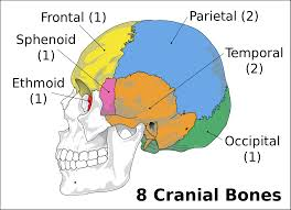
- 1 Frontal bone (forehead region)
- 2 Parietal bones (one on each side)
- 2 Temporal bones (one on each side)
- 1 Occipital bone (back and base of the skull)
- 1 Sphenoid bone (base of the skull, towards the middle)
- 1 Ethmoid bone (between the eyes at the roof of the nasal cavity)
Facial Bones
The face is constructed from 14 facial bones that provide structure, assist in eating, breathing, and expressing emotions, and support sensory organs.
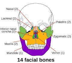
- 2 Nasal bones
- 2 Maxillae (upper jaw)
- 2 Zygomatic bones (cheekbones)
- 2 Palatine bones (back part of hard palate)
- 2 Lacrimal bones (medial wall of eye socket)
- 2 Inferior nasal conchae
- 1 Vomer bone (part of nasal septum)
- 1 Mandible (lower jaw, only movable skull bone)
Key Sub-Concepts Related to Skull Bones
Sutures of the Skull
Sutures are the immovable joints that fuse most skull bones, adding stability and protecting the brain. The major sutures include the coronal (between frontal and parietal), sagittal (between the two parietals), lambdoid (between parietals and occipital), and squamous (between parietal and temporal).
Functions of Skull Bones
- Enclose and protect the brain
- Form the framework of the face
- Anchor teeth and facial muscles
- Assist in breathing, eating, and speaking
Movable vs Immovable Bones
While all skull bones (except the mandible) are fused and immovable, the mandible is connected to the skull by a synovial joint and moves during chewing and speaking. This distinction is frequently tested in NEET exams.
Principles and Relationships in Skull Anatomy
A key anatomical rule for remembering skull bones is: 8 cranial and 14 facial bones, totaling 22 bones. The symmetry of bones (pairs vs unpaired) is also frequently assessed. The arrangement of bones and location of sutures influences cranial capacity and facial structure, directly affecting functions like protection of the brain, mastication, and sensory input. Understanding these relationships is vital for both theory and MCQs in NEET.
Features and Functions of Skull Bones
- Protection: Cranial bones form a hard shell around the brain.
- Support: Facial bones provide structure for facial features and hold teeth.
- Mobility: Only the mandible allows movement for chewing and talking.
- Sensory Support: Bones like ethmoid and nasal conchae are associated with smell.
Why are the 22 Skull Bones Important for NEET?
Knowledge of the 22 skull bones is crucial for NEET as it forms the backbone of human anatomy questions. Understanding the distinction between cranial and facial bones, joint types, and bone functions helps in answering MCQs accurately. This topic often serves as the foundation for complex questions on the nervous system, sense organs, and injuries to the skull. A clear grasp of skull anatomy supports your learning in diverse areas of human physiology and pathology, both of which are central in NEET Biology.
How to Study the 22 Bones of the Skull Effectively for NEET
- Study labeled diagrams like the ones above and regularly test your recall.
- Memorize paired vs unpaired bones using mnemonics (e.g., for cranial: "PEST OF 6" - Parietal, Ethmoid, Sphenoid, Temporal, Occipital, Frontal).
- Understand not just names, but locations, shapes, and how bones connect (sutures).
- Practice NEET-style MCQs that test bone counts, functions, or joint types.
- Revise through short tables or flashcards for rapid recall before exams.
- Group bones by function (protection vs facial structure) to simplify memorization.
Common Mistakes Students Make with the 22 Skull Bones
- Confusing cranial with facial bones or miscounting paired and unpaired bones.
- Forgetting that only the mandible is movable, others are fused by sutures.
- Ignoring special bones like the vomer or ethmoid that aren't immediately visible.
- Not connecting bone function (like protection or mobility) with anatomy.
Quick Revision Points: 22 Skull Bones
- Total skull bones: 22 (8 cranial + 14 facial)
- Cranial bones: 1 frontal, 2 parietal, 2 temporal, 1 occipital, 1 sphenoid, 1 ethmoid
- Facial bones: 2 nasal, 2 maxillae, 2 zygomatic, 2 palatine, 2 lacrimal, 2 inferior nasal conchae, 1 vomer, 1 mandible
- Only the mandible is movable
- Major sutures: coronal, sagittal, lambdoid, squamous
- Skull bones support protection, sensation, chewing, breathing, and facial expression
- Relate diagrams to physical locations for better memory and MCQ solving
FAQs on 22 Bones Of The Skull in NEET Biology
1. What are the 22 bones of the skull in humans for NEET?
The human skull consists of 22 bones, crucial for NEET exams, grouped into cranial and facial bones. These bones protect the brain and form the facial framework.
- Cranial bones (8): Frontal, 2 Parietal, 2 Temporal, Occipital, Sphenoid, Ethmoid
- Facial bones (14): 2 Nasal, 2 Maxillae, 2 Zygomatic, 2 Palatine, 2 Lacrimal, 2 Inferior nasal conchae, 1 Vomer, 1 Mandible
2. How do you remember the bones of the skull for NEET?
Mnemonics help students remember the 22 skull bones for NEET and Biology exams. Consider using these tips:
- Cranial bones (8): 'Old People From Tokyo Eat Spinach Pie' (Occipital, Parietal x2, Frontal, Temporal x2, Ethmoid, Sphenoid)
- Facial bones (14): 'Virgil Can Not Make My Pet Zebra Laugh' (Vomer, Conchae, Nasal x2, Maxilla x2, Mandible, Palatine x2, Zygomatic x2, Lacrimal x2)
3. What is the difference between cranial and facial bones in the skull?
The human skull has cranial and facial bones, each with distinct functions for NEET students.
- Cranial bones (8): Enclose and protect the brain (e.g., frontal, parietal, temporal, occipital, sphenoid, ethmoid).
- Facial bones (14): Shape the face and form the jaw, nose, and orbits (e.g., maxilla, zygomatic, mandible, nasal).
4. Name the paired and unpaired bones of the human skull (NEET syllabus).
Paired and unpaired bones in the skull are a common NEET question.
- Paired cranial bones: Parietal (2), Temporal (2)
- Unpaired cranial bones: Frontal, Occipital, Sphenoid, Ethmoid
- Paired facial bones: Maxilla (2), Zygomatic (2), Nasal (2), Lacrimal (2), Palatine (2), Inferior nasal conchae (2)
- Unpaired facial bones: Mandible, Vomer
5. What is the function of the skull bones?
The 22 skull bones perform vital roles for survival and NEET studies.
- Protect the brain and sensory organs
- Provide structure to the head and face
- Support and anchor facial muscles
- Enable chewing and speech
6. How many bones make up the cranium and face in the human skull?
The human skull has 8 cranial bones and 14 facial bones, totalling 22 bones, as per the NEET syllabus.
- Cranial bones: 8
- Facial bones: 14
7. Which is the only movable bone in the skull?
The mandible is the only movable bone of the human skull, an important NEET fact.
- Also known as the lower jawbone
- Enables chewing and speaking
- All other skull bones are immovable, except the mandible
8. What are the differences between the male and female human skull for NEET?
Male and female skulls show key differences, often highlighted in NEET exams.
- Male skull: Larger, heavier, more pronounced brow ridges
- Female skull: Smaller, rounded forehead, less prominent features
- Mastoid process and external occipital protuberance are more marked in males
9. List the names of the cranial bones in the human skull as per NEET syllabus.
The 8 cranial bones of the human skull for NEET are:
- Frontal
- Parietal (2)
- Temporal (2)
- Occipital
- Sphenoid
- Ethmoid
10. What are the facial bones of the human skull for NEET?
The facial bones in the human skull, needed for NEET, are 14 in all:
- Nasal (2)
- Maxilla (2)
- Zygomatic (2)
- Lacrimal (2)
- Palatine (2)
- Inferior nasal conchae (2)
- Vomer
- Mandible
11. What are the main parts of the skull?
The skull has two main parts, essential for NEET questions:
- Cranium: Encloses and protects the brain
- Facial skeleton: Shapes the face, holds the teeth, and forms orbits
12. Why is the knowledge of skull bones important in NEET Biology?
Understanding skull bones is critical for NEET Biology because it covers foundational anatomy topics.
- Helps answer NEET multiple choice and short answer questions
- Supports knowledge of brain protection, sensory organs, and appearance
- Provides a basis for advanced topics in medicine and dentistry