

What Are the Main Functions of a Microscope in Biology
Microscopes are invaluable instruments that empower students, researchers, and professionals to study the intricate details of the microscopic world. This comprehensive guide explains the function of microscope in clear, simple terms, outlining the microscope parts and functions that are essential for exploring biological specimens, diagnosing diseases, and even solving forensic puzzles.
In this article, we will delve into the various types of microscopes, examine the microscope parts and their specific roles, and highlight some unique insights to enhance your learning experience. Along the way, we will also detail the function of the stage in the microscope and the function of the objective lens in the microscope—two critical components that significantly impact the clarity and precision of the observed image.
Types of Microscopes: Beyond Simple Magnification
Microscopes work on the twin principles of magnification and resolution. While magnification enlarges the image, resolution determines the level of detail visible in the specimen. Understanding the function of a microscope starts with knowing the different types available:
Light Microscopes
Compound Microscope: Equipped with multiple objective lenses (typically ranging from 4x to 100x magnification), this microscope offers high magnification and resolution. It is perfect for studying cells, tissues, and bacteria.
Simple Microscope: Using a single lens, simple microscopes are easy to use and rely on natural light. Though less powerful, they are excellent for basic observations.
Dissection (Stereo) Microscope: Ideal for examining the surface details of larger, solid specimens such as insects or plant parts, these microscopes provide a three-dimensional view.
Electron Microscopes
Transmission Electron Microscope (TEM): Electrons pass through an ultra-thin specimen to form highly detailed images. TEM is essential for viewing internal structures at a very high resolution.
Scanning Electron Microscope (SEM): SEM scans the surface of specimens using an electron beam, making it perfect for analysing textures and surface details.
Additional advanced microscopes, such as scanning probe and scanning acoustic microscopes, continue to push the boundaries of resolution and application.
Also, read Parts of Microscope
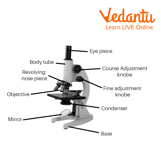
Microscope Parts and Functions: A Closer Look
A deeper understanding of microscope parts and functions is vital for any budding scientist. Below is an overview of the key components and their roles:
Structural Components
Head (Body): Houses the optical elements and is where the function of microscope becomes apparent. It includes the eyepiece and the upper portion of the optical tube.
Arm: Provides support and connects the head to the base. It is designed for comfortable handling.
Base: Supports the microscope and contains the illuminator, a key component in ensuring the sample is well-lit.
Optical Components
Eyepiece: The lens through which the specimen is viewed. It works in tandem with the objective lenses to provide the desired magnification.
Objective Lenses: These are the main lenses with various magnification powers. Understanding the function of objective lenses in the microscope is crucial; they gather light from the specimen and create a magnified image. Each objective lens contributes to the overall magnification and resolution.
Nosepiece: Holds the objective lenses and allows for easy switching between different magnification levels.
Adjustment and Illumination
Fine and Coarse Focus Knobs: Used to adjust the clarity of the image. The coarse knob moves the stage rapidly for initial focusing, while the fine knob refines the image.
Stage: This is the platform where the specimen is placed. The function of stage in microscope is essential—it holds and manoeuvres the slide to view different areas of the specimen. Mechanical stages often come with clips or adjustable knobs to ensure precise movement.
Illuminator: Provides the necessary light for the specimen to be observed. Typically found in the base, modern microscopes offer variable lighting control.
Condenser and Diaphragm: The condenser focuses the light onto the specimen, and the diaphragm (or iris) regulates the amount of light that passes through. Together, they optimise image brightness and contrast.
By understanding these microscope parts, you gain a clearer insight into how each element contributes to the overall microscope parts and functions. This not only improves your practical skills in handling a microscope but also deepens your appreciation of the engineering behind these instruments.
Practical Functions of Microscopes in Science
Microscopes serve a range of functions, each tailored to different scientific and practical needs:
Biological Studies
Cellular and Microbial Analysis: The function of microscope in biology is to reveal the complex structures of cells, viruses, and bacteria. Compound microscopes, with their high magnification capabilities, are invaluable in medical diagnostics and research.
Fungal and Algal Studies: Both simple and compound microscopes help in identifying and studying various forms of fungi and algae, supporting research in ecology and environmental science.
Medical and Forensic Applications
Pathology and Diagnosis: Microscopes enable detailed observation of tissue samples, aiding in the diagnosis of diseases.
Forensic Analysis: High-resolution imaging of fingerprints and other microscopic evidence is made possible by advanced microscopes, proving critical in forensic investigations.
Industrial and Research Uses
Materials Testing: Electron microscopes allow researchers to study the microstructure of materials, assisting in quality control and failure analysis.
Tissue Imaging and Device Testing: These microscopes are also employed in medical research for tissue imaging and in engineering for device testing.
Enhancing Your Microscope Experience: Tips and Unique Insights
To make the most of your microscope, consider these additional tips and insights:
Regular Calibration: Ensure your microscope is calibrated regularly to maintain high resolution and accurate measurements.
Optimal Lighting: Experiment with the diaphragm and condenser settings to achieve the best contrast and brightness. Adjusting the function of stage in microscope properly can further enhance the clarity of your observations.
Maintenance: Clean the lenses and other optical components routinely. Proper maintenance ensures longevity and consistent performance of all microscope parts.
Advanced Techniques: Explore digital imaging techniques that integrate with traditional microscopy to capture, store, and analyse images. This is particularly useful for sharing findings or creating detailed reports.
Safety First: Always handle specimens and slides with care, and follow laboratory safety protocols. This ensures a safe environment while exploring the function of microscope and its various parts.
For those interested in more advanced studies, our related pages on Cell Biology, Microbiology, and Biochemistry provide deeper insights into the microscopic world and how these techniques are applied in modern research.
Recap of Key Microscope Components and Their Functions
Function of Microscope: To magnify and resolve minute details in a specimen, facilitating detailed biological and material studies.
Microscope Parts and Functions: Every part, from the eyepiece to the stage, plays a vital role in creating a clear, magnified image.
Function of Stage in Microscope: Holds and manoeuvres the specimen, allowing for precise observation of different areas.
Function of Objective Lens in Microscope: Gathers light and creates the magnified image, significantly influencing the overall clarity and detail.
Microscope Parts: Includes structural components (head, arm, base) and optical components (eyepiece, objective lenses, condenser), all working together to deliver a complete viewing experience.
Fun Facts About Microscopes
Ancient Beginnings: The first microscopes were invented in the 16th century. Early versions were simple devices that paved the way for the compound microscopes we use today.
Tiny Discoveries: The invention of the microscope has led to monumental discoveries, including the identification of bacteria, which was crucial in understanding diseases and hygiene.
High-Tech Marvels: Modern electron microscopes can magnify objects up to 2 million times, revealing structures as small as individual molecules—a scale unimaginable with traditional light microscopes.
Real-World Applications of Microscopes
Microscopes are not just confined to laboratory use—they have a broad range of real-world applications that impact various industries:
Healthcare: Pathologists use microscopes to examine tissue samples, enabling accurate diagnoses of diseases such as cancer. This directly influences treatment plans and patient care.
Forensic Science: In forensic laboratories, microscopes help in analysing trace evidence such as hair fibres, fibres, and minute residues. This aids in criminal investigations by providing crucial clues.
Environmental Studies: Ecologists utilise microscopes to study microorganisms in water and soil, which helps in assessing environmental health and biodiversity.
Industrial Quality Control: Manufacturers use microscopes for inspecting materials and components at a micro-level to ensure product quality and reliability, particularly in electronics and aerospace.
Education and Research: From school laboratories to advanced research centres, microscopes provide a window into the microscopic world, fostering learning and innovation across scientific disciplines.


FAQs on Functions of Microscope and How It Works
1. What is the main function of a microscope?
The main function of a microscope is to magnify very small objects so they can be seen clearly by the human eye. It helps in studying structures that are too tiny to observe without assistance.
- Enlarges images of cells, tissues, and microorganisms
- Improves visibility through magnification and resolution
- Allows detailed study of biological specimens in laboratories
2. What are the functions of a microscope in biology?
In biology, a microscope is used to observe and analyze microscopic living organisms and cell structures. It plays a crucial role in understanding life at the cellular level.
- Examines cell structure and organelles
- Identifies bacteria, fungi, and protozoa
- Studies tissues in histology
- Supports research in microbiology and pathology
3. How does a microscope work?
A microscope works by using lenses to magnify and focus light on a specimen to produce an enlarged image. The combined action of its lenses increases the apparent size of the object.
- Objective lens magnifies the specimen first
- Eyepiece (ocular lens) further enlarges the image
- Light source illuminates the specimen for clarity
4. What is the function of the objective lens in a microscope?
The objective lens provides the primary magnification of the specimen in a microscope. It determines the level of detail that can be observed.
- Usually available in different powers (e.g., 4×, 10×, 40×, 100×)
- Forms the first enlarged image of the specimen
- Works with the eyepiece to produce total magnification
5. What is the function of the eyepiece in a microscope?
The eyepiece, also called the ocular lens, further magnifies the image formed by the objective lens. It is the lens through which the observer looks.
- Commonly has 10× magnification
- Enlarges the intermediate image
- Helps produce the final visible image
6. What is the function of the condenser in a microscope?
The condenser focuses light onto the specimen to improve image clarity and resolution. It ensures proper illumination for detailed observation.
- Concentrates light from the source
- Enhances contrast and image sharpness
- Essential for high-power magnification
7. Why is a microscope important in studying cells?
A microscope is important because most cells are microscopic and cannot be seen with the naked eye. It allows scientists to study cell structure and function.
- Reveals cell organelles like nucleus and mitochondria
- Helps understand cell division (mitosis and meiosis)
- Supports diagnosis of diseases at the cellular level
8. What are the different types of microscopes and their functions?
Different types of microscopes are designed for specific magnification and research purposes. Each type serves a unique function in biological studies.
- Light microscope: Observes cells and tissues using visible light
- Electron microscope: Provides very high magnification for ultrastructural details
- Compound microscope: Uses multiple lenses for higher magnification
- Stereo microscope: Views larger specimens in three dimensions
9. What is the difference between magnification and resolution in a microscope?
Magnification increases the size of an image, while resolution determines the clarity and detail of that image. Both are essential for effective microscopic observation.
- Magnification: How much larger the specimen appears
- Resolution: Ability to distinguish two close points as separate
- High magnification without good resolution gives a blurry image
10. Can you give an example of how a microscope is used in medical science?
In medical science, a microscope is used to examine blood samples and tissue sections for disease diagnosis. It helps detect abnormalities at the cellular level.
- Identifies bacteria and parasites in infections
- Examines blood cells for disorders like anemia or leukemia
- Studies biopsy samples to diagnose cancer


