


NCERT Exemplar for Class 12 Biology - Human Reproduction - Free PDF Download
MULTIPLE-CHOICE QUESTIONS
1. Choose the incorrect statement from the following:
a. In birds and mammals internal fertilization takes place
b. Colostrum contains antibodies and nutrients
c. Polyspermy in mammals is prevented by the chemical changes in the egg surface.
d. In the human female implantation occurs almost seven days after fertilization.
Ans: Correct Option is c.
Internal fertilization is the type of fertilization during which union of egg and sperm nuclei occurs inside the body of a parent i.e., internally. This occurs in birds, mammals, and reptiles.
Colostrum is the first milk produced after giving birth to a young one by the mother. It is a yellow-colored milk secretion which contains a large number of antibodies viz. IgA and nutrients to nourish the young one. It is a very important meal for a newborn and must not be discarded.
Polyspermy i.e., fertilization of one egg by many sperms is prevented in mammalian eggs by the process of depolarization. The egg, after being fertilized or entered in by one sperm nucleus, gets depolarized at the plasma membrane due to certain reactions. This helps to prevent any additional sperm from entering the egg cell and hence, polyspermy. It is, therefore, not prevented by chemical changes but changes in electrical potentials of the plasma membrane of the egg surface.
In the human female, implantation is bound to occur after 7-10 days of fertilization as the zygote form makes its way through the fallopian tube to uterus and then to the implantation site in the endometrium.
2. Identify the correct statement from the following:
a. High levels of estrogen trigger the ovulatory surge.
b. Oogonial cells start to proliferate and give rise to functional ova in regular cycles from puberty onwards.
c. Sperms released from seminiferous tubules are highly motile.
d. Progesterone level is high during the post ovulatory phase of the menstrual cycle.
Ans: Correct option is d.
The high level of LH or FSH triggers the ovulatory surge. The oogonial cells help to proliferate in the middle of a regular cycle from puberty. The sperms released from seminiferous tubules are pooly motile hence it is also incorrect.The statement d is correct as there is a considerably higher level of the hormone progesterone during the postovulatory phase of the menstrual cycle during which the follicle after rupture turns into corpus luteum. The corpus luteum is a steroidogenic cluster of cells which is yellow in color and secretes progesterone mainly. Hence option d is the correct answer.
3. Spot the odd one out from the following structures with reference to the male reproductive system:
a. Rete testis
b. Epididymis
c. Vasa efferentia
d. Isthmus
Ans: Correct Option is d.
Rete testis is the part or a network of tubules just outside the seminiferous tubules located at the hilum of testis and carries the spermatozoa produced in seminiferous tubules to vasa efferentia.
Epididymis is a part of male reproductive that carries sperms towards the vas deferens from the vasa efferentia of testis. In this, the sperms have very less motility and aren’t mature.
Vasa efferentia is a part of male reproductive system too, which is a carrier of spermatozoa from the rete testis
Isthmus, on the other hand, is a part of the fallopian tubes of female reproductive system constituting the distal portion of an oviduct. It receives the egg after it is released from the ovary by the help of fimbriae.
4. Seminal plasma, the fluid part of semen, is contributed by.
i. Seminal vesicle
ii. Prostate gland
iii. Urethra
iv. Bulbourethral gland
a. i and ii
b. i, ii and iv
c. ii, iii and iv
d. i and iv
Ans: Correct Option is b.
The liquid part of the semen or the seminal plasma is formed by the accessory parts of male reproductive system i.e., seminal vesicles, prostate gland and bulbourethral gland. The urethra however, only acts as a conduit for the movement of semen to the outside during ejaculation. Seminal vesicles provide citric acid, fructose, prostaglandins, and fibrinogen while the prostate gland provides calcium and phosphate ions to the seminal plasma of the males as the sperms pass through or by these glands. Bulbourethral gland secrete alkaline mucus which is a vital secretion for penile lubrication during the act of semen passage. All these three accessory glands make the semen qualitatively good for survival in the female reproductive tract.
5. Spermiation is the process of the release of sperms from:
a. Seminiferous tubules
b. Vas deferens
c. Epididymis
d. Prostate gland
Ans: Correct Option is a.
Spermiation refers to the process of release of spermatozoa from seminal or seminiferous tubules from where these cells are transferred to epididymis via vasa efferentia. The sperms mature in the epididymis of the male reproductive pathway.
6. Mature Graafian follicle is generally present in the ovary of a healthy human female around:
a. 5 – 8 day of menstrual cycle
b. 11 – 17 day of menstrual cycle
c. 18 – 23 day of menstrual cycle
d. 24 – 28 day of menstrual cycle
Ans: Correct option is b.
Graafian follicle is the fully grown secondary oocyte which is present in the stroma of the ovary and on maturation, it bursts leading to the release of secondary oocyte from the ovary which is then transferred to the fallopian tubes. The Graafian follicle ruptures only after its complete maturation and a mature follicle in the ovary can be seen at arounf 11-17 day of the menstrual cycle of a human female which is just before the middle of the cycle when ovulation i.e., Graafian rupture occurs.
7. Acrosomal reaction of the sperm occurs due to:
a. Its contact with zona pellucida of the ova
b. Reactions within the uterine environment of the female
c. Reactions within the epididymal environment of Androgens produced in the uterus
Ans: Correct option is a.
Acrosomal reaction is referred to the release of various contents of acrosome viz. hyaluronidase, corona penetrating enzymes, etc. when the acrosome at the head of a spermatozoa comes in contact with the zona pellucida of the ova, an outer layer of the egg. The acrosomal contents help the spermatozoa penetrate zona pellucida and corona radiata and reach plasma membrane of the ova.
8. Which one of the following is not a male accessory gland?
a. Seminal vesicle
b. Ampulla
c. Prostate
d. Bulbourethral gland
Ans: Correct Option is b.
Male accessory glands are the secondary or accessory glands in a male reproductive system which even though secondary to the sperm producing structures, play a major role in the preparation of a qualitatively and chemically correct semen. The male accessory glands include prostate, seminal vesicle and bulbourethral glands. The ampulla is a part of fallopian tube or oviduct of a female reproductive system.
9. The spermatogonia undergo division to produce sperms by the process of spermatogenesis. Choose the correct one with reference to above.
a. Spermatogonia have 46 chromosomes and always undergo meiotic cell division.
b. Primary spermatocytes divide by mitotic cell division.
c. Secondary spermatocytes have 23 chromosomes and undergo second meiotic division.
d. Spermatozoa are transformed into spermatids
Ans: Correct Option is c.
In testis, on the inner walls of seminiferous tubules lie diploid cells called spermatogonia which divide by mitosis. Some of these cells are primary spermatocytes and thereafter undergo meiosis or reduction division to form haploid daughter cells called secondary spermatocytes having only one set of chromosomes. Each of these secondary spermatocytes’ form two spermatozoa of haploid ploidy by second meiosis and this is, in brief, the entire process of spermatogenesis. According to this, option c stands correct.
10. Match between the following representing parts of the sperm and their functions and choose the correct option.
a. A-ii, B-iv, C-i, D-iii
b. A-iv, B-iii, C-i, D-ii
c. A-iv, B-i, C-ii, D-iii
d. A-ii, B-i, C-iii, D-iv
Ans: Correct Option is b.
A spermatozoan or a sperm cell can be divided into four different parts according to its anatomy:
The head contains the nucleus and is an elongated structure with acrosome at the tip of the head. The head mainly contains the genetic material in its nucleus so it matches with iv.
Middle piece consists of the neck and the central body or region of the sperm. The neck contains two nuclei whereas the body, which is cylindrical contains mitochondria, responsible for providing energy to the sperm when inside the female reproductive tract. Thus, this matches with iii.
Acrosome is present at the tip of a sperm cell and contains hydrolyzing enzymes which help it to penetrate zona pellucida i.e., it matches with i.
Tail is long, thin structure containing cytoplasm and helps in moving the sperm through the female reproductive tract thus it provides motility to the sperm and hence it matches with ii.
11. Which among the following has 23 chromosomes?
a. Spermatogonia
b. Zygote
c. Secondary oocyte
d. Oogonia
Ans: Correct Option is c.
23 chromosomes would mean only one set of chromosomes is present i.e., the cell being asked in question should be diploid. Among the given options, only the secondary oocyte is a haploid structure hence, this is the answer.
12. Match the following and choose the correct options:
Options:
a. A-ii, B-i, C-iii, D-iv
b. A-iii, B-iv, C-ii, D-i
c. A-iii, B-i, C-ii, D-iv
d. A-ii, B-iv, C-iii, D-i
Ans: Correct Option is b.
Trophoblast, the outer layer of cells formed by the cells oriented and organized towards the outermost side of blastocyst, is the layer which helps in fetal implantation and matches with option iii.
Cleavage is just another name for a mitotic division which occurs in a zygote, in a rapid fashion and efficiently. This matches with iv.
Inner cell mass refers to the mass of cells lying inside a blastocyst towards a side which later differentiate to form the embryo and its various parts. This, hence, matches with ii.
Implantation is the process of attachment and embedding of the embryo at blastocyst stage into the uterine endometrium achieved with the help of trophoblast layer. This matches with i.
13. Which of the following hormones is not secreted by human placenta?
a. hCG
b. Estrogens
c. Progesterone
d. LH
Ans: Correct Option is d.
Human placenta secretes a number of hormones which lead to various changes in the mother but are primarily secreted to maintain pregnancy and develop the embryo. The LH hormone is the one required for follicle maturation and does not play any role in maintaining the pregnancy and hence is not produced by the placenta.
14. The vas deferens receives duct from the seminal vesicle and opens into urethra as:
a. Epididymis
b. Ejaculatory duct
c. Efferent ductule
d. Ureter
Ans: Correct option is b.
The vas deferens collects sperms from epididymis and while travelling towards the urethra receives a duct from the seminal vesicle to form an ejaculatory duct into the urethra.
15. Urethral meatus refers to the:
a. Urinogenital duct
b. Opening of vas deferens into urethra
c. External opening of the urinogenital duct
d. Muscles surrounding the urinogenital duct
Ans: Correct Option is c.
The word meatus means an opening of a canal in any natural body or otherwise, therefore, urethral meatus would mean the opening of the urogenital duct through which both urine and semen pass towards the outside in human males.
16. Morula is a developmental stage:
a. Between the zygote and blastocyst
b. Between the blastocyst and gastrula
c. After the implantation
d. Between implantation and parturition
Ans: Correct Option is a.
After fertilization, a diploid zygote is formed which undergoes subsequent cleavages to form 2, 4, 8 and then 16 celled stage embryo, the daughter cells of which are known as blastomeres. These divisions are mitotic in nature and occur rapidly dividing the zygote right when it starts moving from oviduct to the uterus. At the 8 celled stage, the embryo is called morula and then it divides to form blastocyst, 18-celled stage. Thus, morula is a developmental stage of embryo between the zygote and the blastocyst.
17. The membranous cover of the ovum at ovulation is:
a. Corona radiata
b. Zona radiata
c. Zona pellucida
d. Chorion
Ans: Correct Option is a.
At the time of ovulation, a fully mature graafian follicle is present in the ovary containing an ovum within. This ovum has various layers of covering produced by the surrounding follicular cells or glycoproteins by the ovum itself. The membranous covering however, is the corona radiata produced or secreted by follicular cells.
18. Identify the odd one from the following:
a. Labia minora
b. Fimbriae
c. Infundibulum
d. Isthmus
Ans: Correct Option is a.
The labia minora is a part of external female genitalia and represents the paired folded tissue present beneath the labia majora and surrounds the opening of vagina.
Fimbriae, infundibulum and isthmus are all parts of the oviduct or fallopian tube consisting of the internal parts of the female reproductive system.
VERY SHORT ANSWER TYPE QUESTIONS
1. Given below are the events in human reproduction. Write them in correct sequential order.
Insemination, gametogenesis, fertilization, parturition, gestation and implantation.
Ans: The correct sequential order for the events occurring in human reproduction is:-
Gametogenesis - Insemination - Fertilization - Implantation - Gestation - Parturition.
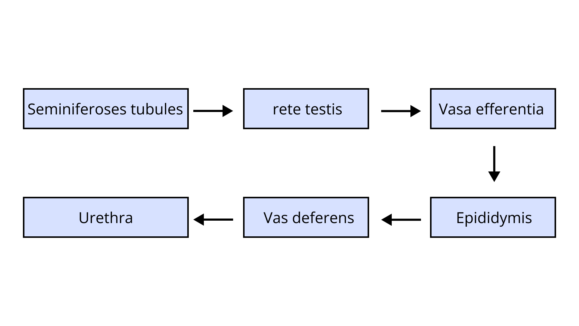
2. The path of sperm transport is given below. Provide the missing steps in blank boxes
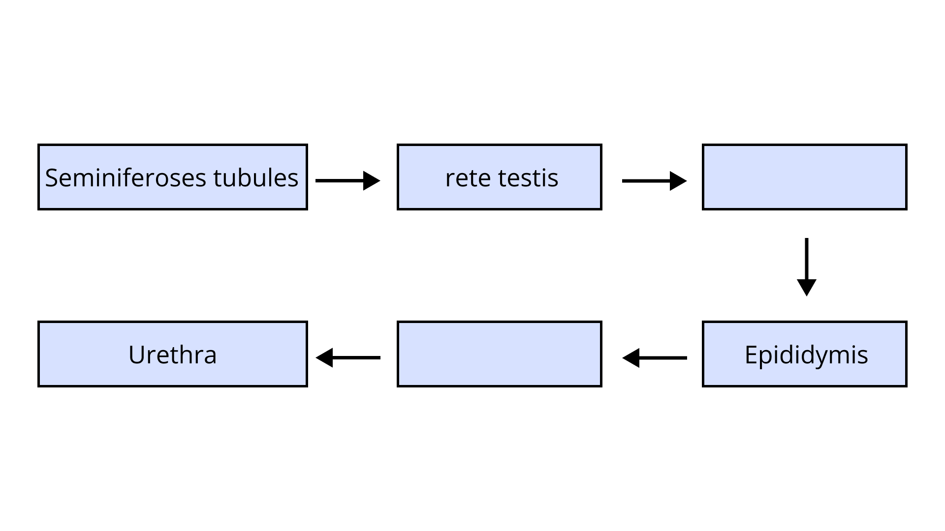
Ans: The path of transport of spermatozoa is respectively from seminiferous tubules to rete testis leading to vasa efferentia which takes the sperms to epididymis and then Vas deferens and, finally, urethra from where these reach outside.
3. What is the role of the cervix in the human female reproductive system?
Ans: Cervix, a part of the birth canal in the female reproductive structure is the part present at the ends of the uterus and beginning of vagina i.e., it acts as a connection between the two. It is the lowest part of the uterus and covers the end of it with strong muscles that keep the opening closed generally. The major role of the cervix is to act as a pathway for passage of menstrual blood in a menstruating female. Its other important function can be the making of the birth canal during parturition. The cervix gets dilated during delivery of the newborn and helps deliver the newborn by creating a passage from the uterus to vagina.
4. Why are menstrual cycles absent during pregnancy?
Ans: The high levels of various pregnancy hormones, especially progesterone, suppress the release of gonadotropins from anterior pituitary by the negative feedback mechanism. The absence of gonadotropins leads to absence of development or maturation of follicles in the ovary therefore, no ovulation occurs and hence no new menstrual cycle is seen as long as pregnancy continues.
5. Female reproductive organs and associated functions are given below in column A and B. Fill the blank boxes.
Ans: Female reproductive organs and associated functions are -
6. From where the parturition signals arise-mother or foetus? Mention the main hormone involved in parturition.
Ans: Parturition refers to the delivery of a newborn or fully developed fetus after the pregnancy has been completed. The first signals of parturition arise from the fully-developed embryo itself. It is called fetal-ejection reflex and causes the first initial contraction of the uterus which triggers the mother’s body’s mechanism for parturition. After this initial reflex, a massive release of oxytocin occurs which causes uterine contractions which acts as positive feedback for more release of oxytocin and a positive feedback loop is thus set till the newborn is delivered.
7. What is the significance of epididymis in male fertility?
Ans: The epididymis is a structure for carrying the spermatozoa from vasa efferentia to the vas deferens. The important role of this structure is the maturity sperms achieve while traveling through the epididymis including maturity, increased motility and fertilizing capacity. In the initial part, it helps in movement of sperms till the time they become capable of moving by segmental and peristaltic contractions at various intervals which push the spermatozoa away from the testes and towards the vas deferens.
8. Give the names and functions of the hormones involved in the process of spermatogenesis. Write the names of the endocrine glands from where they are released.
Ans: GnRH is Gonadotropin releasing hormone, secreted by the hypothalamus, which causes release of gonadotropins LH (Luteinizing hormone) and FSH (Follicle stimulating hormone) from the anterior pituitary gland.
LH acts on Leydig cells and leads to activation of synthesis of androgens in testes which further lead to initiation of spermatogenesis.
FSH, on the other hand, acts on nurse cells or Sertoli cells to stimulate secretion of some factors involved in the process of spermatogenesis.
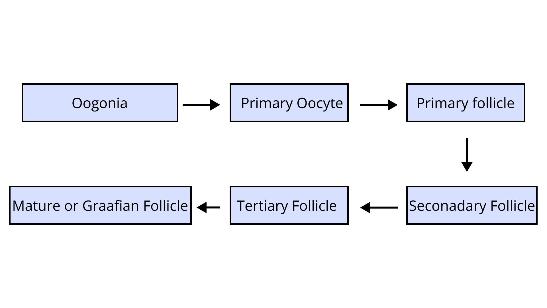
9. The mother germ cells are transformed into a mature follicle through series of steps. Provide the missing steps in the blank boxes.
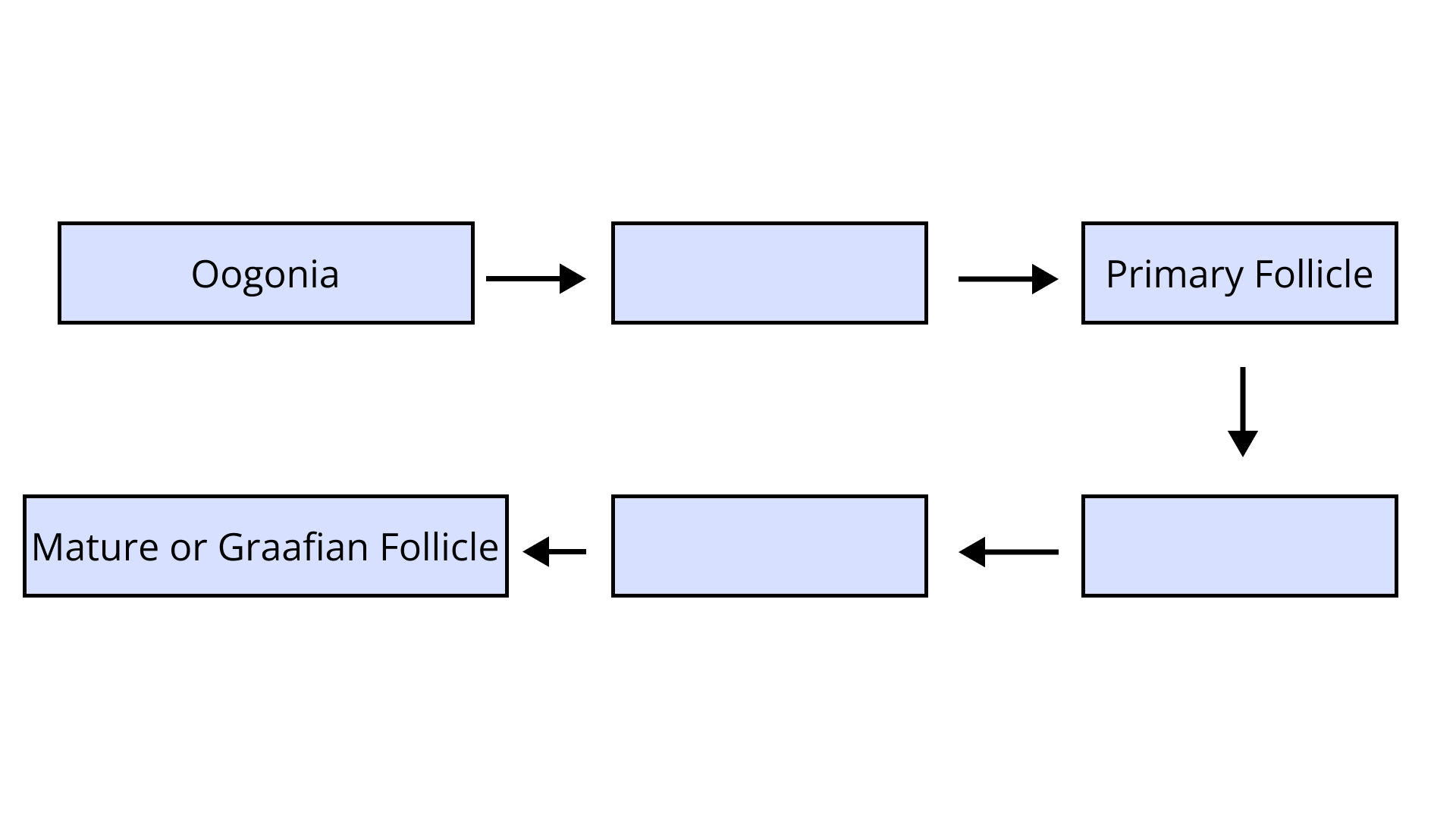
Ans: The mother germ cells or oogonia are transferred into formation of a mature or a graafian follicle by passing through various stages and divisions:
First, a primary oocyte is formed which becomes the primary follicle which further forms a secondary and then a tertiary follicle until the graafian follicle is achieved. This mature follicle ruptures and releases secondary oocytes out of the ovary and causes ovulation.
10. During reproduction, the chromosome number (2n) reduces to half (n) in the gametes and again the original number (2n) is restored in the offspring, what are the processes through which these events take place?
Ans: The restoration of a diploid number of chromosomes in the zygote through combination of two haploid gametes is a constant stability in the reproduction state. For a zygote to be diploid and same as the parent, the gametes fusing to form it must be haploid as n+n would lead to 2n. The diploid germ cells in a parent undergo reduction division or meiosis to achieve a haploid state(n) which occurs in formation of all gametes. The chromosome number is reduced to half in this.
The restoration of diploid ploidy in a zygote occurs through fertilization of egg (n) with sperm(n).
11. What is the difference between a primary oocyte and a secondary oocyte?
Ans: The difference is as given below:
12. What is the significance of ampullary–isthmic junction in the female reproductive tract?
Ans: At the junction of ampulla and isthmus, the parts of a fallopian tube or an oviduct, the ovum is located when a sperm can come and fertilize it i.e., it is the primary site of fertilization in an ideal scenario of fertilization. It is at this junction that the sperm nucleus and ovum are united to form a zygote and the zygote travels a predetermined path to the uterus for implantation within the required time.
13. How does zona pellucida of ovum help in preventing polyspermy?
Ans: The moment one spermatozoan comes into contact with the ovum or the egg, the zona pellucida undergoes some cortical changes which makes this layer impenetrable for other sperms. Thus, no more than one sperm can fertilize the ovum i.e., polyspermy is prevented.
14. Mention the importance of LH surge during menstrual cycle.
Ans: The LH surge in the menstrual cycle has an extremely important role and is essential for a fully functioning menstrual cycle in a female. The LH surge occurs around mid of the menstrual cycle i.e., on the 14th day and causes the rupture of graafian follicle due to which the secondary oocyte is released from the ovary causing ovulation.
15. Which type of cell division forms spermatids from the secondary spermatocytes?
Ans: The type of cell division during which the secondary spermatocytes from spermatids is the reduction division or meiosis in which the chromosome number is halved leading to production of haploid gametes.
SHORT ANSWER TYPE QUESTIONS
1. A human female experiences two major changes, menarche and menopause during her life. Mention the significance of both the events.
Ans: In a human female, the two major changes menarche and menopause are both related to the menstrual cycle of the female. While menarche represents the onset of menstruation, occurring when a female acquires puberty and gets her first menses, the menopause is an event marking the cessation of menstrual cycle. Generally, menarche occurs around 9-15 years of age and begins the age of reproductive activeness in the female i.e., the female is now a woman of reproductive age under government regulations. On the other hand, menopause occurs at around 45-55 years of age when the woman stops having menses and the bleeding stops. From here on, the female cannot give birth to a child through natural means. The eggs are deteriorated or already used up when menopause arrives.
2. a) How many spermatozoa are formed from one secondary spermatocyte?
Ans: A primary sperm cell first goes through its meiosis or the reduction division to form two identical and haploid cells called secondary spermatocytes with a ploidy of n=23 chromosomes each. This secondary spermatocyte then undergoes a second meiosis to give rise to four identical and haploid sperm cells (n). Each of these four sperm cells differentiate to produce spermatozoa.
b) Where does the first cleavage division of zygote take place?
Ans: First division or cleavage which is a mitotic division in a zygote begins approximately 30 hours after fertilization has occurred. It is seen when the zygote moves through the isthmus of the oviduct towards the uterus for implantation.
3. Corpus luteum in pregnancy has a long life. However, if fertilization does not take place, it remains active only for 10-12 days. Explain.
Ans: Corpus luteum is a yellowish mass formed by the leftover graafian follicle after ovulation has occurred in the ovary and the secondary oocyte released. This corpus luteum secretes progesterone in very high quantities which is used to maintain the uterine endometrial lining as the thick, glandular lining. In the presence of fertilization, zygote is formed in the oviduct which hence, does not pass out of the uterus, and gets implanted instead. This leads to longer life of corpus luteum as the newly formed zygote cannot produce progesterone on its own and the hormone is required to keep the endometrium healthy for the zygote. This function is satisfied by the persisting corpus luteum.
4. What is fetal ejection reflex? Explain how it leads to parturition?
Ans: Parturition is the process of delivery of a newborn or a fetus, in simple words but for parturition to occur, many other processes are responsible and help to make a successful childbirth. Fetal ejection reflex is one of those important initial processes which is observed at the time of delivery. It is the initial mild contraction of the uterus which is caused by the placenta after the baby is fully developed in the womb. This reflex leads to release of oxytocin from the maternal pituitary gland which further intensifies the contractions leading to parturition finally.
5. Except for the endocrine function, what are the other functions of placenta?
Ans: Placenta serves the endocrine function primarily by releasing many hormones, mainly progesterone but placenta has many other non-endocrine functions as well:
It acts as a structural and functional connection between the developing embryo and the body of the mother.
It supplies nutrition, oxygenated blood, antibodies and various other things to the fetus in the womb.
It also helps in taking away the waste products or excretory material from the fetus and removes the waste from the body of embryo including carbon dioxide.
6. Why do doctors recommend breastfeeding during the initial period of infant growth?
Ans: Breast milk especially in the first 6 months after birth of a newborn is the exclusive feed that should be provided to the infant. Colostrum produced during the initial few days after delivery of the baby is extremely healthy for the baby and helps in building immunity, providing nutrition and strengthening the newborn. Breast milk has antibodies, required nutrients and various other elements required for an infant. This is the reason a newborn should be given breast milk unless otherwise advised by a medical professional.
7. What are the events that take place in the ovary and uterus during the follicular phase of the menstrual cycle?
Ans: There are a number of events that occur in the ovaries and uterus during the follicular phase of the menstrual cycle:
(a) One of the many primary follicles lying in the ovary develops to become a fully mature Graafian follicle.
(b) Endometrium in the uterus is proliferated, thick and glandular to receive a zygote in case fertilization occurs.
(c) Growing follicles when developing and undergoing maturation during the follicular phase produce a lot of estrogen which peak right before the middle of the cycle when it leads to LH and FSH surge which causes ovulation.
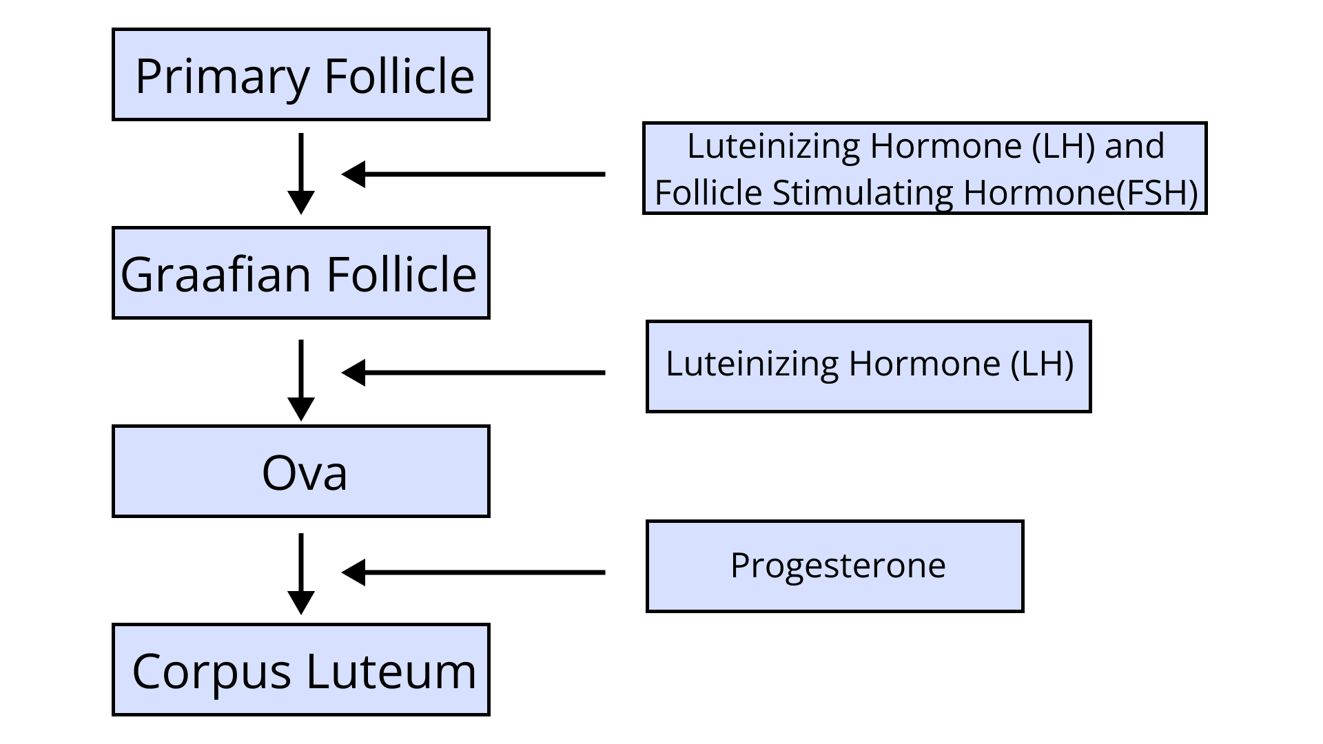
8. Given below is a flow chart showing ovarian changes during menstrual cycle. Fill in the spaces giving the name of the hormones responsible for the events shown.
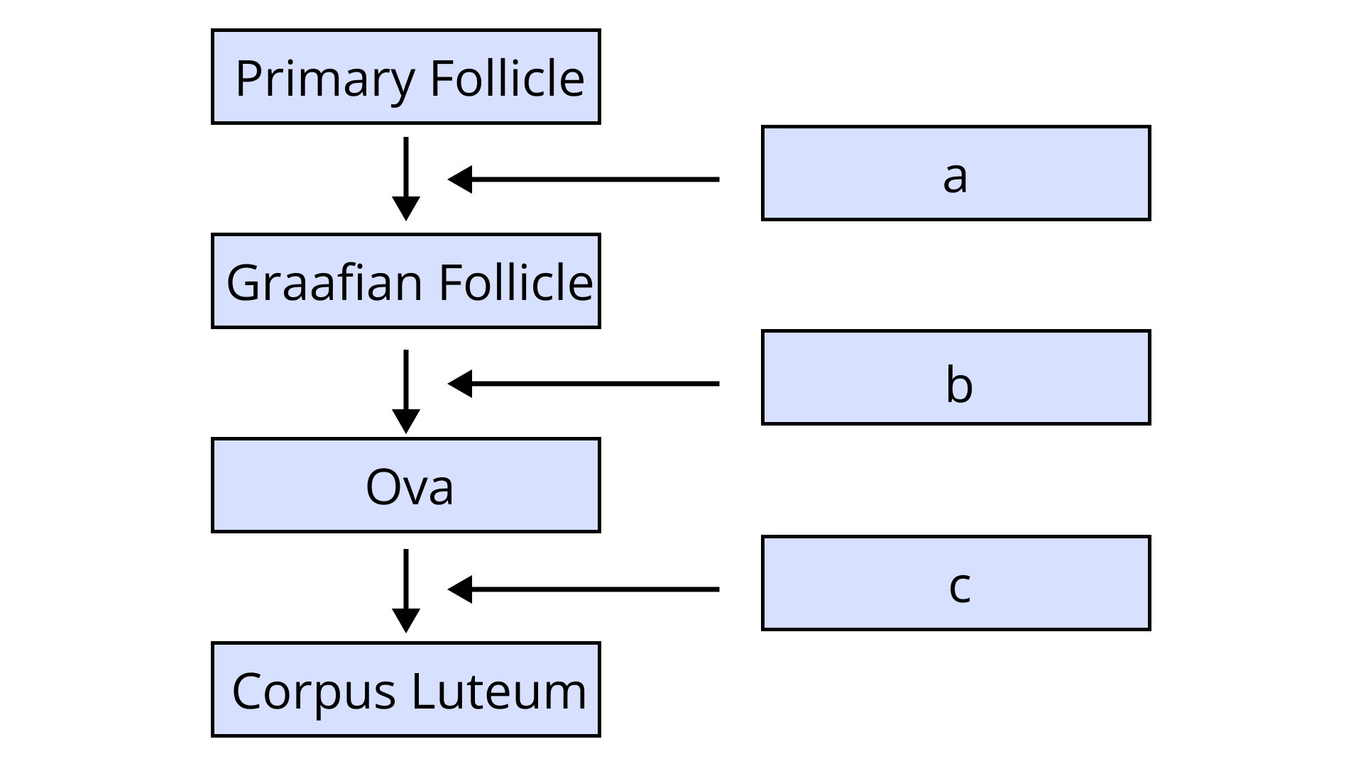
Ans:
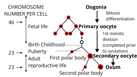
9. Give a schematic labelled diagram to represent oogenesis (without descriptions)
Ans:
10. What are the changes in the oogonia during the transition of a primary follicle to Graafian follicle?
Ans: Various changes occur in the oogonia as they transition from a primary follicle into a mature graafian follicle containing secondary oocyte:
(a) Each of the primary oocytes is surrounded by a single layer of granulosa cells called theca and this is known as the primary follicle.
(b) This primary follicle becomes surrounded by another layer of granulosa cells and a new theca if formed making it the secondary follicle.
(c) Secondary follicles soon develop to form the tertiary follicle which is characterized by a fluid-filled cavity; the antrum. At this stage, the primary oocyte inside the follicle undergoes second meiosis to form the secondary oocyte and a small polar body on the periphery.
(d) Tertiary follicle then transforms into a mature Graafian follicle during this phase. A new layer called the zona pellucida can then be seen surrounding the newly formed secondary oocyte.
LONG ANSWER QUESTIONS
1. What roles do pituitary gonadotropins play during follicular and ovulatory phases of menstrual cycle? Explain the shifts in steroidal secretions.
Ans: The two pituitary gonadotropins are FSH (Follicle Stimulating Hormone) and LH (Luteinizing hormone) playing vital roles during follicular and ovulatory phases of menstrual cycles. The hormone, FSH, stimulates the release of secondary oocytes from a mature graafian follicle and leads to growth of endometrium as well. It leads to development and maturation of the graafian follicle lying in the ovary hence it is more important for the follicular phase. The more important part of ovulation is LH surge which is 6-10 times more than the FSH surge and occurs around about the 14th day of the cycle leading to the rupture of graafian follicle and hence ovulation. This also marks the beginning of the next phase, luteal phase.
The shift in various steroidal secretions can be explained as regards to the changes going on in the ovary of a human female. As the follicles are maturing and developing, levels of estrogen are on the higher end which peaks just some time before the 14th or the ovulation day. By this time, follicles have undergone quite some maturation and then the FSH and LH begin to rise while the estrogen starts to decline. FSH and LH lead to ovulation or graafian rupture following which the oocyte is out of the ovary. The remaining mass left is called yellowish corpus luteum which starts secreting progesterone mainly and some amounts of estrogen. This progesterone secreted maintains the endometrial lining of the uterus and makes it ready for fertilization. In case of failure of that, after a few days, the corpus luteum starts to degenerate and the progesterone secretion decreases leading to shedding of endometrium of the uterus called menstruation.
2. Meiotic division during oogenesis is different from that in spermatogenesis. Explain how and why?
Ans: Meiosis during oogenesis i.e., development of ova is quite different from the meiotic division occurring during spermatogenesis in many ways:
Unlike in spermatogenesis, the reduction division in females begins intra-uterine i.e., within the fetus itself.
The primordia inside the two ovaries of females undergo the first part of meiosis to form primary oocyte and it occurs till the 20th week of intra-uterine life following which no new primary oocytes are formed. So, the number of eggs a female can make during her lifetime are limited, unlike the sperms in males, which begin at puberty and last till old age.
In spermatogenesis, all the phases of meiosis are completed once initiated and do not have any pauses whereas in females, meiosis in the uterus gets paused at the prophase 1 stage. The later stages are completed after puberty.
Where in spermatogenesis, a complete haploid spermatozoa nucleus is formed after a complete meiosis and before ejaculation, in females, the secondary oocyte is released from the ovary which forms an egg only if fertilization occurs.
Unlike spermatogenesis, 2 polar bodies are formed in a complete meiosis cycle of females.
The following may be some plausible reasons for these patterns of oogenesis and spermatogenesis differences:
In males, a large number of gametes has to be produced to ensure fertilization whereas in females, one single egg can suffice.
A lesser exposure to environment is endured by an egg from the female body than a spermatozoa which therefore have to be produced in larger numbers to ensure survival.
The lesser number of eggs and the pauses in meiosis help conserve the resources of female energy which are used in embryonic development and helping uterine proliferation.
3. The zygote passes through several developmental stages till implantation, describe each stage briefly with suitable diagrams.
Ans: The zygote is formed at the ampulla-isthmus junction of the fallopian tube when the union of secondary oocyte and spermatozoa occurs. The zygote undergoes various divisions and changes before it reaches the uterus for implantation.
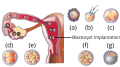
The above figure shows the various stages of development of a zygote as it passes through the oviduct and uterus:
(a) The zygote divides into a two-celled stage via division or 1st cleavage while the zygote is still in the isthmus of the oviduct.
(b) Several more mitotic divisions or cleavages occur in the 2 celled stage to form a 2, 4, 8 and finally a 16-celled stage of the zygote. The 16 celled stage is called the morula and various cells formed after cleavage are called blastomeres.
(d) Morula then changes into a blastocyst after a few more divisions and this stage contains a fluid filled cavity in the embryo. The blastomeres become arranged and line up into an outer layer of cells called the trophoblast and an inner mass of cells. The fluid filled cavity is called blastocoel.
(e) Implantation of the embryo occurs at this blastocyst stage by the help of trophoblast layer which embeds itself into the uterine endometrium.
4. Draw a neat diagram of the female reproductive system and label the parts associated with the following (a) production of gamete, (b) site of fertilization (c) site of implantation and, (d) birth canal.
Ans:
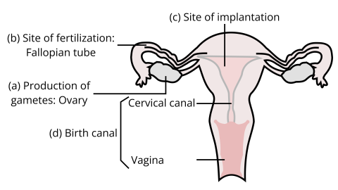
a) Production of gamete is done in the ovaries lying on either side of the uterus. At one time, only one ovary releases the gamete.
b) Site of fertilization is the ampulla-isthmus junction in the oviduct or the fallopian tube of a human female.
c) Site of implantation in a human female is the uterine endometrium. However, ectopic pregnancies are fairly common where zygote gets implanted on the wrong site.
d) Birth canal is the cervical plus vaginal canal of a female i.e., the canal through which a grown fetus passes during its birth to come out of the womb.
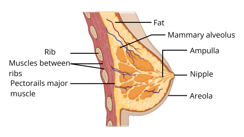
5. With a suitable diagram, describe the organization of the mammary gland.
Ans: The mammary glands are paired structures meant mainly for the production of milk or newborn in a female after pregnancy has been achieved. These structures contain variable amounts of glandular tissue and huge amounts of fat. The glandular tissue of each breast is divided into about 15-10 mammary lobules which contain a group of cells called alveoli, the milk secreting cells. The milk secreted or made by these alveoli is stored in lumen of the alveoli until its transfer is required. This milk by the alveoli is transferred next to the mammary ducts into which the lumen or cavities of alveoli directly open. Many of these mammary ducts join together to form a lactiferous duct which opens up on the nipple or the areola. It is this opening through which a newborn sucks the milk out.


FAQs on NCERT Exemplar for Class 12 Biology Chapter-3 (Book Solutions)
1. What are the three stages of parturition according to NCERT Exemplar for Class 12 Biology ?
Parturition simply means childbirth. Childbirth involves a lot of steps which include the pregnancy, (during which a child grows inside a woman’s uterus or womb). Childbirth is known as labor. Pregnant Humans go into labor roughly nine months after getting pregnant. The three stages of parturition are as follows -
Dilation- The 1st stage of parturition starts with the start of labor. It continues until the cervix is completely dilated.
Expulsion- The 2nd stage of parturition begins at the time of full dilation and continues until birth.
Placental- The 3rd stage of parturition begins after childbirth and ends with the delivery of the afterbirth.
2. What happens during spermatogenesis according to NCERT Exemplar for Class 12 Biology ?
Spermatogenesis is the complete process of sperm cell development. The rounded and immature sperm cells go through the successive meiotic and mitotic divisions, also called spermatocytogenesis, and a metamorphic change, also called spermiogenesis, to eventually produce the spermatozoa. The process of Spermatogenesis happens to create mature Male gametes, which then later fertilize Female gametes to create a zygote which is a single-celled organism. This results in cell division and multiplication to create a mature fetus. For a healthy offspring, the number of total chromosomes must be maintained properly across the body as any failure can lead to many abnormalities.
3. What is the process of fertilization according to NCERT Exemplar for Class 12 Biology?
The process of Reproduction in all Humans is carried out phase-wise as mentioned below:
Pre-fertilization – Gametes are formed and transferred
Fertilization- The formation of a zygote after a sperm fertilizes the egg takes place in this phase
Post-fertilization- The Mitotic division of the zygote leads to the formation of an embryo. This reproductive stage is also referred to as embryogenesis.
These are the Reproduction phases of Human fertilization. To know more about Human Reproduction, Class 11 students can check out Vedantu's Official site and Youtube Channel.
4. Describe the Female reproductive system?
The Female accessory ducts are constituted by the oviducts, vagina, and uterus.
The section which is nearer to the ovary is a funnel-shaped infundibulum that contains the fimbriae (finger-like projections facilitating the assimilation of ovum after the ovulation).
The infundibulum directs to the wider section of the oviduct, known as the ampulla.
The last section of the oviduct is called the isthmus, has a narrow lumen joining the uterus.
The external genitalia of Females comprises – mons pubis, labia minora, labia majora, hymen, and clitoris.
The uterus is also called the womb.
The cervical cavity, known as the cervical canal which goes on to form the birth canal along with the vagina.
5. Describe the Male reproductive system?
The Male reproductive system is positioned in the pelvis region of the Male body and comprises a pair of testes in addition to the accessory glands, ducts, and external genitalia.
A pouch-like structure known as the scrotum encloses the testes located outside the abdominal cavity
Each testis has close to 250 testicular lobules. These lobules comprise one to three seminiferous tubules wherein the sperms are produced. The lining of these tubules includes the 2 types of cells – Male Germ Cells and Sertoli cells.
The exterior of these tubules consists of spaces containing blood vessels and Leydig cells.
Male sex accessory ducts comprise, vasa efferentia, rete testis, epididymis, and vas deferens.
The urethra eventually opens externally to the urethral meatus
The external genitalia of the Male is the penis is covered by the foreskin which is a loose fold of skin.