


Revision Notes on Locomotion and Movement for NEET 2025 - Free PDF Download
Introduction
Movement is a feature shared by all living organisms. It constitutes the various key aspects of living organisms, ranging from protoplasmic motion in a cell or unicellular organisms to organ movement in complex organisms.
Locomotion refers to the movement that results in a location change.
Types of Movements:
There seem to be three types of movement in a cell and organ. They are as follows:
Amoeboid Movement: Similar to pseudopodia in amoeba, ameboid movement can be seen in macrophages, leukocytes, and even cytoskeletal microfilaments.
Ciliary and Flagellar Movement: Ciliary motion in the tracheal epithelial lining, reproductive tract, and so on. Flagellar movement is seen in sperm.
Muscular Movement: Muscles account for the majority of movement in a complex organism. Breathing, heart function, digestion, appendage movement, and locomotion are all carried out by various muscles of our body. The synchronised movement of the skeletal, neural, and muscular systems is referred to as locomotion.
Muscle
Muscles develop from the germinal layer of the mesoderm.
Muscular tissues have distinct characteristics including contraction, excitation, extension, elasticity, and so on.
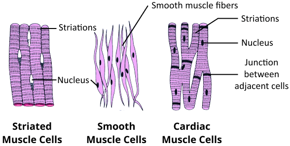
We are all aware that there are three types of muscles:
Cardiac muscles are striated and involuntary muscles found in the heart.
Visceral muscles are nonstriated and smooth. They are involuntary too and support different internal organs as well as participate in functions including digestion and reproduction.
Skeletal muscles are striated and voluntary muscles that control locomotion and appendage movement.
Types of Muscles
Muscle Fibre and Sarcomere Anatomy
Skeletal muscles are present in the animal's body most abundant.
They are composed of bundles of muscle fibres surrounded by connective tissue.
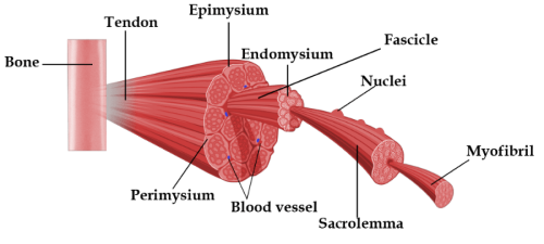
Structure of a Skeletal Muscle Fibre
Fascicles (Muscle Bundles): A muscle, such as a biceps, is made up of numerous muscle bundles (fascicles) that are held together by fascia, a connective tissue layer. Several muscle fibres are found in each fascicle.
Muscle Fibres: Muscle fibres are long cells. They are grouped into fascicles. Muscle fibres have the following characteristics:
Skeletal muscle fibres are all long, cylindrical, and striated.
It is syncytium, which means it has many nuclei.
The plasma membrane of the muscle fibre is known as the sarcolemma.
Sarcoplasm is the muscle fibre's cytoplasm.
The sarcoplasmic reticulum is the muscle fibre's endoplasmic reticulum. It is the Ca2+ storage facility.
Myofibrils: Multiple myofibrils run lengthwise and adjacent to each other in the sarcoplasm of each muscle fibre. Myofibrils' alternating dark and light bands bring the muscle a striated look. Each myofibril is made up of even simplified structures known as myofilaments.
Myofilaments: In a myofibril, there are two kinds of myofilaments. Thin filaments and others that are thick. Muscle contraction requires the attachment of thin and thick filaments.
Actin Filaments (Thin Myofilaments):
Thin filaments, also known as Actin filaments, are composed of three types of proteins.
It is made up of two polymeric filamentous or 'F' actin filaments that are wound around each other. They are globular or G-actin monomer polymers.
Tropomyosin filaments coil lengthwise around actin filaments.
Troponin, which is found at specific points on tropomyosin, protects the active binding zones for myosin.
Troponin and tropomyosin govern actin and myosin filament binding and thus play a significant role in muscular contractions.
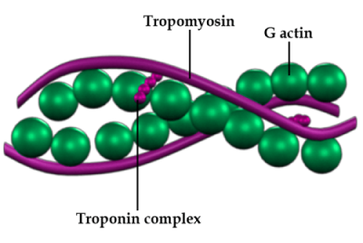
Structure of Actin (thin) Filament
Myosin Filament (Thick Myofilaments):
Myosin is used to make thick filaments or Myosin filaments.
It is a polymeric protein made up of monomeric units of the protein meromyosin.
It is composed of three parts: a tail, a short arm or neck, and a globular head.
At regular times, the head and cross arm extends from the filament.
The globular head comprises ATP and actin-binding sites.
The enzyme ATPase is activated by the head.
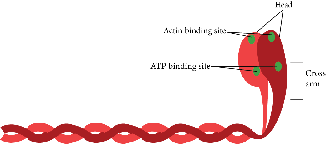
Structure of Myosin (Thick) Filament
Sarcomere:
A sarcomere is a muscle contraction functional unit.
A myofibril is made up of hundreds of sarcomeres that are linked end to end.
Sarcomeres are the fundamental unit of muscle contraction. It is made up of actin and myosin filament repeating units in a particular order.
Sarcomeres are merged at the ends by filaments that interweave to form the 'Z' line. It is a sarcomere's limiting membrane.
At regular intervals, actin or thin filaments are linked to the Z-line.
Between the thin filaments are thick or myosin filaments. They are kept together by a really thin fibrous membrane called the 'M' line.
Actin and myosin filaments span each other in a particular pattern, resulting in muscle striation. They form three kinds of bands.
The 'I' band or isotropic band, is the light band and is made up of actin filaments connected by two adjacent sarcomeres. Thick and thin filaments do not overlap in this case.
The 'A' band or anisotropic band, is the dark band, which contains both actin and myosin filaments that overlap.
The 'H' zone is the myosin filaments' centre portion, where actin filaments do not coincide with thick (myosin) filaments.

Sarcomere
So, a sarcomere is a basic muscle unit composed of thick and thin myofilaments. Myofibril contains hundreds of sarcomeres. A muscle fibre is made up of multiple myofibrils. Fascicles are bundles of muscle fibres. A muscle is made up of multiple muscle bundles that are encased in a fascia.
Muscle Contraction Mechanism
Actin and myosin filaments slide over each other to cause muscle contraction.
Andrew and Hugh Huxley proposed the sliding filament model.
Sliding increases the overlap of thick (myosin) and thin (actin) filaments and causes the sarcomere to shorten and the muscles contract as a result of this.
Steps of Muscle Contraction:
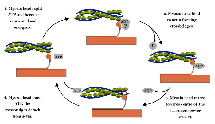
Steps of Muscle Contraction
The brain or spinal cord (CNS) sends a signal to motor neurons to initiate muscle contraction.
The neural signal induces the neurotransmitter acetylcholine to be released at the neuromuscular junction's synaptic cleft. Acetylcholine binds to receptors on muscle fibres, causing sarcolemma depolarization.
The generated action potential propagates through the muscle fibre. The sarcoplasmic reticulum releases Ca2+ ions into the sarcoplasm. Another protein, dystrophin, regulates Ca2+ release. (NOTE: The dystrophin coding gene is the human body's longest gene.)
Ca2+ ions interact with troponin and alter its conformation. Myosin-binding areas on actin filaments become visible.
The myosin head also has an ATP binding site, where ATP binds. The myosin head's ATPase activity catalyses ATP hydrolysis. The cocked (energised) myosin head binds to actin's active binding sites, creating a cross bridge.
Following the attachment, the myosin head releases phosphate, resulting in the 'power stroke.' Myosin filaments deform and pull actin filaments towards the sarcomere's centre, causing the sarcomere and muscle to shorten. During the process, ADP is released.
ATP is also responsible for myosin head detachment.
The process is repeated in the existence of sufficient Ca2+ ions.
Muscle Relaxation
When the neural signal stops, Acetylcholinesterase is a protein that deactivates acetylcholine in the synaptic cleft. Muscle fibres enter a state of rest.
Ca2+ ions are returned to the sarcoplasmic reticulum.
In the complete lack of Ca2+ ions, the troponin-tropomyosin complex re-covers the actin filaments' myosin-binding sites.
Sarcomere Z-line comes back to its original position, and muscles relax.
Muscle Fatigue
ATP drives muscle contraction.
ATP is obtained by muscle fibres from the backup of creatine phosphate and glycogen. Under normal conditions, glycogen is turned into glucose, which is then used to produce ATP during cellular respiration.
Muscles require a lot of energy when doing strenuous exercise. The body could not substitute the oxygen demand and glucose is degraded anaerobically.
As a result, lactic acid builds up, causing muscle fatigue.
Rigour Mortis, or short-term muscle stiffening after death, is caused by ATP depletion as cellular respiration terminates. ATP is necessary for the dissociation of the myosin head; in the absence of ATP, the cross-bridges in the muscles that were contracting remain intact. It aids in calculating the death time.
Muscle fibres are classified into two types based on the quantity of oxygen-binding pigment which is myoglobin, present in them.
Red fibres, also known as aerobic muscles, are reddish and contain too much myoglobin and mitochondria.
White fibres (anaerobic muscles) are pale or white and have less myoglobin and mitochondria, but much more sarcoplasmic reticulum.
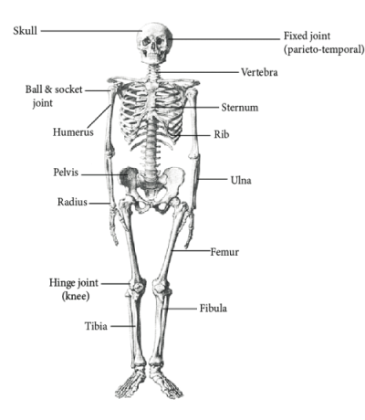
Skeletal System
The skeletal system serves as our body's structural framework and aids in movement as well as locomotion.
It shields the internal organs from harm. Our skeletal system is made up of different types of connective tissues, such as bones and cartilage.
A human being has 206 bones. Because of the presence of Calcium salts in the matrix, bones are hard, whereas cartilage consists of chondroitin salts.
The human skeleton is divided into two sections: the axial skeleton (80 bones) and the appendicular skeleton (126 bones).
The axial skeleton (80 bones) includes the skull, vertebral column, ribs, and sternum.
The appendicular skeleton has 126 bones consisting of the pectoral and pelvic girdles, as well as the limbs.
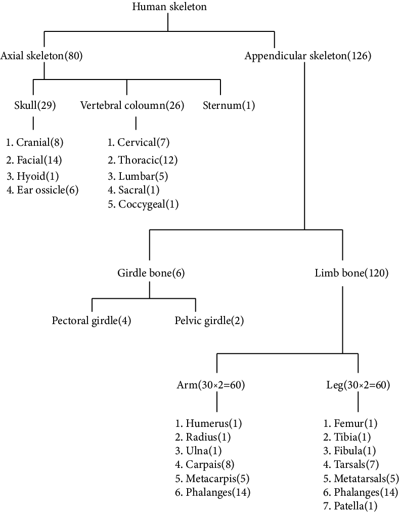
Flow Chart of all Bones Present in the Human Body
Human Skeleton
Joints
Joints exist between bones as well as between a bone and cartilage. Joints facilitate movement and locomotion.
Joints are classified into three types:
Fibrous joints are immobile joints found in the cranium.
Cartilaginous joints have limited movement, for example, vertebrae in the spine have an intervertebral disc between two vertebrae.
Synovial joints are mobile joints. They have a joint cavity filled with liquid between two bones that allows for significant movement. The following are the main synovial joints:
Pivot joint (between 1st and 2nd cervical vertebrae Atlas and Axis)
Socket and ball joint (shoulder)
Hinge joint (knee, elbow)
Gliding joint (carpals)
Saddle joint (between carpal and metacarpal)
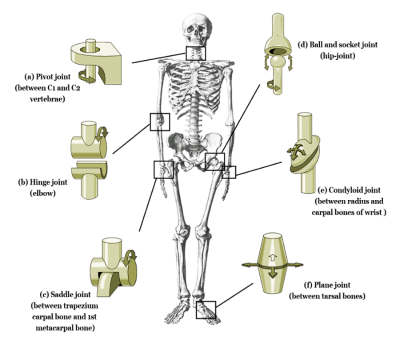
Types of Joints Found in the Human Body
Disorders of the Muscular and Skeletal System
Tetany is caused by a calcium deficiency. Spasms occur as a result of continued contraction of involuntary muscle as Ca ions carry back to the sarcoplasmic reticulum.
Tetanus or lockjaw is a bacterial disease caused by Clostridium tetani. The bacteria's toxin mimics acetylcholine and unites the receptors on muscle fibres permanently, causing painful contractions.
Myasthenia Gravis is a type of autoimmune disorder. Antibodies are made against acetylcholine, resulting in muscle weakness and paralysis.
Duchenne Muscular Dystrophy is an X-linked recessive genetic disorder. The gene that codes for dystrophin protein is faulty. Muscle degeneration progresses, resulting in difficulty breathing and death. Males are disproportionately affected.
Osteoarthritis develops as a result of an injury, infection, or another disease that causes joint inflammation.
Rheumatoid Arthritis is an example of an autoimmune disease that affects joints and causes inflammation as a result of the body's immune system attacking the joints.
Gout is a metabolic disease caused by an increase in uric acid levels. Deformities, pain, and inflammation are caused by the accumulation of uric acid crystals in the joints.
Osteoporosis is an age-linked disease that is caused by demineralisation and a low oestrogen level. It causes decreased bone mass, which leads to bone weakness and frequent fractures.
Points to Remember:
Movement is a requirement for all living things. Animals exhibit a variety of movements including protoplasmic movement, ciliary movements, fin, limb, and wing movements, among others.
Locomotion is defined as a voluntary movement that causes an animal to change its location. Animals generally migrate in search of food, mate, shelter, a breeding site, a better climate, or to protect themselves.
Human body cells move in amoeboid, ciliary, and muscular ways.
Coordination of muscular activities is required for locomotion and several other movements.
Our body contains three types of muscles.
Skeletal muscles connect to skeletal elements. They have a striated appearance and are voluntary.
Visceral muscles are nonstriated and involuntary muscles found in the interior lining of visceral organs.
Cardiac muscles are the heart muscles. The appearance of these muscles is striated and branched. Thy are involuntary in function.
Muscles are excitable, contractile, extensible, and elastic.
The basic unit of muscle is the muscle fibre. Myofibrils are parallelly organised in each muscle fibre. Each myofibril contains numerous serially organised functional units called sarcomeres.
Each sarcomere has a central 'A' band of thick myosin filaments and two half 'I' bands of thin actin filaments on each side, denoted by 'Z' lines.
Actin and myosin are contractile polymerised proteins. A protein-troponin masks the active sites present for myosin on resting actin filament. Myosin head contains ATPase, as well as ATP binding sites and actin active sites.
A motor neuron sends a signal to a muscle fibre, which causes an action potential to be generated in it. Ca2+ is released from the sarcoplasmic reticulum as a result of this. Ca2+ causes actin to bind to the myosin head, forming a cross bridge. These cross bridges cause the actin filaments to slide over the myosin filaments, allowing contraction. Ca2+ is then returned to the sarcoplasmic reticulum, where it inactivates actin. Cross bridges are destroyed, and muscles relax.
Muscle fatigue results from repeated stimulation. Muscles are categorised as red or white fibres based on the amount of myoglobin, a red pigment, in them.
Our skeletal system is made up of bones and cartilages. The skeletal system is divided into two parts: axial and appendicular. The axial skeleton is made up of the skull, vertebral column, ribs, and sternum. The appendicular skeleton is made up of limb bones and girdles.
Fibrous, cartilaginous, and synovial joints develop between bones or between bones and cartilage. Synovial joints facilitate the significant movements and thus play an important role in locomotion.
Importance of Biology Locomotion and Movement for NEET
As we have mentioned earlier, this chapter introduces the students to different concepts related to Locomotion and movement in organisms. It covers the meaning and definition of Movement and Locomotion. Students can understand how these terms are different from one another by studying the Locomotion and Movement notes. The chapter further delves into the different types of movements in organisms and goes on to explain each one in detail. There are mainly three types of movement namely Ciliary Movement, Amoeboid Movement, and Muscular Movement.
The locomotion and Movement chapter further elaborates on the Muscular Movement type. In this section, students will get familiar with the concept of Muscles and Skeletons. They will learn about the muscular and skeletal systems of different organisms. The chapter contains labelled diagrams for an easy understanding of the system. They can learn about the different parts associated with these systems such as the Joints and Ribs which are important for movement in animals. Further elaborations are provided on disorders related to the Skeletal and Muscular systems in detail.
Very important concepts are covered in this chapter and with the help of Locomotion and Movement notes PDF, students can easily understand the concept of the chapter. They can learn how different organisms move from one place to another and the role of other organs in movement.
Benefits of Vedantu’s Notes for NEET Locomotion and Movement
1. Expertly Crafted Notes:
Vedantu’s revision notes are meticulously curated by subject matter experts, ensuring a comprehensive and accurate overview of Locomotion and Movement topics.
2. Concise Explanations:
The notes offer a concise and precise explanation of various subjects within the Locomotion and Movement chapter, facilitating efficient understanding for NEET preparation.
3. Practical Information:
Students can gather practical information encompassing different movement types and the varied organs involved in the process of movement and locomotion.
4. Efficient Study Source:
Created with the aim of providing a convenient and efficient study source, Vedantu’s revision notes are tailored to meet the specific needs of students preparing for the NEET entrance examination.
5. Easy Download and Reference:
The notes are easily downloadable, allowing students quick access to valuable study material. Convenient reference enables seamless integration into study routines.
6. Interactive Elements:
Students can engage with the content by answering important questions embedded in the notes. This interactive feature enhances comprehension and knowledge retention.
7. Doubt Clarification:
Use the notes as a resource to clarify doubts on any topic within the Locomotion and Movement chapter. The detailed explanations aid in addressing uncertainties effectively.
Prepare for NEET with Locomotion and Movement Notes
You have the chance to download Locomotion and Movement Class 11 notes for NEET PDF from Vedantu. Don’t miss out on the opportunity to download these notes and include them in your study material. Students can learn difficult concepts easily with the help of these important notes. This can be a huge help in NEET preparation for sure.
Conclusion
Vedantu’s expertly crafted Locomotion and Movement revision notes offer students a clear and concise understanding of key biology concepts. These notes, created by subject matter experts, serve as an efficient study companion for NEET preparation. Students can easily download, reference, and interact with the material, gaining practical insights into movement types and organ functions. With the inclusion of important questions, doubt clarification becomes straightforward. Embrace these notes as an invaluable tool to simplify complex topics, ensuring a solid foundation for success in the NEET entrance examination. Elevate your learning journey with Vedantu’s accessible and effective study resources.
Other Important Links
NEET Biology Revision Notes - Chapter Pages

FAQs on Locomotion and Movement for NEET Notes 2025 - Free PDF Download
1. Which tissues and body cells in human beings display the amoeboid movement?
The leukocytes and macrophages display the amoeboid movement. Also, some cytoskeletal components like microfilaments exhibit the same movement.
2. Which systems are responsible for the process of locomotion taking place in organisms?
Locomotion happens in organisms due to the contribution of both the Skeletal as well as the neural systems.
3. Is movement different from locomotion?
Yes, movement can be defined as the displacement of any particular body part of an organism from one place to another. But locomotion is the actual displacement of the organism itself from one place to another.
4. What are the names of the middle ear bones?
Incus, Stapes, and Malleus are the bones of the middle ear.
5. Why should I use Vedantu’s Locomotion and Movement notes for NEET preparation?
Vedantu’s expertly curated notes provide a concise explanation of key topics, offering practical insights and serving as a convenient study resource for effective NEET preparation.
6. How can I benefit from Vedantu’s revision notes?
Easily download, refer to, and engage with the notes, which include important questions for clarity, making your study process efficient and comprehensive.