


Revision Notes on Cell: The Unit of Life for NEET 2025 - Free PDF Download
Cell The Unit of life Class 11 Notes for NEET 2025
Introduction
The cellular organisation is found in all living things.
Some organisms, such as Bacteria, Amoeba, Paramecium, Chlamydomonas, etc, are made up of only one cell, thus, are called unicellular.
There are numerous other organisms, such as plants, animals, and humans, that are formed of millions of cells and are called multicellular.
All living beings have cells that serve as structural and functional units.
With his primitive microscope, Robert Hooke (1665) discovered hollow cavities like compartments in a quite thin slice of cork and named them ‘cellulae’ or ‘cells’.
Anton Von Leeuwenhoek was the first to see and describe a living cell.
The nucleus in a cell was later discovered and named by Robert Brown.
J. E. Purkinje coined the term protoplasm to describe the living substance found inside the cell.
Cell Theory
Cell theory is credited by two scientists: a German botanist, M.J. Schleiden (1838), and a British zoologist, T. Schwann (1839).
Schleiden proposed that all tissues are composed of individual cells. However, Schwann said that all plants are composed of cells. Then, they jointly developed the concept of "all living organisms are created by cells and cell products".
The cell theory was developed by Schleiden and Schwann, but it did not clarify how new cells were developed.
Viruses, viroids, and prions are obligate parasites that deny the cell theory (subcellular in nature).
Rudolf Virchow (1855) was the first to explain that the new cells produced from the pre-existing cells (Omnis cellula-e cellula).
To give the cell theory a complete form, he modified Schleiden and Schwann's hypothesis.
According to cell theory,
All living organisms are made up of cells and cell products.
All cells develop from pre-existing ones.
Protoplasmic membrane-bound cell organelles do not survive within or even outside the protoplasm.
All cells have the same basic structure and metabolic responses.
The genetic information is saved as DNA in the nucleus' chromosomes.
An Overview of Cell
Every cell has a compact membrane-bound structure called the nucleus, which houses the chromosomes composed of the genetic material i.e. DNA.
Cells with membrane-bound nuclei are referred to as eukaryotic, whereas cells without a membrane-bound nucleus are referred to as prokaryotic.
Endoplasmic reticulum (ER), Golgi body, lysosomes, mitochondria, microbodies, and vacuoles are membrane-bound distinct structures found in eukaryotic cells. Such membrane-bound organelles are not found in prokaryotic cells.
The size, shape, and activities of cells vary greatly.
Mycoplasmas are only 0.3 µm long which are the smallest cells, whereas bacteria can be 3 to 5 µm long.
Human red blood cells have a diameter of about 7.0 µm when compared to other multicellular organisms.
Nerve cells are among the longest cells in the body.
Cells are classified as prokaryotic or eukaryotic based on the nature of their nucleus.
Prokaryotic Cell
Bacteria, Blue-green algae (Cyanobacteria), Mycoplasma, and PPLO are examples of unicellular organisms (Pleuropneumonia-like Organisms).
Bacillus (rod-shaped), coccus (spherical), vibrio (comma-shaped), and spirillum (spiral) are the four basic shapes of bacteria.
A cell wall made up of peptidoglycans surrounds the cell membrane in all prokaryotes. The cytoplasm is the fluid matrix that fills the cell.
The genetic material (DNA) is circular in shape and naked that is not contained within a nuclear envelope.
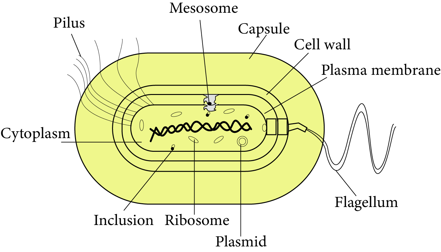
Structure of Bacteria
In addition to genomic DNA which is a single chromosome or circular DNA, several bacteria have small circular DNA called plasmids, which induce certain unique phenotypic characteristics, such as antibiotic resistance.
Plasmid DNA is used to track the transformation of bacteria with foreign DNA.
Mesosomes are circular coiled bodies that exist on the plasma membrane and are simply inner foldings of the plasma membrane. Because they consist of respiratory enzymes such as oxidases and dehydrogenases, they aid in respiration. They aid in the formation of cell walls, the replication of DNA, and the distribution of DNA to daughter cells.
Cell Envelope and Modifications
The cell envelope is a closely packed three-layered structure that includes the outermost glycocalyx, the cell wall, and the plasma membrane.
Bacteria are categorised into two groups based on differences in the cell envelope and how they respond to Gram's staining procedure:
Those that take up the gram stain are Gram-positive bacteria like Streptococcus, Bacillus, Staphylococcus, Mycobacterium, Streptomyces, etc.
Those that do not are Gram-negative bacteria like Salmonella, Escherichia coli, Pseudomonas, Rhizobium, Helicobacter, etc.
Outside the cell wall of many bacteria is a thick slime capsule composed of polysaccharides and nitrogenous compounds. It provides resistance to phagocytosis and antibiotics.
The plasma membrane is semi-permeable and interacts with the surrounding environment. This membrane has a similar structure to that of eukaryotes.
Other membranous modifications into the cytoplasm of some prokaryotes, such as cyanobacteria, are called chromatophores and contain pigments such as chlorophyll-a, beta-carotene, myxoxanthophyll, myxoxanthin C-phycocyanin, and C-phycoerythrin.
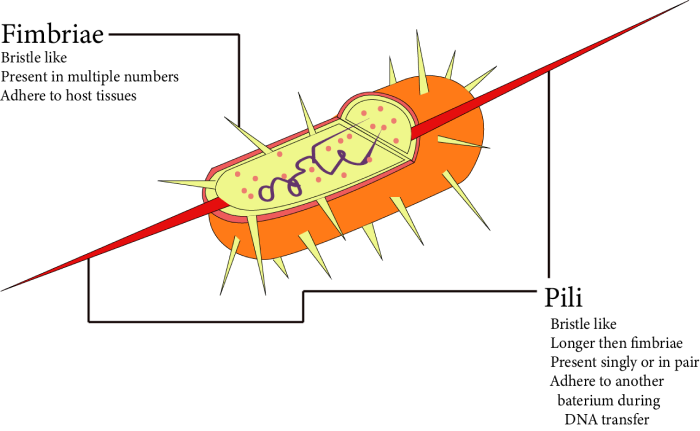
Fimbriae and Pili
Bacterial cells can be motile or stationary. If they are motile, they have flagella, which are thin filamentous extensions of their cell wall.
The bacterial flagellum is made up of three parts: the filament, the hook, and the basal body. The filament is the longest segment, extending from the surface of the cell to the outside.
In addition to flagella, pili and fimbriae are surface structures of the cell that do not contribute to motility.
Ribosomes and Inclusion Bodies
The existence of a large number of ribosomes (the 70s) causes the cytoplasm to be granular.
A polyribosome is formed when several ribosomes connect to a single mRNA. Protein synthesis occurs at these sites.
Reserve materials, such as phosphate granules, glycogen granules, and cyanophycean granules, are deposited in the cytoplasm as inclusion bodies.
Gas vacuoles are observed in photosynthetic bacteria that are blue, green, purple, and green.
Eukaryotic cell
Protists (single-celled eukaryotes), plants, animals, and fungi are all eukaryotes.
There is a well-organised nucleus. It contains hereditary information that is surrounded on all sides by a nuclear envelope.
Plant and animal cells, for example, are not all identical eukaryotic cells.
Cell Membrane
Every living cell is surrounded on all sides by a plasma membrane, which is a thin, elastic, regenerative, and selectively permeable membrane.
Cell Membrane’s Composition
Lipoproteins, lipids, and proteins are the major components that makeup 60% of the plasma membrane.
Proteins supply mechanical strength and are in charge of transporting various substances. Proteins can also function as enzymes.
Depending on the form of cell and organism, lipids may account for 28-79 percent of the total.
Plasma membrane lipids are classified into three types: phospholipids, glycolipids, and sterols. The sterol contained in the membrane can be cholesterol (from animals), phytosterol (from plants), or ergosterol (from plants) (microorganisms).
Carbohydrates account for 2% to 10% of the total. Plasma membrane carbohydrates are covalently attached to both lipid and protein elements.
Singer and Nicolson, in 1972, proposed a modified model of cell membrane structure that is widely accepted as the fluid mosaic model.
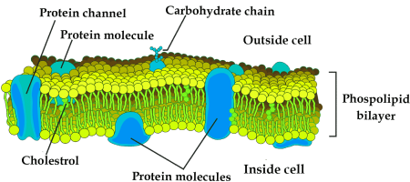
Fluid-mosaic Model of Plasma Membrane.
According to this model, the quasi-fluid appearance of lipids allows for lateral protein movement within the whole bilayer.
The fluidity of the plasma membrane is due to the presence of phospholipids, which are high in unsaturated fatty acids.
Phospholipids are the most important component of cell membranes because they provide the membrane with a structural framework.
Lipids are amphipathic, which means they have an asymmetric structure with a polar hydrophilic head and a non-polar hydrophobic tail.
Some lipids have sugar chains connected to their polar heads on the outside and are thus known as glycolipids.
The membrane contains two types of proteins: peripheral or extrinsic proteins and intrinsic or integral proteins. Peripheral proteins help to communicate cells with its adjacent cells whereas integral proteins help to transport polar molecules across the plasma membrane.
Transport Across Cell Membrane
Many molecules can move quickly across the membrane without using any energy, which is known as passive transport.
Neutral solutes like water, oxygen and carbon dioxide can move freely across the membrane via simple diffusion in the direction of concentration gradient, i.e. from higher concentration to lower concentration.
Water diffusion across a selectively or semi-permeable membrane is referred to as osmosis.
Because polar molecules cannot cross the non-polar lipid bilayer, they must be transported across the membrane by a carrier protein.
A few ions or molecules are shipped across the membrane in the opposite direction of their concentration gradient, from lower to higher concentration. This type of transport is an energy-dependent process that uses ATP and is known as Active transport. For example, the Sodium-Potassium ion pump.
Bulk transport is the movement of large amounts of micromolecules, macromolecules, and food particles across a membrane. It is associated with the formation of carrier vesicles or transport vesicles.
Functions of Cell Membrane
Cell membrane provides mechanical strength and serves as a protective layer.
The plasma membrane is in charge of transporting materials, molecules, ions, and so on.
Gases like oxygen and carbon dioxide diffuse across the plasma membrane via simple and facilitated diffusion.
Differences Between Active and Passive Transport are Given Below:
Cell Wall
Robert Hooke discovered the cell wall while studying the cell walls in cork tissue.
It is the rigid, protective, non-living, and supportive layer found on the outside of all plant cells, bacteria, Cyanobacteria, and some protists. It does not exist in animal cells.
Cell Wall’s Composition
The cell wall of algae is composed of cellulose, galactans, mannans, and minerals such as calcium carbonate, whereas the cell wall of other plants is composed of cellulose, hemicellulose, pectins, and proteins.
The primary wall of a young plant cell is capable of growth, which slowly diminishes as the cell matures, and the secondary wall is constituted on the cell's inner side.
The outermost region of the lamella is found as a common connecting layer between two cells.
It's made from calcium and magnesium pectate.
Numerous plasmodesmata or cytoplasmic fibres are present in the pit that connects the cytoplasm of two cells.
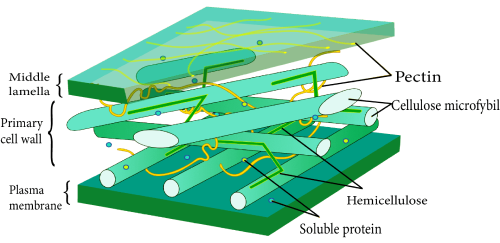
Plant Cell Wall Structure
Functions of Cell Wall:
The cell wall not only gives the cell shape and protects it from mechanical injury and infection, but it also aids in cell-to-cell interaction and acts as a barrier to unwanted macromolecules.
Plant cells, unlike animal cells, can withstand a wide range of environmental variations due to their cell walls.
Cytoplasm
Cytoplasm
The cytoplasm is a substance found around the nucleus or inside the cell membrane that contains various organelles and inclusions.
It accounts for roughly half of the cell's volume and contains approximately 90% water.
It contains ions as well as biomolecules including sugar, amino acids, nucleotides, tRNA, enzymes, and vitamins.
Plasmids, lysosomes, sphaerosomes, peroxisomes, glyoxysomes, mitochondria, ribosomes, centrosomes, flagella, and cilia are examples of cytoplasmic organelles.
Endoplasmic Reticulum
Endoplasmic reticulum (ER), Golgi complex, lysosomes, and vacuoles are all components of the endomembrane system.
The mitochondria, chloroplasts, and peroxisomes are not considered endomembranes because their functions are not integrated with the above components.
Endoplasmic Reticulum (Er)
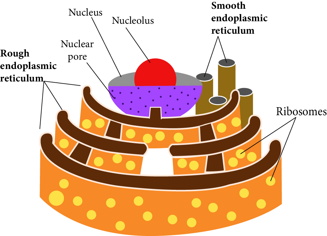
Endoplasmic Reticulum
The endoplasmic reticulum (ER) is a web or reticulum of small tubular structures scattered throughout the cytoplasm.
The ER is found in nearly all eukaryotic cells. However, it is absent in a few cells, including ovum, embryonic cells, as well as mature RBCs. It is also missing from prokaryotic cells. It is less developed in quick-dividing cells.
The ER is composed of three parts. A single membrane connects all three structures.
Cisternae (cisterna): These are sac-like structures that are flattened and unbranched. They are arranged in parallel stacks (piles). They contain ribosomes.
Vesicles: These are vacuole-like elements that are scattered throughout the cytoplasm. Ribosomes are also present in these.
Tubules: Broader, tubular, branched elements that are mostly found near the cell membrane. Ribosomes are not present. These are more prevalent in lipid-forming cells.
Rough Endoplasmic Reticulum (RER) refers to the endoplasmic reticulum with ribosomes on its surface.
They appear smooth in the absence of ribosomes and are known as Smooth Endoplasmic Reticulum (SER).
RER is frequently found in cells that are deeply engaged in protein synthesis as well as secretion.
The smooth endoplasmic reticulum is the primary site of lipid synthesis. SER synthesises lipid-like steroidal hormones in animal cells.
Functions
Specific protein synthesis and secretion via Golgi bodies.
It provides a surface for steroids, cholesterol, ascorbic acid, visual pigments, and hormones such as testosterone and oestrogen.
ER promotes glycogenolysis in liver cells and aids in the purification of many toxic substances and drugs.
The ER is a component of the cell's cytoskeleton which is spread out like a net and gives mechanical support as well as shape to the cell.
Golgi Apparatus
Camillo Golgi (1898) discovered densely stained reticular frameworks near the nucleus for the first time.
These are made up of several flat, disc-shaped vesicles or cisternae ranging in diameter from 0.5m to 1m. These are stacked one on top of the other and were named Golgi complex.
It can be found in all eukaryotic cells. These are called "dictyosomes" in plants because they are scattered unevenly in the cytoplasm.
These are lacking in bacteria, blue-green algae, RBCs, bryophyte, pteridophyte spermatozoa, and sieve tube cells of phloem in angiosperms.
The Golgi body is composed of four main parts:
Cisternae (cisterna): The Golgi apparatus is composed of cisternae, which are stack or flat sac-like structures.
Each cisterna's margins are gently curved. The dictyosome's cisternae are made up of forming faces (F-faces) or cis faces.
The trans face of the maturing face (M-face) consists of the cisternae at the concave end.
The forming face is found near the nucleus or the endoplasmic reticulum.
Typically, the maturing face is oriented toward the plasma membrane. It is the Golgi body's functional unit.
Tubules: These create a complex network after cisternae are fenestrated.
Secretory vesicles: These are small components, each about 40 mm in diameter, found along the convex surface of cisternae's edges. These vesicles are smooth and coated.
Vacuoles: These are cisternae that have been expanded and modified to construct vacuoles.
Functions of Golgi Body:
Because the Golgi body's primary function is secretion, it is the largest of the secretory cells.
Glycolipidation is the introduction of oligosaccharides to lipids to create glycolipids.
Protein glycosylation is the introduction of carbohydrates to generate glycoproteins.
Promotes the creation of primary lysosomes.
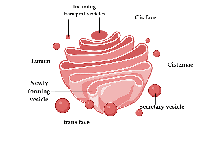
Golgi Apparatus
Lysosomes
Lysosomes are microscopic cytoplasmic vesicular structures surrounded by a single membrane that are engaged in intracellular digestive activities and contain hydrolytic enzymes. These are commonly referred to as "suicidal bags."
Christian de Duve (1955), a Belgian biochemist, found these in liver cells and named them "pericanalicular dense bodies."
Except for mammalian RBCs, these are missing in prokaryotes but found in all eukaryotic animal cells.
The isolated lysosomal vesicles were discovered to be extremely rich in nearly all kinds of hydrolytic enzymes called hydrolases like lipases, proteases, carbohydrases, etc, that are optimally active at acidic pH.
These enzymes can break down carbohydrates, proteins, lipids, and nucleic acids.
Lysosome Classification:
Lysosomes are classified into four types based on their contents:
Primary lysosomes: A recently created lysosome is made up entirely of enzymes. Its enzymes are most likely inactive.
Secondary lysosomes: When digestible material enters a primary lysosome, the latter is known as a secondary lysosome, phagolysosome, or digestive vacuole.
Residual bodies/Tertiary lysosomes: The residual bodies/ tertiary lysosome is a secondary lysosome that contains indigestible matter.
Autolysosomes/Autophagosomes: A cell's own organelles, like mitochondria and the ER, can be digested. This is known as autophagy. Lysosome acid hydrolases digest the organelles, giving rise to the term autophagosome.
Functions of Lysosomes:
Sperm lysosomes contain enzymes that break the limiting membrane of the egg, such as hyaluronidase.
Lysosomes act as cell division triggers or start cell division by metabolising repressor molecules.
They also consume carcinogens.
Vacuoles
Spallanzani firstly found the vacuole in plants. The tonoplast is a non-living reservoir surrounded by a differentially/ selectively permeable membrane.
These are made up of water, minerals, and anthocyanin pigments.
Some protozoans contain contractile vacuoles that expand due to fluid accumulation or collapse due to expulsion from the cell.
Sap vacuoles, contractile vacuoles, and gas vacuoles are examples of vacuoles.
Functions of Vacuoles:
The vacuole maintains the cell's osmotic relationship, which aids in water absorption. The sap concentrations in the vacuole cause turgidity and flaccidity in cells.
Mitochondria
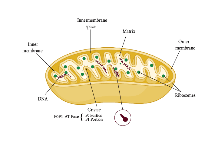
Mitochondria
The mitochondria can be found singly or in groups, and their shape and size (0.5 micrometre to 0.2 micrometre) vary from cell to cell and are dependent on the physiological activity of the cell.
Mitochondria are considered semi-autonomous organelles because they have their own protein synthesis machinery that is not controlled by the nucleus.
The mitochondrion is a double membrane-bound structure, with the outer and inner membranes distinctly dividing its lumen into two aqueous compartments, namely the outer and inner compartments.
The inner compartment is referred to as the matrix. The outer membrane serves as the organelle's continuous limiting boundary.
In the direction of the matrix, the inner membrane creates a set of infoldings known as cristae. Cristae expands the surface area of the inner me.
Oxysomes contain a molecule of ATPase enzyme and are thus in charge of ATP synthesis.
ATP formation consumes energy, thus, it is an endergonic reaction. These fundamental particles are also known as F0 - F1 particles.
The matrix also contains a single circular DNA molecule, just a few RNA molecules, ribosomes (the 70S), and protein synthesis elements. Fission is the process by which mitochondria divide.
Functions of Mitochondria:
Mitochondria are known as the "powerhouse" of the cell because they produce 95 percent of the ATP. This energy is generated as a result of the breakdown of food molecules, which includes glycolysis, oxidative decarboxylation, as well as oxidative phosphorylation (including Krebs cycle and respiratory chain).
Intermediate molecules of cell respiration are used in the synthesis of steroids, chlorophyll, cytochromes, and other compounds.
These are also the sites of amino acid biosynthesis.
The electron transport system is found in mitochondria.
Plastids
Plastids are self-contained organelles that contain DNA, RNA, ribosomes, and a double membrane envelope.
Although Haeckel (1865) discovered plastids, Schimper coined the term (1883).
De Von Wettstein described a well-organised system of grana and stroma in the plastid of a normal barley plant.
The idea of quantasomes was introduced by Park and Biggins (1964).
Plastids are classified into three types: leucoplasts, chromoplasts, and chloroplasts.
Leucoplasts:
These are colourless plastids that are found near the nucleus in non-green cells and have internal lamellae.
These primarily store food materials and are found in cells that are not exposed to sunlight, such as seeds, underground roots, stems, tubers, rhizomes, and so on.
There are three kinds of leucoplasts:
Amyloplast: Starch grains are synthesised and stored.
Elaioplast: Also called Lipidoplast or Oleoplast. These are the cells that store lipids and oils.
Aleuroplasts: Also called proteinoplasts. Proteins are stored in aleuroplasts.
Chromoplasts:
Chromoplasts are colored plastids that are not green.
Because of the presence of carotenoids, these plastids are colored red, orange, yellow, and so on.
These can be found in the petals and fruits.
Chloroplasts:
Sachs discovered the chloroplast and Schimper named it.
Because of the presence of chlorophyll, a green pigment, chloroplasts are green plastids.
These are abundant in green leaves and green shoot parts.
These capture solar energy, which is then used to produce food. As a result, these are referred to as photosynthesis sites.
It has a two-membrane structure. The membranes are both smooth. The inner membrane is comparatively less permeable than the outer membrane but contains more proteins, particularly carrier proteins.
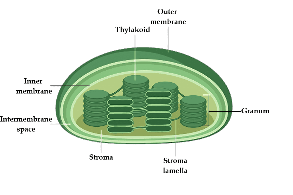
Chloroplast
The periplastidial space is the intermembrane space. A matrix is present inside the membranes, which is separated into two parts:
Grana: The chloroplast's inner plastidial membrane is folded inside to produce a series of connected membranous sheets called lamellae, that form a number of oval-shaped sealed sacs called thylakoids. Quantasomes (photosynthetic units) are tiny spherical para-crystalline bodies found on the inner side of the thylakoid membrane.
Stroma: Stroma is a clear, proteinaceous, and watery substance. This section contains the dark reaction of photosynthesis. Stroma is nearly filled with the "RuBisCO" enzyme (approximately 15% of the overall enzyme, protein), and this enzyme accepts carbon dioxide. Carbohydrates are formed as a result of CO2 assimilation.
Pigments of Chloroplasts:
Chlorophyll-a: Most commonly found in green plants.
Chlorophyll-b
Chlorophyll-c
Chlorophyll-d
Carotenoids: These are of two types:
Carotenes: It is derived from vitamin-A. These are of various types like alpha-carotene, beta-carotene, and gamma-carotene, but beta-carotene is the most popular one.
Xanthophylls: It is yellow in colour. Fucoxanthin is an example.
Functions of Chloroplasts:
It is the location of photosynthesis for both light and dark reactions.
In granum, water is photolyzed and NADP is reduced to NADPH2.
Photophosphorylation via cytochrome b6f, plastocyanin, and plastoquinone, among others.
They are starch storage facilities or sugar synthesis factories.
Ribosomes
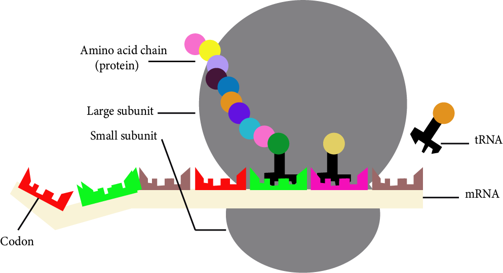
Ribosomes Forming Proteins after Reading the Codon of mRNA
Ribosomes are the shortest known organelles without a membrane, consisting of ribonucleoprotein particles attached to the RER or floating independently in the cytoplasm and serving as protein synthesis sites.
During protein synthesis, they get attached to the mRNA and help in synthesising proteins.
Types of Ribosomes:
There are two types of ribosomes - 70S and 80S (S stands for Svedberg unit).
70S ribosomes are found in prokaryotes, mitochondria, and eukaryotic plastids. 70S ribosome has one larger subunit of 50S and a smaller subunit of 30S.
Eukaryotic cytoplasm contains 80S ribosomes. 80S ribosome has one larger subunit of 60S and a smaller subunit of 40S.
Functions of Ribosomes:
Ribosomes are also known as cell protein factories.
The enzyme peptidyl transferase is found in the large subunit of the ribosome and aids in protein synthesis.
Cytoskeleton
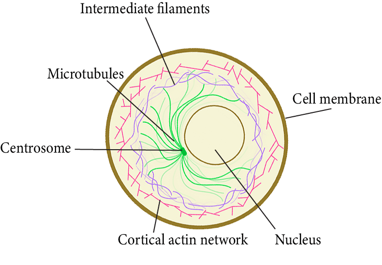
Cytoskeleton
To support the interconnected system of membranes in a eukaryotic cell, a structure of fibrous protein components became necessary.
These elements combine to form the cell's cytoskeleton. There are three kinds of these:
1. Microtubules:
Microtubules are electron-microscopic entities that are only found in eukaryotic cellular structures such as cilia, flagella, centrioles, basal bodies, astral fibres, and spindle fibres.
These are primarily composed of tubulin protein.
Functions:
These are cytoskeleton components that aid in cell shape and mechanical integrity.
Cilia and flagella microtubules aid in locomotion.
Asters' microtubules and spindle fibres formed in the mitotic apparatus aid in the movement of chromosomes to opposite poles during cell division.
2. Microfilaments:
These are non-contractile, long, thin, cylindrical proteins located only in the eukaryotic cytoplasm. These are found in microvilli, muscle fibres called myofilaments, and other places.
Prokaryotes, on the other hand, lack these. These are primarily composed of actin, a contractile protein.
Functions:
Microfilaments are cytoskeleton components that influence cell shape, motility, and cell division during development.
The microfilaments direct the movement of molecules and organelles within the cell.
3. Intermediate Filaments:
These are structural elements found in the cytoplasm of eukaryotic cells. These are not found in mammalian RBCs.
The IFs are about 10 nm thick and slightly larger than the microfilaments.
These are solid, unbranched structures made up of non-motile structural proteins like keratin, desmin, and vimentin.
Functions:
These are part of the cytoskeleton, which facilitates the liquid cytosol and keeps the cell's shape.
These provide myofibrils with support, which is required for contraction.
Cilia and Flagella
Cilia (singular: cilium) and flagella(singular: flagellum) are hair-like extensions of the cell membrane.
Cilia are tiny structures that act like oars, causing the cell or surrounding fluid to move.
Flagella are relatively longer and are in charge of cell movement.
- Both cilia and flagella have structurally similar parts, including a basal body, rootlets, a basal plate, and a shaft.
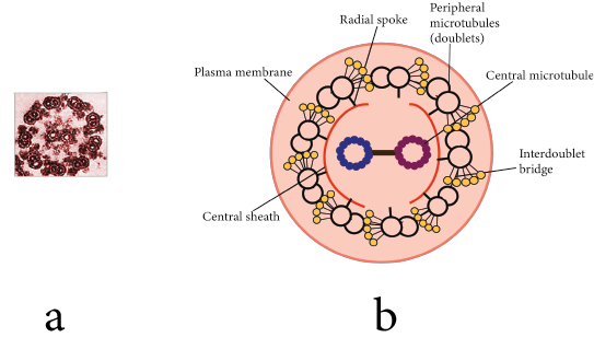
Electronic Micrograph & Structure of Cilia and Flagella
Basal Body: The basal body is found in the cytoplasm beneath the plasma membrane. The structure is closely related to centrioles, which are composed of nine triplets of microtubules.
Rootlets: These are made of microfilament and support the basal body.
Basal Plate: The basal plate is dense and located above the plasma membrane.
Shaft: The projecting hair-like part of cilia and flagella that remains outside the cytoplasm. It has 9 microtubule doublets with radial symmetry. These are known as axonemes. There are 11 fibrils in each axoneme, 9 on the periphery and 2 in the centre. The pattern is known as the 9 + 2 pattern.
The central tubules are made of dynein protein, whereas the peripheral microtubules are made of tubulin protein.
Functions of Cilia and Flagella:
These aid in locomotion, cleaning, respiration, circulation, and feeding, among other things.
These are sensitive to light, temperature, and contact.
Centrosome and Centrioles
Centrosomes are organelles that typically contain two cylindrical structures known as centrioles.
Both centrioles in a centrosome are perpendicular to each other and have a cartwheel-like organisation.
They are composed of nine tubulin peripheral fibrils that are evenly spaced. The peripheral fibrils are all triplets. The adjacent triplets are linked as well.
The central part of the centrioles, known as the hub, is also proteinaceous and is linked to tubules of the peripheral triplets via radial spokes made of protein.
It has a pattern of (9 + 0).
The centriole is not protected by a membrane.
Functions of Centrosome:
During cell division, centrioles aid in the organisation of spindle fibres and astral rays.
They supply basal bodies, which give rise to flagella and cilia.
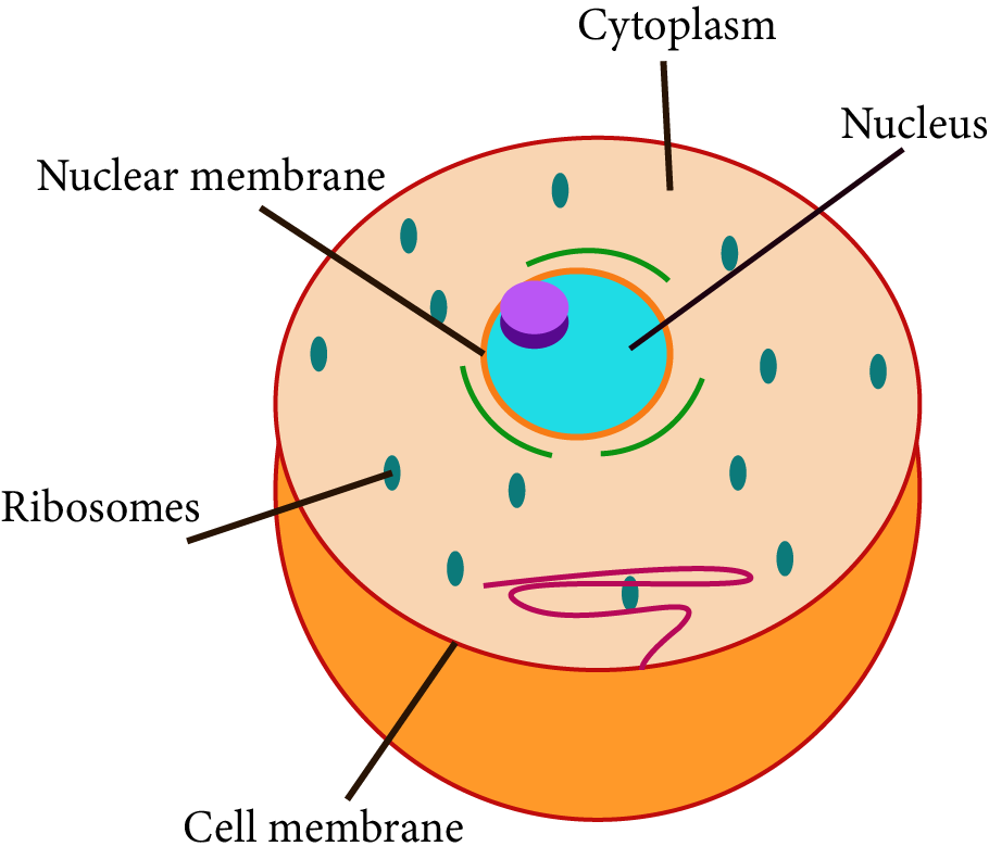
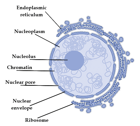
Nucleus
The nucleus is the most important component of the cell, directing and controlling all cellular functions.
Robert Brown first described the nucleus as a cell organelle in 1831.
Some mature cells, such as erythrocytes in many mammals and sieve tubes in vascular plants, even lack a nucleus.
The Following Structures are Found in A Typical Nucleus:
1. Nuclear Membrane:
The nuclear membrane, also known as the nuclear envelope, is made up of two parallel membranes separated by a space of about 10 to 50 nm known as the perinuclear space, which serves as a barrier between the substances found inside the nucleus and those found in the cytoplasm.
The outer membrane is usually consistent with the endoplasmic reticulum and contains ribosomes.
It is punctured by minute pores formed by the merging of its two membranes.
Functions:
Small organic molecules, Inorganic ions, ribosomal subunits, RNAs, and proteins can pass through nuclear pores.
It maintains the nucleus's shape.
2. Nucleolus:
The nucleolus is a prominent, darkly stained circular body located in the nucleoplasm.
It is made up of a large number of ribosomal proteins as well as ribosomal RNA.
It is commonly linked to the nucleolar organiser region (NOR) of nucleolar chromosomes.
Functions:
It is the site of rRNA biogenesis and also keeps rRNA.
It is essential for spindle formation when the cell divides.
Nucleus Having Genetic Material (DNA) Enclosed in Nuclear Membrane
3. Nucleoplasm:
Inside the nuclear membrane, it is a clear, homogeneous, semifluid, colloidal ground substance.
It aids in the maintenance of the nucleus' shape, as well as the formation of spindle proteins of ATP, NAD, DNA, RNAs, and ribosomal subunits.
4. Nuclear Matrix:
It is a fine web of protein-containing fibrils that runs throughout the nucleus.
Functions:
It aids in the preservation of the nucleus' shape.
It acts as a chromatin anchor.
5. Chromatin Fibres/ Nuclear Chromatin:
These are thread-like structures that are evenly distributed throughout the nucleoplasm.
These are only seen during the "interphase stage." Chromatin is made up of DNA and some basic proteins known as histones, non-histone proteins, and RNA.
Functions:
Chromatin holds genetic information.
It creates chromosomes to ensure that genetic information is distributed evenly during cell division as well as reproduction.
These are hereditary DNA-protein structures formed by the compaction of chromatin fibres for equal distribution of DNA during cell division as well as reproduction.
Chromosomes:
Chromosomes are hereditary DNA-protein structures formed by the compaction of chromatin fibres.
Every chromosome has a primary constriction, known as the centromere, on which disc-shaped processes known as kinetochores are present.
The chromosomes are divided into four types based on the centromere position:
Metacentric: A centromere in the middle of the metacentric chromosome forms two equal arms of the chromosome.
Sub-metacentric: The centromere on the sub-metacentric chromosome is closer to one end of the chromosome, giving rise to one shorter and one long arm.
Acrocentric: The centromere is located near the end of an acrocentric chromosome, forming one super short and one enormously long arm.
Telocentric: There is a terminal centromere on the telocentric chromosome.
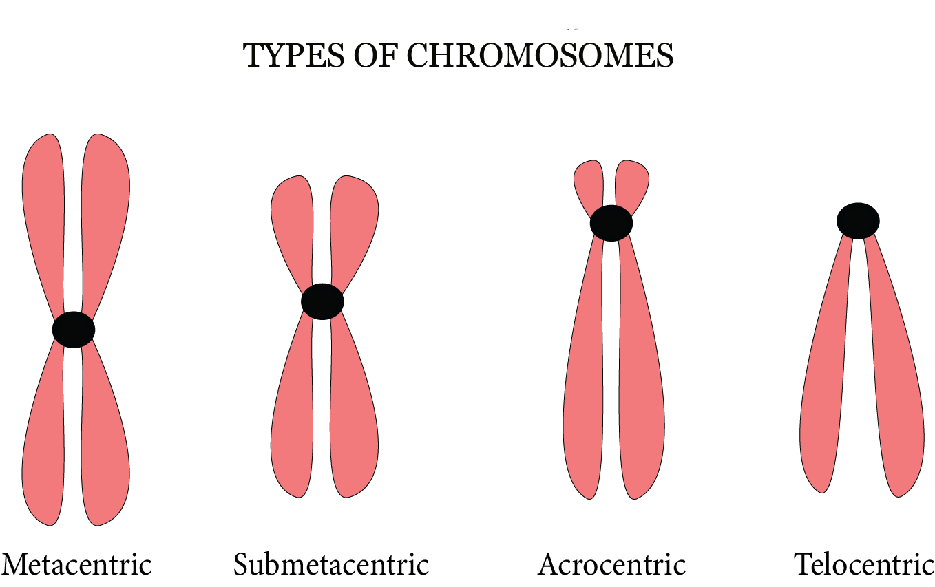
Types of Chromosomes
A few chromosomes may have non-staining secondary constrictions in a consistent location. This creates the illusion of a tiny segment known as the satellite.
Role of Satellite chromosome: Chromosome satellites have recurring, heterochromatic DNA sequences. The terminals of chromosomal sequences are trimmed during chromosome replication. Because the satellites are nonsense sequences, they are lost during replication, preserving the useful euchromatic DNA near the chromosome's core.
Microbodies
Microbodies are membrane-bound minute vesicles found in both plant and animal cells that comprise various enzymes.
Sphaerosomes
These are found in all plant cells involved in lipid synthesis and storage, including the endosperm and cotyledon of oil seeds.
These include hydrolytic enzymes such as protease, ribonuclease, phosphatase, esterase, and others.
Peroxisomes:
Peroxisomes were given this name because they contain peroxide-creating enzymes (oxidases) as well as peroxide destruction enzymes (catalases).
These are found in green photosynthetic cells. Peroxisomes are found in vertebrates, invertebrates, and protozoans such as Paramecium.
Their membrane is permeable to amino acids, uric acids, and other substances. They contain four H2O2 metabolism enzymes which are catalase, peroxiredoxin, ascorbate peroxidase, and glutathione peroxidase-like enzymes.
Glyoxysomes:
In fungi, some protists, and forming fatty seeds, insoluble lipid food deposits must be converted into soluble sugars. These are not found in animal cells.
These are surrounded by a single membrane and consist of enzymes for glycolic acid metabolism via the glyoxylate cycle.
These also contain enzymes for the beta-oxidation of fatty acids, which results in the production of acetyl CoA. It is metabolised to produce carbohydrates in the glyoxylate cycle.
Lomasomes:
These are sac-like structures located in the haustoria of fungal hyphae as well as spore-generating structures, algal cells, and some cells of higher plants.
Key Points to Remember
All organisms are composed of cells or cell aggregates.
Cells differ in shape, size, and activity/function.
Cells and thus organisms can be classified as eukaryotic or prokaryotic determined by the existence of a membrane-bound nucleus and other organelles.
A cell membrane, nucleus, and cytoplasm are all components of a typical eukaryotic cell.
Plant cells have a cell wall that extends beyond the cell membrane.
The plasma membrane is selectively permeable, allowing several molecules to pass through.
The endomembrane system consists of the ER, the Golgi complex, lysosomes, and vacuoles.
All cell organelles serve distinct but distinct functions.
The centrosome and centriole combine to form the basal body of cilia and flagella, which aid in locomotion.
During cell division, centrioles in animal cells also form spindle apparatus. Nucleoli and chromatin fibres are found in the nucleus. It not only regulates organelle activity but also plays an important role in heredity.
The endoplasmic reticulum is made up of tubules or cisternae. They come in two varieties: rough and smooth. The ER aids in the transport of substances as well as the synthesis of proteins, lipoproteins, and glycogen.
The Golgi body is a flattened sac-like membranous organelle. Cell secretions are loaded into them and transported away from the cell.
Lysosomes are single-membrane structures that contain enzymes for the digestion of all macromolecules.
Ribosomes play a role in protein synthesis. These can be found freely in the cytoplasm or are linked with the ER.
Mitochondria contribute to oxidative phosphorylation and the production of adenosine triphosphate. They are connected by a double membrane; the outer membrane is smooth, while the inner one folds into several folds called cristae.
Plastids are pigmented organelles found only in plant cells.
Chloroplasts are concerned with trapping light energy in plant cells, which is required for photosynthesis. In the plastid, the grana is the location of light reactions but the stroma is the site of dark reactions. The chloroplasts that are green contain chlorophyll, however, the chromoplasts that are other colours may contain pigments such as carotene and xanthophyll.
The nuclear envelope, a double membrane arrangement with nuclear pores, surrounds the nucleus. The nucleoplasm and chromatin material are enclosed by the inner membrane.
As a result, the cell is the structural as well as the functional unit of life.
Importance of NEET Revision Notes Class 11 Biology Cell The Unit of Life
Plants and animals have cells that are substantially different. The absence of cell walls in animal cells is one of the most significant differences between the two. The egg or female ovum is the largest cell in the human body.
Students will learn about different cells, including Prokaryotic Cells, Eukaryotic Cells, and Cell Organelles. According to Matthias Schleiden and Theodor Schwann, cells make up all living organisms, generating new cells from pre-existing cells.
By using Cell the Unit of Life NEET notes, you will be able to comprehend the concepts covered in this chapter and answer questions relevant to this chapter. The chapter will give you the clear picture about cell and other functions.
Benefits of Using Cell the Unit of Life Class 11 Notes for NEET
Using notes by subject matter experts of Vedantu, students will be able to prepare for the NEET exam and learn more about the cell function. It will further help in understanding the topic well. Vedantu's subject experts use simple language and explained things that makes easy to prepare.
Understand the meaning and significance of the cell and its units. Refer to Vedantu's review notes for cell the unit of life NEET notes to easily absorb the concepts completely.
Solve the problems relating to these concepts to prepare for the NEET test. Follow the instructions in the revision notes for the NEET preparation.
Download the Cell the Unit of Life NEET notes PDF
Complete your study materials by downloading the free Cell Unit of Life class 11 notes pdf for this chapter. By using the PDF file, students can easily learn about the chapter and the important concepts before the exam. Students can easily download Cell the Unit of Life class 11 notes pdf and can access it anytime without the need of searching the notes several times. This way you can revise your notes anytime and build your knowledge about the chapter to prepare for the NEET exam.
NEET Biology Revision Notes -Chapter Pages
Other Important Links

FAQs on Cell The Unit of life Class 11 Notes for NEET 2025
1. What are the functions of mitochondria?
Mitochondria are actually membrane-bound cell organelles producing the majority of the chemical energy required by the cell's metabolic activities.
2. What is the size of the cell?
The size, life span, and cellular activity of cells vary greatly; for example, Mycoplasma (the tiniest cell) or PPLOs (Pleuro-Pneumonia Like Organisms) are just 0.3 Jim long, whereas bacteria are about 3-5 Jim long.
3. What is flagella and its functions?
The flagellum is a motility organelle helping in the movement and chemotaxis. Flagella has various distinct activities that vary between bacteria and its bacterial life cycle.
4. What is the shape of a cell?
The form of the cells varies as well. Polygonal, disc-like amoeboid, thread-like, cuboid, or irregular amoeboid, are some of the common shapes. The shape of a cell is varies depending on the function it performs. You can learn about the cell unit and sizes using notes from Vedantu for free.