


Introduction to Locomotion and Movement
Movement is a fundamental characteristic of living organisms, enabling them to interact with their environment. While all living beings exhibit movement, locomotion specifically refers to the voluntary displacement of an organism from one place to another. In humans, locomotion is primarily facilitated by the musculoskeletal system, whereas in other animals, structures like cilia, flagella, and pseudopodia assist in movement. Understanding locomotion and movement is crucial for NEET aspirants, as it forms the foundation of topics like muscle physiology, joint mechanics, and skeletal functions. This guide will explore the types, mechanisms, and importance of movement and locomotion in biological systems.
Important Concepts
Locomotion and Movement
Locomotion is the movement of an organism away from its original place.
Movement can occur with or without an organism moving away from its original place.
Types of Movement
Locomotion in Humans
Human locomotion is a result of the complex interplay between muscles, bones, and joints. The skeletal system provides structure, while the muscular system enables movement through contraction and relaxation.
1. The Human Skeletal System
Composed of 206 bones that provide support and leverage for movement.
Divided into axial skeleton (skull, vertebral column, rib cage) and appendicular skeleton (limbs, girdles).
Joints like ball-and-socket, hinge, pivot, and saddle joints facilitate movement.
2. The Muscular System
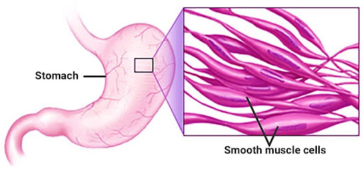
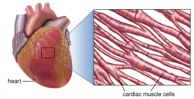
Consists of skeletal, smooth, and cardiac muscles.
Skeletal muscles are responsible for voluntary movements.
Sliding Filament Theory explains muscle contraction:
Actin and myosin filaments slide past each other, shortening the sarcomere.
ATP and calcium ions play a crucial role in contraction and relaxation.
3. Coordination of Movement
Controlled by the nervous system (brain, spinal cord, and motor neurons).
Reflex actions, voluntary movements, and postural control are regulated by neural pathways.
The cerebellum and motor cortex coordinate precision in movements.
Muscles
Muscle is a specialised tissue of mesodermal origin, and about 40-50% of the bodyweight of a human adult is made of muscles. It is classified on the basis of location, appearance, and nature of regulation of their activities.
On the basis of location, muscles are of three types as shown below:
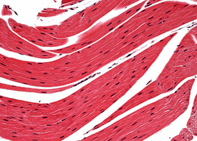
Skeletal Muscles and Contraction
Each organised skeletal muscle is made of a number of muscle bundles or fascicles held together by a common collagenous connective tissue layer: Fascia.
Each muscle fibre is lined by a plasma membrane: Sarcolemma enclosing the sarcoplasm.
The endoplasmic reticulum, i.e., the sarcoplasmic reticulum of the muscle fibres is the storehouse of Ca2+ ions.
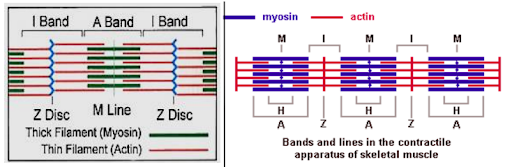
The characteristic feature of the muscle fibre is the presence of a large number of parallelly arranged filaments in the sarcoplasm, i.e., myofilaments or myofibrils.
Each myofibril has an alternate dark and light bands on it and its striated appearance is due to the distribution pattern of two important proteins – Actin and Myosin.
The light bands are called I-band or Isotropic band and contain actin whereas the dark band called ‘A’ or Anisotropic band contains myosin and is located in the centre of the sarcomere.
An extremely thin and comparatively dense Z-line (Krause's membrane) separates sarcomeres. It's right in the middle of the I-band.
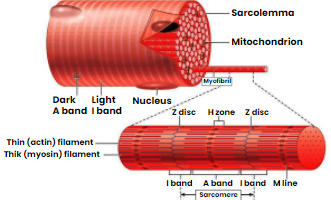
At the centre of the A-band, a comparatively less dark zone (not overlapped by thin filaments) is present: H-zone (Hensen zone).
In the centre of the H-zone, M-line is presently formed by threads that connect the myofilaments.
Structure of Contractile Protein
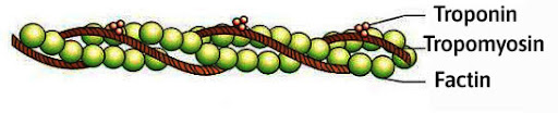
Each actin (thin) filament is made up of two helically wound 'F' (filamentous) actins. A polymer of monomeric 'G' (Globular) actins makes up each 'F' actin.
Tropomyosin, a protein, has two filaments that run close to the 'F' actins throughout its length.
Troponin, a complex protein, is found at regular intervals on tropomyosin.
Troponin subunits obscure the active binding sites for myosin on actin filaments in the resting state.
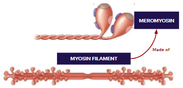
Each thick filament of myosin is also a polymerised protein. Meromyosins are a group of monomeric proteins that form a thick filament.
Each meromyosin contains two major components: a globular head with a short arm and a tail; the former is known as the heavy meromyosin (HMM), while the latter is known as the light meromyosin (LMM).
Mechanism of Muscle Contraction
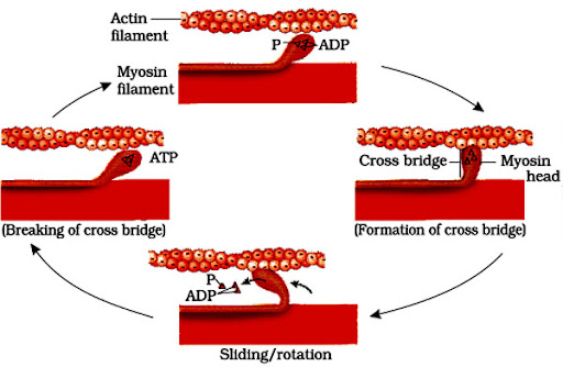
The sliding filament theory states that the contraction of a muscle fibre is caused by the sliding of thin (actin) filaments across thick (myosin) filaments.
A signal from the central nervous system (CNS) to a motor neuron causes muscle contraction.
The neuromuscular junction, also known as the motor endplate, is the point where a motor neuron meets the sarcolemma of a muscle fibre.
When a neuronal signal reaches this junction, a neurotransmitter (Acetylcholine) is released, causing the sarcolemma to create an action potential.
The action potential travels along with the muscle fibre, causing calcium ions to be released into the sarcoplasm. Increased Ca2+ levels cause calcium to bind to a troponin subunit (present on actin filaments). The masking of active sites for myosin is removed due to this binding.
The myosin head now binds to the exposed active sites on actin, generating a cross bridge, using the energy from ATP hydrolysis.
The connected actin filaments are subsequently pulled towards the centre of the 'A' band by the development of cross-bridges.
The Z-line is pulled inwards by the actin filaments as they pull towards the centre of the A-band.
The sarcomere shortens or contracts as a result of this. The length of the I-band decreases when the actin filaments are dragged inwards.
The myosin-containing A-band maintains its length.
The ADP and Pi are then released, and the myosin returns to its relaxed condition. When a fresh ATP binds, the cross-bridge is broken.
The myosin head hydrolyses this ATP, and the cross-bridge creation and contraction cycle proceeds.
Difference Between Red and White Muscle Fibres
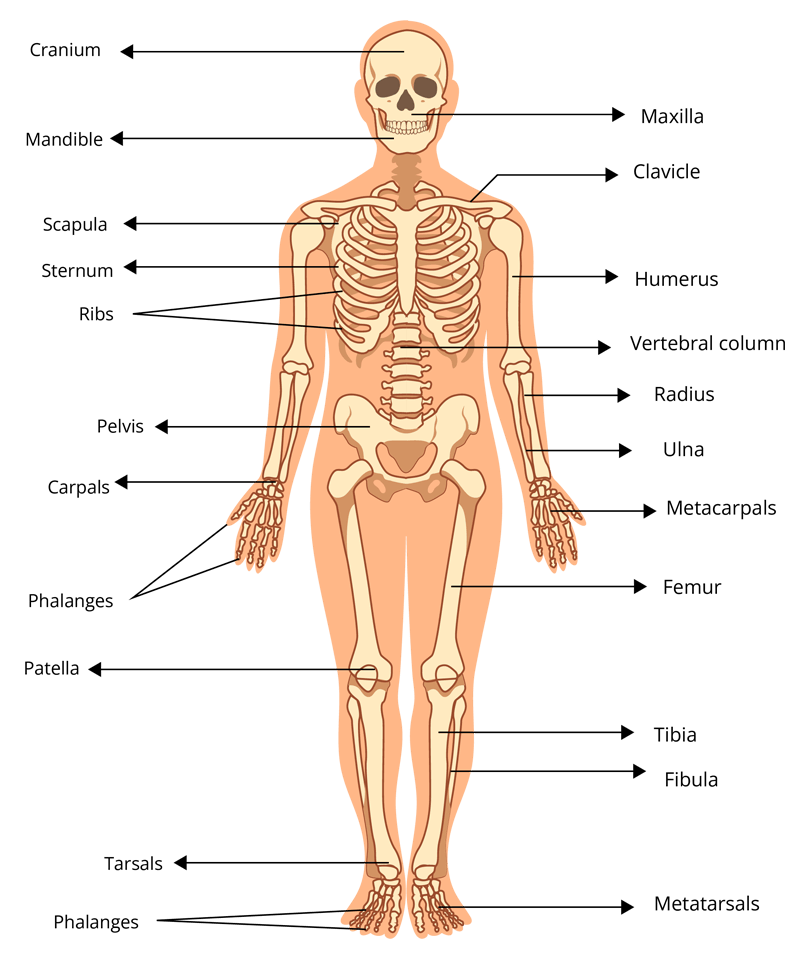
Human Skeletal System
A structure of bones and a few cartilages make up the skeletal system.
Bone and cartilage are connective tissues with specific functions. Due to calcium salts, the former has a very hard matrix, whereas the latter has a slightly flexible matrix due to chondroitin salts.
Function of the Skeletal System
Support - It gives support to the other softer body parts.
Protection - It protects the delicate body parts.
Muscle attachment - It provides attachment for large muscles.
Movement - It helps in bringing about movements.
Body form - It forms a typical body form of an individual animal.
Blood cell formation - RBCs, WBCs, and platelets are produced by the bone marrow.
Mineral reservation - It maintains the calcium and phosphorus levels of the blood.
Helps in breathing and hearing - Cartilages of the larynx, trachea, sternum, and ribs are helpful in breathing and ear bones of the middle ear transmit the sound vibrations from the tympanic membrane to the internal ear.
Difference Between Axial Skeleton and Appendicular Skeleton
Different Types and Functions of the Axial Skeleton
Different Types and Functions of the Appendicular Skeleton
Joints
All forms of movements need the use of joints. The points of contact between bones, or between bones and cartilages, are referred to as joints. According to mobility, they are classified into three types:
Fibrous Joints (fixed or immovable joints)
There is no movement possible with this type of joint. The flat skull bones fuse end-to-end with the help of dense fibrous connective tissues in the form of sutures to form the cranium in this type of joint.
Cartilaginous Joints (slightly movable joints)
In this, the bones involved are joined together with the help of cartilages. It permits limited movement. For example; the vertebrae (intervertebral discs) and the pubic symphysis of pubis.
Synovial Joints (freely movable joints)
Synovial joints are characterised by the presence of a fluid filled synovial cavity between the articulating surfaces of two bones. It aids locomotion and a variety of other movements.
Types of Synovial Joints
Disorders of Skeletal and Muscular System
Myasthenia gravis
Myasthenia gravis is an autoimmune disorder affecting the neuromuscular junction leading to fatigue, weakening and paralysis of skeletal muscle.
Muscular dystrophy
Muscular dystrophy is a genetic disorder that involves progressive degeneration of skeletal muscles. It is caused due to a defect in the dystrophin gene that is responsible for keeping muscles intact.
Tetany
Tetany is characterised by rapid contractions or spasms in the muscles, caused due to low Ca2+ ion concentration in the body fluid.
Arthritis
Arthritis is a condition in which one or more joints swell and become tender. The most common symptoms of arthritis are joint pain and stiffness, which often worsen with age.
Osteoporosis
This is an age-related disorder, caused due to decrease in bone mass. It increases the chance of fractures. A decrease in oestrogen levels contributes to osteoporosis.
Gout
It is due to a defect in purine metabolism that causes an excess of uric acid and its salts (urates). In gout, the level of uric acid is raised in the blood. When crystals of uric acid salts (e.g., sodium urate) accumulate in the joints causing gouty arthritis. Urates in excess can cause stones in the kidneys.
Paget's Disease
It is caused by abnormal bone resorption by abnormal osteoclasts. It is characterised by irregular thickening and softening of bones, resulting in the deformation of bones.
Solved Examples/Problems from the Chapter
1. Which of the following 'labels' in the figure below shows the presence of the acromion process?
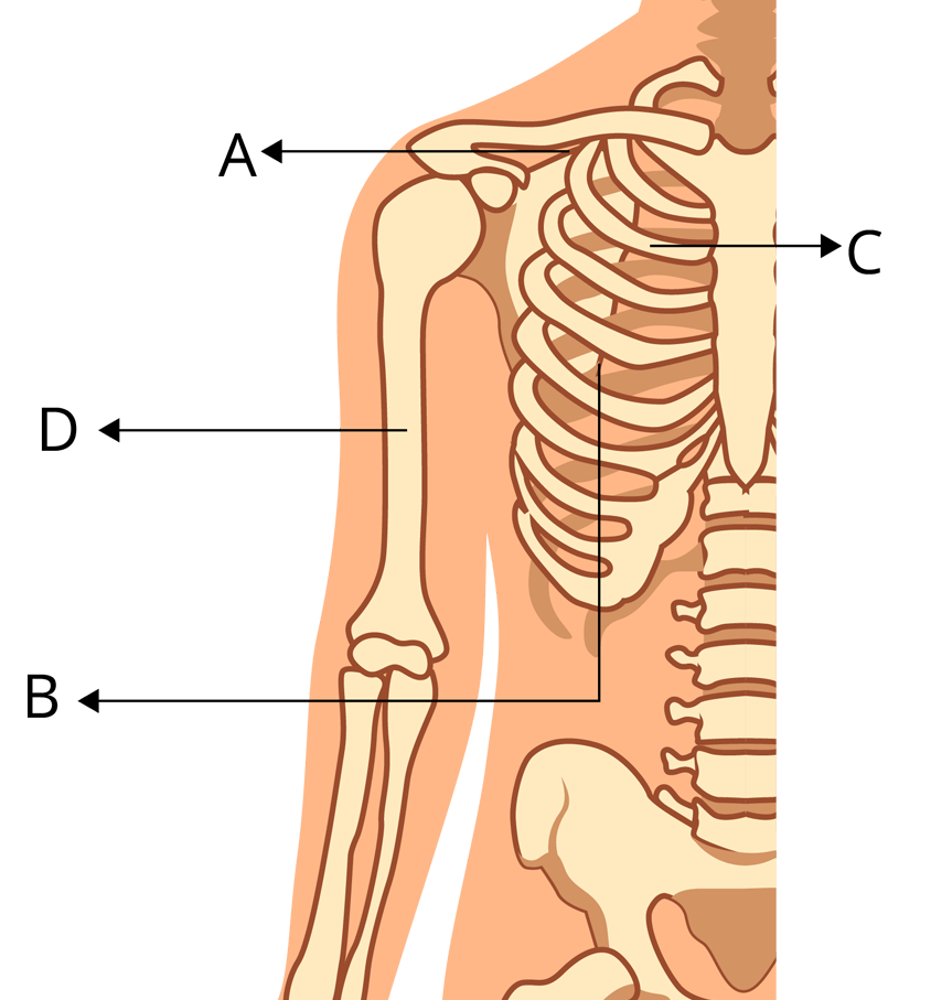
Ans: The labelling of the given diagram is
(A) Clavicle
(B) Scapula
(C) Rib
(D) Humerus
Key to remember:
Clavicle- collar bone
Scapula- shoulder blade
Humerus- funny bone
2. Give the role of calcium ions in the contraction and relaxation of muscles.
Ans: Calcium ions play a major role in the contraction and relaxation of muscles. Calcium ions are mainly stored in the sarcoplasmic reticulum (during muscle relaxation) and are released from the sarcoplasm during muscle contraction. The overall role of calcium can be understood by the events given below-:
When a skeletal muscle is excited and an action potential travels along the T tubule, the concentration of calcium ions increases and calcium ions are released from sarcoplasm. These calcium ions attach to troponin, causing tropomyosin to move away from the myosin-binding sites on actin due to a conformational shift. Once these binding sites are free, myosin heads bind to them to form cross-bridges which result in muscle contraction. The decrease in calcium ion concentration in the sarcoplasmic reticulum causes tropomyosin to slide back and block the myosin-binding sites on actin which causes the muscle to relax.
Key to remember:
Concentration of calcium increases- contraction
Concentration of calcium decreases- relaxation
3. Following is the figure of actin (thin) filaments. Identify parts A, B, C in the given diagram.
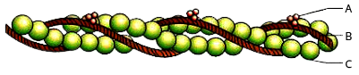
Ans: A. Troponin, B. Tropomyosin, C. F-actin
According to sliding filament theory, the contraction of muscle fibres is the result of sliding of thin (actin) filament over the thick (myosin) filaments.
Tropomyosin (regulatory protein) is one of the important proteins that play an important role in muscle contraction and it wraps around the actin and prevents myosin from grabbing. It does not allow the muscles to contract without the signal. Troponin is present at the regular interval on the tropomyosin. During the resting stage of the muscle fibre, tropomyosin partially covers the myosin-binding site on the actin filaments
Solved Problems of Previous Year Questions
1. Select the correct option.
a. 11th and 12th pairs of ribs are connected to the sternum with the help of hyaline cartilage.
b. Each rib is a flat thin bone and all the ribs are connected dorsally to the thoracic vertebrae and ventrally to the sternum.
c. There are seven pairs of vertebrosternal, three pairs of vertebrochondral and two pairs of vertebral ribs.
d. 8th, 9th and 10th pairs of ribs articulate directly with the sternum.
Ans: c. There are seven pairs of vertebrosternal, three pairs of vertebrochondral and two pairs of vertebral ribs.
Vertebrosternal ribs which are also called true ribs are joined to the thoracic vertebrae on the dorsal side and connected to the sternum on the ventral side by hyaline cartilage. The first seven pairs of ribs are true ribs.
The eighth, ninth, and tenth pairs of ribs do not articulate directly with the sternum but are joined by hyaline cartilage to the seventh ribs. These are vertebrochondral ribs, sometimes known as false ribs.
Only the first seven pairs of ribs are ventrally attached to the sternum; the last two pairs of ribs, 11 and 12, are called floating ribs because they are not connected ventrally.
Trick to remember:
True ribs- Vertebrosternal ribs
False ribs- vertebrochondral ribs
Floating ribs- last two pairs of ribs
2. The pivot joint between atlas and axis is a type of
a. Fibrous joint
b. Cartilaginous joint
c. Synovial joint
d. Saddle joint
Ans: c. Synovial joint
A synovial joint is a form of pivot joint. The synovial joints allow for considerable movement as the area between the articulating surfaces of the two bones is filled with synovial fluid.
Trick to remember: Synovial joint- allow for movement
Practice Questions
1. The _______ muscle is in charge of pulling the lower jaw, head, and tongue together.
a. Backwards.
b. Maximus
c. Retractor
d. Abductor
Ans: c. Retractor
The retractor muscle is the muscle that pulls the body component in the opposite direction.
2. During muscle contraction:
a. ATP is broken down
b. ATP is formed
c. GTP is broken down
d. None of the above
Ans: a. ATP is broken down
ATP is broken down into ADP and phosphate, releasing energy that is stored in the myosin head, and later used for myosin and actin binding, resulting in muscle contraction.
Key point to remember: ATP breakdown- release of energy
Conclusion
This Locomotion and Movement topic is written keeping in mind the students who are preparing for NEET 2025 or will prepare in the future. It covers all of the major concepts of the chapter and provides clear explanations, making it ideal for quick and successful study and revision. It covers key ideas, concepts, and problems from previous year's NEET question papers, as well as NEET practice tests and Biology NCERT books. Make sure to test your understanding and level of preparedness by attempting the practice question on your own.
Essential Study Materials for NEET UG Success
NEET Important Chapter - Locomotion and Movement

 Share
ShareFAQs on NEET Important Chapter - Locomotion and Movement
1. What are the names of the chapters that hold more weightage in NEET?
Human Physiology, Biological Classification, Molecular Basis of Inheritance, and Biomolecules had the highest weightage from the previous year's NEET paper analysis.
2. What is the number of questions that are asked from Human Physiology in NEET?
The number of questions that come from this unit are 13.
3. For NEET, how many MCQs should I solve per day?
As you will have to solve 180 MCQs in 180 minutes without losing pace and concentration, it is advisable that you practice at least 250 MCQs in 250 minutes per day.
4. What is locomotion? How is it different from movement?
Locomotion refers to the movement of an organism from one place to another, e.g., walking, running, flying.
Movement is a general term for any motion of body parts, e.g., blinking of eyes, heart contraction, or movement of limbs.
5. What are the different types of movements in the human body?
Amoeboid movement – Movement using pseudopodia (e.g., WBCs, macrophages).
Ciliary movement – Found in the respiratory tract and fallopian tubes.
Muscular movement – Involves muscle contraction and relaxation (e.g., walking, running).
6. What are the three types of muscles?
Skeletal muscles – Voluntary, striated muscles attached to bones (e.g., biceps, triceps).
Smooth muscles – Involuntary, non-striated muscles found in the digestive tract, blood vessels, etc.
Cardiac muscles – Striated, involuntary muscles found in the heart.
7. What is the structural unit of muscle fibers?
The structural and functional unit of muscle is the sarcomere, present between two Z-lines. It consists of actin and myosin filaments.
8. What is the difference between actin and myosin filaments?
Feature | Actin Filament | Myosin Filament |
Size | Thin | Thick |
Protein | Actin, Troponin, Tropomyosin | Myosin |
Function | Forms thin filaments, provides binding sites for myosin | Forms thick filaments, has heads for cross-bridge formation |
9. Explain the sliding filament theory of muscle contraction.
The sliding filament theory states that muscle contraction occurs when actin filaments slide over myosin filaments, shortening the sarcomere.
ATP is required for this process.
Calcium ions (Ca²⁺) and troponin-tropomyosin complex play key roles.
10. What are the components of a skeletal system?
Axial skeleton – Skull, vertebral column, ribs, and sternum.
Appendicular skeleton – Pectoral girdle, pelvic girdle, and limb bones.