


What is the QRS Complex?
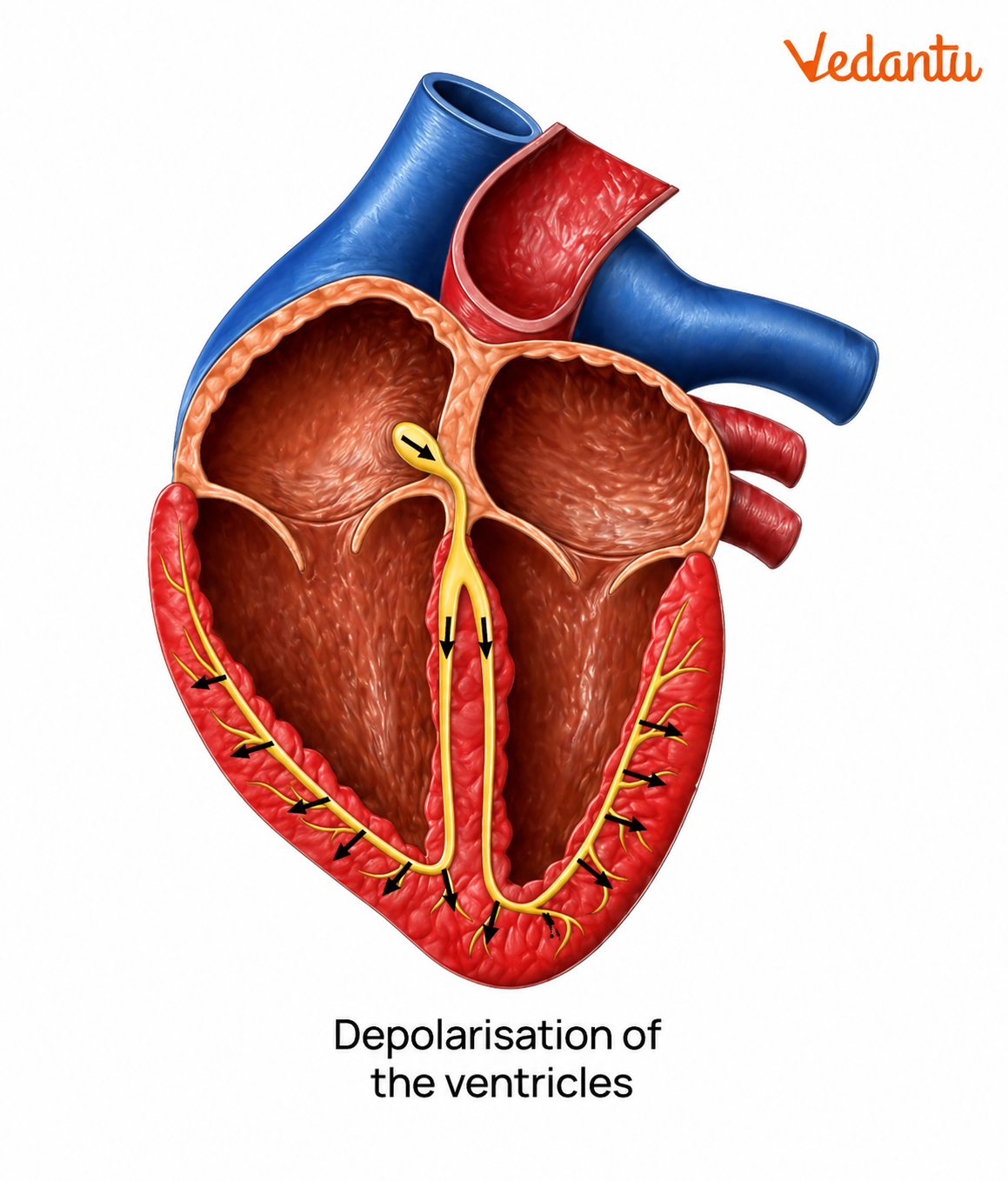
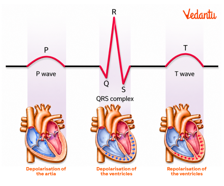
The QRS complex in an ECG is the most prominent and easily visible part of an electrocardiogram tracing. It represents the electrical activity of the ventricles, specifically the depolarisation of the right and left ventricles, which leads to ventricular contraction and pumping of blood.
In simple terms, the QRS complex shows how efficiently the ventricles are activated and how well the heart is functioning at a muscular level.
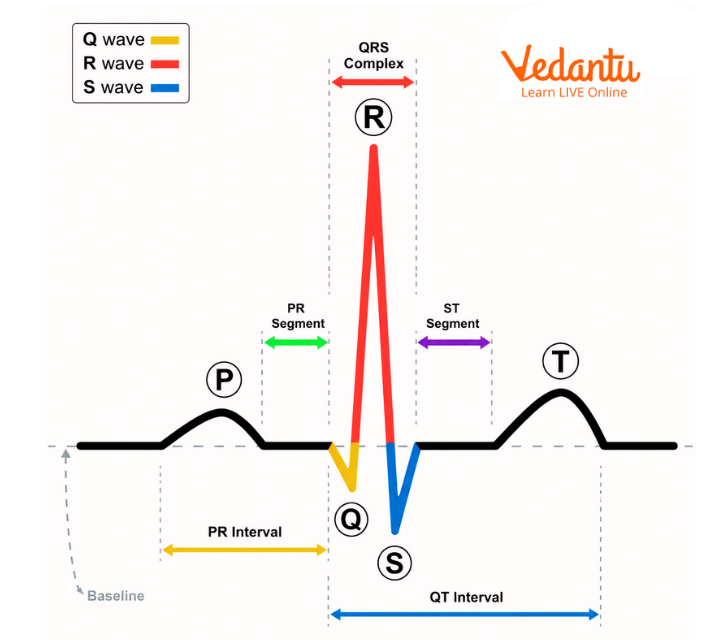
The QRS complex consists of three components:
Q wave – first downward deflection after the P wave
R wave – upward deflection
S wave – downward deflection following the R wave
These waves occur rapidly and together represent a single electrical event in the heart.
Key Points:
Signals originate from the conduction system (Bundle of His and Purkinje fibres)
Both ventricles depolarise almost simultaneously
This results in a strong contraction to pump blood to the lungs and body
The QRS complex is larger than the P wave because the ventricles have more muscle mass
QRS Complex Duration and Normal Range
1. Normal QRS Complex Duration
Typical duration: 0.08 to 0.10 seconds (80–100 ms)
Slight variation may occur depending on age and physiology
Children usually have slightly shorter durations
2. Measurement:
Start: Beginning of Q wave (or end of PR interval)
End: End of S wave
A normal duration indicates efficient conduction through the ventricles.
3. Narrow QRS Complex
A narrow QRS complex is one in which the duration is within the normal range (less than 100 ms).
4. Characteristics:
Efficient and rapid conduction
Normal functioning of the Bundle of His and the Purkinje fibres
Seen in healthy individuals
5. Clinical Meaning:
Indicates that electrical impulses are traveling through the normal conduction pathway
Usually associated with normal sinus rhythm
Wide QRS Complex
A wide QRS complex occurs when the duration exceeds 0.10–0.12 seconds.
Causes:
Bundle branch block
Ventricular arrhythmias
Electrolyte imbalance (e.g., hyperkalemia)
Ventricular hypertrophy
Delayed conduction in ventricles
Clinical Significance:
A widened QRS suggests that electrical impulses are not travelling normally, leading to delayed ventricular activation.
Components of the QRS Complex Explained
1. Q Wave
Small negative deflection
Represents septal depolarization
Normal if small and short
Deep or wide Q waves may indicate a previous myocardial infarction
2. R Wave
First positive deflection
Represents main ventricular depolarisation
Its amplitude increases across chest leads
3. S Wave
Negative deflection after the R wave
Represents final phase of ventricular depolarisation
QRS Complex Formation (Electrical Basis)
The QRS complex is formed due to rapid electrical conduction through:
Bundle of His
Right and Left Bundle Branches
Purkinje fibers
This system ensures that both ventricles contract almost simultaneously, producing an efficient heartbeat.
QRS Complex Abnormalities
Changes in the QRS complex help diagnose several heart conditions.
1. Prolonged QRS Duration
Indicates delayed conduction
Seen in bundle branch blocks
2. Increased Amplitude
Suggests ventricular hypertrophy
3. Pathological Q Waves
Indicate past myocardial infarction
4. RSR′ Pattern
Seen in bundle branch block
Extra upward deflection (R′ wave)
5. Poor R Wave Progression
May indicate anterior myocardial infarction
Can also occur due to improper ECG placement
QRS Complex in ECG Interpretation
The QRS complex is one of the most important tools in ECG analysis.
It helps in diagnosing:
Arrhythmias
Ventricular hypertrophy
Conduction defects
Myocardial infarction
Electrolyte disturbances
It is also used to determine the heart's electrical axis and to assess overall cardiac health.
QRS Complex Amplitude and Significance
Amplitude refers to the height of the QRS waves.
Normal Values:
R and S waves vary across leads
Excessively high amplitude may indicate hypertrophy
Clinical Use:
Detects enlargement of heart chambers
Helps identify cardiac stress conditions
R Wave Progression in QRS Complex
Normally:
R wave increases from lead V1 to V5
S wave decreases progressively
Abnormal Patterns:
Poor progression → myocardial infarction
Reverse progression → conduction defects
Importance of QRS Complex
The QRS complex plays a critical role in understanding heart function.
Reflects ventricular activity
Indicates conduction efficiency
Helps detect serious heart diseases
Essential in emergency cardiac diagnosis
Quick Summary Table
Insights from Experts?
The QRS complex on an ECG is a key indicator of how well the heart's ventricles are functioning. A normal QRS complex shows efficient electrical conduction, while abnormalities like a wide QRS complex or unusual patterns can signal serious cardiac conditions.
Understanding the QRS complex duration, structure, and abnormalities is essential for accurate ECG interpretation and early diagnosis of heart diseases.
Related Topics and Internal Links
FAQs on QRS Complex in ECG Explained | Duration, Normal Range, Types, and Abnormalities
1. What is QRS complex in ECG?
The QRS complex is the main spike seen in an ECG. It represents the electrical activity of the ventricles, showing their contraction (depolarization) when blood is pumped out of the heart.
2. What is QRS normal range?
The normal QRS duration is 80–100 milliseconds (0.08–0.10 seconds).
3. What is V1, V2, V3, V4 in ECG?
V1, V2, V3, and V4 are chest leads placed on the front of the chest. They record electrical activity from the front and septal parts of the heart.
4. What is the basic QRS complex?
The QRS complex consists of three waves:
Q wave (downward)
R wave (upward)
S wave (downward)
Together, they represent ventricular depolarization and contraction.
5. What if QRS is high in ECG?
A high QRS amplitude usually means the ventricles are enlarged or thickened, a condition called ventricular hypertrophy.
6. How to read QRS complexes in ECG?
After the P wave:
First downward wave = Q wave
Upward wave = R wave
Downward wave after R = S wave
These waves together form the QRS complex.
7. What is L1, L2, L3, and L4 in ECG?
L1, L2, L3, and L4 refer to different positions where ECG electrodes are placed on the chest to record heart activity.
8. What are the 4 waves of the ECG?
The main ECG waves are:
P wave
QRS complex (Q, R, S waves)
T wave
U wave (sometimes present)
9. What do aVR, aVL, and aVF stand for?
These are augmented limb leads:
aVR: Right arm
aVL: Left arm
aVF: Left foot
They show electrical activity from different angles of the heart.
10. What is an abnormal QRS?
A QRS complex longer than 120 milliseconds is considered abnormal and usually indicates a delay in ventricular conduction.
11. What diseases affect the QRS complex?
Conditions that affect the QRS complex include:
Bundle branch block
Ventricular tachycardia
Ventricular fibrillation
Wolff-Parkinson-White (WPW) syndrome
Ventricular enlargement
12. Can anxiety affect an ECG?
Yes, anxiety can affect ECG results. It can increase heart rate and cause temporary changes like palpitations or irregular rhythms, which may appear abnormal.