


Structure, Function & Location of Transitional Epithelium in NEET
The concept of transitional epithelium is essential in biology and helps explain real-world biological processes and exam-level questions effectively. This tissue type is frequently tested in NEET exams due to its unique features and functional importance in the urinary system.
Understanding Transitional Epithelium
Transitional epithelium refers to a specialized, multi-layered epithelial tissue found mainly in the urinary system. Its chief characteristic is the ability to stretch and change its appearance, adapting to organ expansion and contraction. This concept is important in areas like urothelium structure, urinary bladder adaptation, and difference from stratified epithelium.
Structure of Transitional Epithelium
Transitional epithelium shows three main types of cells arranged in layers:
- Basal layer: Deepest cells, usually cuboidal or columnar, attached to the basement membrane.
- Intermediate (middle) layer: Multiple layers of polygonal or pear-shaped cells, capable of rapid division.
- Apical (superficial) layer: Large, dome or umbrella-shaped cells on top, which flatten when stretched.
These layers allow for significant expansion and contraction, distinguishing transitional epithelium from other epithelial types.
Function of Transitional Epithelium
Transitional epithelium serves unique roles:
- Stretchability: Enables organs like the urinary bladder to expand without tissue damage.
- Impermeable barrier: Protects underlying tissues from urine and toxins.
- Rapid adaptation: Cells change shape instantly as organ fills or empties.
Location and Examples
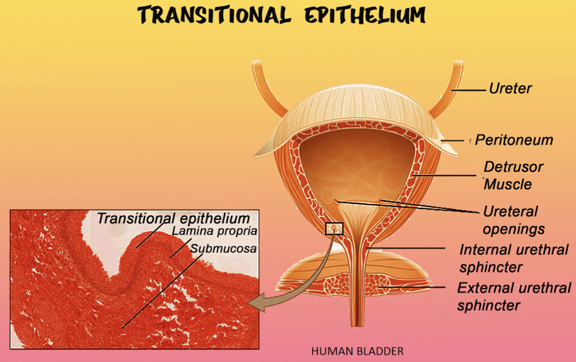
Transitional epithelium is characteristically present in:
- Urinary bladder – classic NEET question focus
- Ureters (tubes linking kidneys to bladder)
- Upper part of urethra
- Renal pelvis
Its function closely matches the physiological needs of these organs.
Here’s a helpful table to understand transitional epithelium vs other epithelia better:
Transitional Epithelium Comparison Table
| Feature | Transitional Epithelium | Stratified Squamous | Simple Cuboidal |
|---|---|---|---|
| Layers | Multiple (3–6) | Many | Single |
| Top Cell Shape | Dome/umbrella (when relaxed) | Flat (squamous) | Cube |
| Stretchable? | Yes | No | No |
| Key Locations | Urinary tract | Skin, esophagus | Tubules (kidney) |
Practice Questions
- What is the main function of transitional epithelium in the urinary bladder?
- List two locations where transitional epithelium is found.
- Draw and label a diagram of transitional epithelium showing umbrella cells.
- How does transitional epithelium differ from stratified squamous epithelium?
Common Mistakes to Avoid
- Confusing transitional epithelium with stratified squamous in MCQs.
- Not labeling umbrella cells or missing layers in the diagram during NEET exam.
- Assuming transitional epithelium is keratinized – it is not!
Real-World Applications
The concept of transitional epithelium is used in fields like medicine, especially in urinary tract health and in identifying certain cancers. Understanding this tissue type also helps in interpreting histology slides and diagnosing urinary diseases. Vedantu helps students relate such topics to practical examples in daily life and enhances exam confidence with targeted questions.
In this article, we explored transitional epithelium, its special structure, core functions, examples, and ways to avoid common errors. To learn more, keep practicing diagrams and difference tables, and check out related biology concepts for NEET with Vedantu.
Related Internal Links for Deeper Learning
FAQs on Transitional Epithelium Explained for NEET
1. What is transitional epithelium in NEET?
Transitional epithelium is a type of stratified epithelial tissue found primarily in the urinary bladder, ureters, and urethra. It is specialized to allow stretching and contraction as the volume of urine changes, protecting underlying tissues from toxic urine components.
2. Where is transitional epithelium found?
The transitional epithelium is mainly located in the lining of the urinary bladder, ureters, and parts of the urethra. It is also present in the prostatic ducts of the male reproductive system. These locations require a tissue that can accommodate volume changes without damage.
3. How to identify transitional epithelium under a microscope?
Under a microscope, the transitional epithelium can be identified by its multiple layers of cells with varying shapes: basal cells are usually cuboidal or columnar, while the superficial (apical) layer contains large umbrella cells that are dome-shaped when relaxed and flattened when stretched.
4. What is the function of transitional epithelium in the urinary bladder?
The transitional epithelium serves two main functions in the urinary bladder: impermeability to prevent urine leakage and protect underlying tissues, and stretchability to accommodate changes in bladder volume without damage, allowing the organ to expand and contract efficiently.
5. What differentiates transitional from stratified epithelium?
While both are types of stratified epithelium, transitional epithelium is unique because its apical cells change shape (from dome-shaped to flat) when stretched, which stratified epithelium generally does not. Transitional epithelium is specialized for organs that undergo volume changes, unlike stratified squamous or cuboidal epithelium.
6. What is the difference between transitional and squamous epithelium?
Transitional epithelium is multi-layered with the ability to stretch, and its superficial cells are dome-shaped when relaxed. Squamous epithelium cells are generally flat and thin and provide a smooth, protective surface but lack the ability to stretch like transitional epithelium.
7. Can transitional epithelium be keratinized?
No, transitional epithelium is typically non-keratinized. It maintains a moist environment suitable for urinary tract lining and does not undergo keratinization like some stratified squamous epithelia. This is an important distinction to avoid common NEET misconceptions.
8. Why do students often confuse transitional with stratified squamous epithelium in diagram questions?
Students confuse these because both are multi-layered epithelia. However, transitional epithelium has distinct umbrella cells and cell shape changes upon stretching. Remembering the location (urinary system) and the function (stretchability) helps differentiate them easily in diagrams.
9. What are umbrella cells in transitional epithelium?
Umbrella cells are large, dome-shaped cells found on the apical surface of transitional epithelium. They form a protective layer that shields underlying cells from the toxic effects of urine and enable the epithelium to stretch and contract without damage.
10. How to avoid mixing location-based questions like ureter vs urethra in MCQs?
Focus on the function and histology differences: the ureters and urinary bladder are lined by transitional epithelium to allow expansion, while the urethra (especially in males) may have different epithelium types toward the distal end. Memorizing organ-specific lining and function is key to accuracy.