


Human Urinary System Diagram: Labelled Parts and How to Remember Them for NEET
The concept of human urinary system diagram is essential in biology and helps explain real-world biological processes and exam-level questions effectively. Learning to accurately draw, label, and understand the human urinary system diagram is a common requirement for NEET and class 11 Biology students.
Understanding Human Urinary System Diagram
Human urinary system diagram refers to the visual representation of the organs involved in filtering blood and excreting waste products as urine in humans. This concept is important in areas like excretory system diagram, kidney and bladder function, and process of urine formation. Knowing this diagram helps you label organs, understand functions, and solve NEET MCQs with confidence.
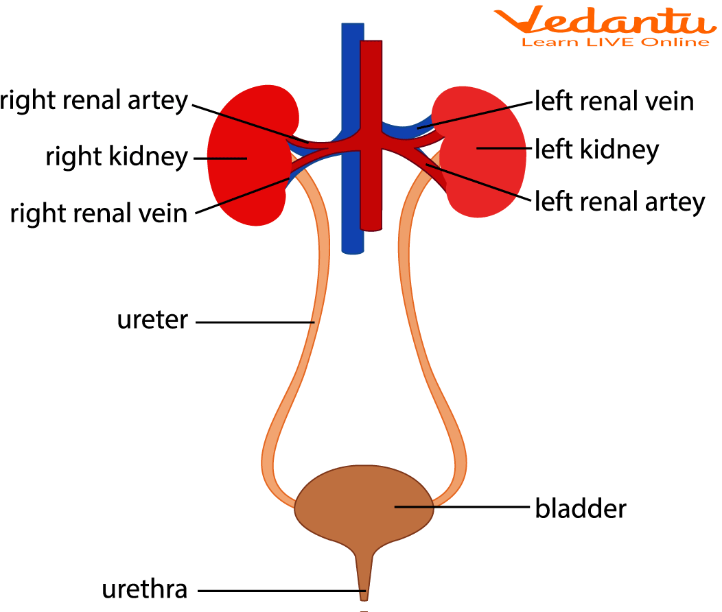
Key parts to always label in the human urinary system diagram:
- Kidneys (Left and Right)
- Ureters
- Urinary bladder
- Urethra
- Renal arteries and veins (optional for NEET fact-based diagrams)
Parts of the Human Urinary System and Their Functions
The human urinary system consists of four major organs. Each plays a distinct role in filtering blood and removing wastes:
- Kidneys: Filter blood, remove waste, maintain water and electrolyte balance.
- Ureters: Transport urine from kidneys to bladder.
- Urinary Bladder: Temporarily stores urine until excretion.
- Urethra: Tube for passage of urine outside the body.
Mechanism of Human Urinary System Diagram
The basic mechanism involves:
- Filtration of blood in the kidneys (in nephrons)
- Urine collection from nephrons into collecting ducts
- Passage through ureters to urinary bladder
- Storage and finally excretion via the urethra
Summary Table – Human Urinary System Parts & Functions
| Organ | Main Function |
|---|---|
| Kidneys | Filter blood, form urine |
| Ureters | Carry urine to bladder |
| Urinary Bladder | Store urine |
| Urethra | Excrete urine from body |
Steps of Urine Formation (NEET Focus)
There are three main steps in urine formation, which are commonly asked in NEET exams:
- Glomerular Filtration: Blood is filtered in the kidney nephrons.
- Reabsorption: Useful substances and water are reabsorbed into the blood.
- Secretion: Additional waste and excess ions are secreted into the urinary tubule.
Diagram Drawing Tips for NEET
- Start with two bean-shaped kidneys at the top, keeping right kidney slightly lower.
- Draw slender ureters from each kidney, curving down to the bladder.
- Sketch a balloon-like urinary bladder at the pelvis (center-bottom).
- Complete with a straight narrow urethra below the bladder.
- Label each part clearly and avoid merging ureter and urethra.
- Mnemonic: KUBU - Kidneys, Ureters, Bladder, Urethra.
Differences: Male vs Female Human Urinary System Diagram
| Feature | Male | Female |
|---|---|---|
| Urethra Length | ~20 cm (long, through penis) | ~4 cm (short, above vaginal opening) |
| Urine & Semen Pathway | Common (shared in penis) | Separate from reproductive tract |
| Infection Risk | Lower (longer urethra) | Higher (short urethra) |
Practice Questions
- Draw and label a human urinary system diagram. Name all parts.
- State one function for each urinary system organ.
- How is the mechanism of urine formation linked to homeostasis?
- What is the difference in urinary system structure between males and females?
Common Mistakes to Avoid
- Confusing ureter and urethra in labels.
- Forgetting to show the right kidney slightly lower due to liver position.
- Neglecting the correct sequence: kidney → ureter → bladder → urethra.
- Misplacing opening of ureters into the bladder.
Real-World Applications
The concept of human urinary system diagram is used in fields like medicine, renal physiology, surgery, and pathology (e.g. kidney stones, urinary tract infections). Vedantu helps students relate such topics to practical examples—such as dialysis or urinalysis in healthcare—in daily life and medical careers.
Related topics to help you master more NEET diagrams and reinforce urinary system concepts:
- Human Excretory System
- Kidney Diagram
- Nephron
- Urinary Bladder
- Homeostasis
- Modes of Excretion
- Difference between Egestion and Excretion
- Kidney Stones – Symptoms
In this article, we explored human urinary system diagram, its key processes, real-life significance, and how to solve questions based on it. To learn more and build confidence, keep practicing diagrams and biology revision with Vedantu.
FAQs on Human Urinary System Diagram – Parts, Functions & NEET Tips
1. What is the human urinary system in NEET?
The human urinary system is a vital biological system for excretion and maintenance of homeostasis. It primarily consists of the kidneys, ureters, urinary bladder, and urethra. For NEET, it is important to understand its structure, function, and the role it plays in removing metabolic wastes and regulating fluid balance.
2. How do I memorize the urinary system diagram quickly?
Memorizing the urinary system diagram can be simplified using these tips:
• Learn the four main parts: kidneys, ureters, bladder, and urethra.
• Use mnemonics like "KUBU" to remember the order.
• Practice drawing the diagram stepwise: sketch kidneys first, add ureters, then bladder and urethra.
• Associate functions alongside each part to reinforce recall.
• Revise with labeled diagrams and use flashcards for rapid review before exams.
3. What are the 7 parts of the human urinary system?
The seven main parts of the human urinary system to remember for NEET are:
1. Kidneys (filter blood)
2. Ureters (carry urine to bladder)
3. Urinary bladder (stores urine)
4. Urethra (excretes urine)
5. Renal cortex (outer kidney layer)
6. Renal medulla (inner kidney region)
7. Nephrons (functional units for filtration)
4. How do you draw a human urinary system for NEET?
To draw the human urinary system accurately:
1. Begin with the pair of bean-shaped kidneys on either side of the spine.
2. Sketch the ureters as thin tubes extending downwards from each kidney.
3. Draw the urinary bladder as a hollow sac lower in the abdomen.
4. Add the urethra exiting from the bladder.
5. Label each part clearly.
Use simple shapes and follow proportional sizes as per NEET guidelines.
Practice stepwise drawing with mnemonics to help under exam pressure.
5. What is the function of each urinary system part?
Each part of the urinary system performs specialized roles:
• Kidneys: Filter blood to remove wastes and balance electrolytes.
• Ureters: Transport urine from kidneys to bladder.
• Urinary bladder: Temporarily stores urine.
• Urethra: Conducts urine outside the body.
• Nephrons: Carry out filtration, reabsorption, and secretion.
• Sphincter muscles: Control urine flow and prevent leakage.
This knowledge helps answer NEET MCQs on structure-function relationships.
6. Is there a difference in the urinary system diagram for males and females?
Yes, there are key differences between the male and female urinary systems:
• The male urethra is approximately 8 inches long and transports both urine and semen.
• The female urethra is shorter, about 1.5 inches long, and only conducts urine.
• Male urinary system includes the prostate gland, which surrounds the urethra near the bladder.
These distinctions influence susceptibility to conditions like urinary tract infections (UTIs) and are often tested in NEET.
7. Why do students often mislabel ureter and urethra in NEET diagrams?
Students commonly confuse the ureter and urethra because of similar spelling but different functions:
• The ureter is a long tube connecting the kidneys to the bladder, transporting urine.
• The urethra is the tube from the bladder to outside the body, releasing urine.
To avoid such mistakes, memorize their positions and specific functions separately.
Label practice with multiple diagrams can reinforce correct identification.
8. What silly mistakes happen in urinary system diagram labelling?
Common labeling errors in the urinary system diagram include:
• Mixing up the left and right kidney positions.
• Confusing ureter with urethra.
• Incorrectly placing the prostate gland.
• Omitting or mislabeling the renal cortex and medulla.
• Not showing the correct connections between parts.
Careful revision and use of accurate NEET-standard diagrams help prevent these mistakes.
9. How to avoid missing the right-left kidney orientation?
To remember the correct kidney orientation in diagrams:
• Recall that the right kidney is slightly lower due to the liver’s position.
• Use the shape of the kidneys — the hilum (entry/exit point for vessels and ureter) faces medially.
• Label kidneys with orientation words like "right" and "left" explicitly.
• Practice drawing with anatomical landmarks, such as the spine and ribs.
This helps avoid errors during NEET diagram labeling and MCQ preparation.
10. Why is the diagram asked so frequently in NEET?
The human urinary system diagram is frequently asked in NEET because:
• It is fundamental to understanding human excretion and homeostasis.
• It integrates knowledge of anatomy and physiology tested in both theory and MCQs.
• Diagram questions assess students’ ability to visualize and accurately reproduce biological systems.
• It links to related topics like urine formation, hormone secretion, and pathologies.
Hence, mastering this diagram is essential for NEET success.
11. How to recall kidney cortex, medulla, and nephron in one glance?
To quickly recall the kidney cortex, medulla, and nephron:
• Visualize the cortex as the outer protective layer.
• Picture the medulla as the inner pyramidal region with collecting ducts.
• Remember nephrons as tiny filtering units spanning cortex and medulla.
• Use mnemonic phrases like "Cortical Cover, Medullary Middle, Nephron Network".
• Practice labelled diagrams highlighting these parts enhances fast recall under exam pressure.
12. What are the key hormones secreted by the kidneys?
The kidneys secrete and activate vital hormones important in physiological regulation:
• Erythropoietin (EPO): Stimulates red blood cell production in bone marrow.
• Renin: Regulates blood pressure through the renin-angiotensin system.
• Calcitriol: Active form of vitamin D, crucial for calcium absorption and bone health.
Understanding these hormones is critical for grasping kidney functions for NEET exams.