


What Is Leucosolenia? Habitat, Body Structure, and Feeding Explained
Quick Facts About Leucosolenia
| Feature | Details |
|---|---|
| Common Name | Leucosolenia |
| Scientific Group | Phylum Porifera |
| Type | Marine Sponge |
| Body Plan | Asconoid (simplest sponge type) |
| Habitat | Shallow marine waters |
| Diet | Microscopic food particles |
| Movement | Non-motile (fixed in one place) |
| Reproduction | Sexual and Asexual |
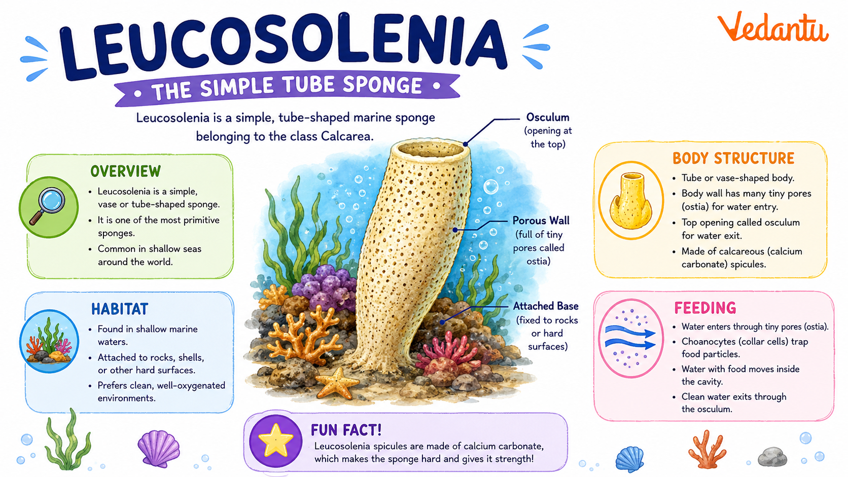
Appearance and Physical Characteristics
- Small, delicate, tube-shaped body.
- Usually white or pale in colour.
- Body wall contains tiny pores called ostia.
- Has a large opening at the top called osculum.
- Body supported by needle-like structures called spicules.
- Lacks true tissues and organs.
Leucosolenia Habitat and Distribution
- Found in shallow marine waters.
- Usually attached to rocks, shells, or seaweed.
- Common in coastal regions.
- Prefers clean, well-oxygenated seawater.
Diet and Feeding Habits
- Leucosolenia diet consists of tiny plankton and organic particles.
- It is a filter feeder.
- Special cells called choanocytes (collar cells) help trap food.
- Water current is created by the beating of tiny flagella.
1. Water enters through ostia.
2. Food particles are trapped by choanocytes.
3. Clean water leaves through the osculum.
Behaviour and Lifestyle
- Non-motile (fixed in one place).
- Lives alone or in small colonies.
- Does not have a nervous system.
- Responds slowly to environmental changes.
Leucosolenia Life Cycle
- Asexual Reproduction: By budding or fragmentation.
- Sexual Reproduction: Produces sperm and eggs.
- Larval Stage: Free-swimming larva settles on a surface.
- Adult Stage: Develops into a tube-shaped sponge.
What Makes Leucosolenia Special?
Importance and Role in Nature
Amazing Leucosolenia Facts
- Belongs to the phylum Porifera, meaning “pore bearer.”
- Its body is full of microscopic pores.
- Can regenerate lost parts.
- Has no brain or heart.
- Water circulation is essential for its survival.
- It is one of the earliest forms of animal life.
Fun Facts for Kids
FAQs on Leucosolenia Sponge: Characteristics, Structure, and Marine Role
1. What is Leucosolenia?
Leucosolenia is a simple, tube-shaped marine sponge that lives in the ocean.
- It belongs to the group Porifera (sponges).
- It has a soft, white, and branching body.
- It is one of the simplest multicellular animals.
- It shows the basic asconoid body structure.
2. Where is Leucosolenia found?
Leucosolenia is found in shallow marine waters around the world.
- Lives in oceans and seas.
- Attaches to rocks, shells, and corals.
- Common in coastal areas.
- Prefers clean, saltwater habitats.
3. What type of body structure does Leucosolenia have?
Leucosolenia has an asconoid canal system, which is the simplest sponge body plan.
- Body is shaped like a small tube.
- Has a central cavity called spongocoel.
- Water enters through tiny pores called ostia.
- Water leaves through a large opening called osculum.
4. How does Leucosolenia feed?
Leucosolenia feeds by filtering tiny food particles from water.
- It is a filter feeder.
- Water enters through ostia.
- Special cells called choanocytes (collar cells) trap food.
- Food includes tiny plankton and microorganisms.
5. How does Leucosolenia reproduce?
Leucosolenia reproduces both sexually and asexually.
- Asexual reproduction by budding.
- Sexual reproduction by producing eggs and sperm.
- Fertilization occurs in water.
- Forms a free-swimming larva before attaching to a surface.
6. Is Leucosolenia harmful to humans?
Leucosolenia is harmless to humans and is not dangerous.
- It does not sting or bite.
- It is non-poisonous.
- It plays a helpful role in cleaning water.
- Important for marine ecosystems.
7. Why is Leucosolenia important in biology?
Leucosolenia is important because it helps scientists study early animal evolution.
- Represents one of the simplest animals.
- Shows basic cellular organization.
- Helps understand multicellularity.
- Used in zoology and marine biology studies.
8. What are the main characteristics of Leucosolenia?
Leucosolenia has simple features that define it as a sponge.
- Body is radially symmetrical.
- Lacks true tissues and organs.
- Has tiny pores for water flow.
- Supported by calcium carbonate spicules.
9. What is the size and shape of Leucosolenia?
Leucosolenia is small and has a thin, branching shape.
- Usually a few centimeters long.
- White or pale in color.
- Forms tube-like structures.
- Often grows in small colonies.
10. What group does Leucosolenia belong to?
Leucosolenia belongs to the phylum Porifera and class Calcarea.
- Kingdom: Animalia
- Phylum: Porifera
- Class: Calcarea
- Type: Calcareous sponge