


What Is a Fluke? Types, Infection Symptoms, and Prevention
A fluke is a type of parasitic flatworm that lives inside the bodies of animals and humans. Though small and simple in structure, flukes have a complex life cycle and amazing survival abilities. They belong to a group of worms called trematodes and are mostly found in freshwater environments. Learning about fluke characteristics, fluke habitat, and fluke life cycle helps us understand how parasites survive and why hygiene and clean water are important.
Quick Facts About Fluke
| Feature | Details |
|---|---|
| Common Name | Fluke |
| Scientific Class | Trematoda |
| Animal Group | Flatworms (Phylum Platyhelminthes) |
| Body Shape | Flat and leaf-like |
| Habitat | Inside animals; freshwater environments (during early stages) |
| Diet | Absorbs nutrients from host’s body |
| Size | Usually a few millimetres to a few centimetres |
| Examples | Liver fluke, Blood fluke, Lung fluke |
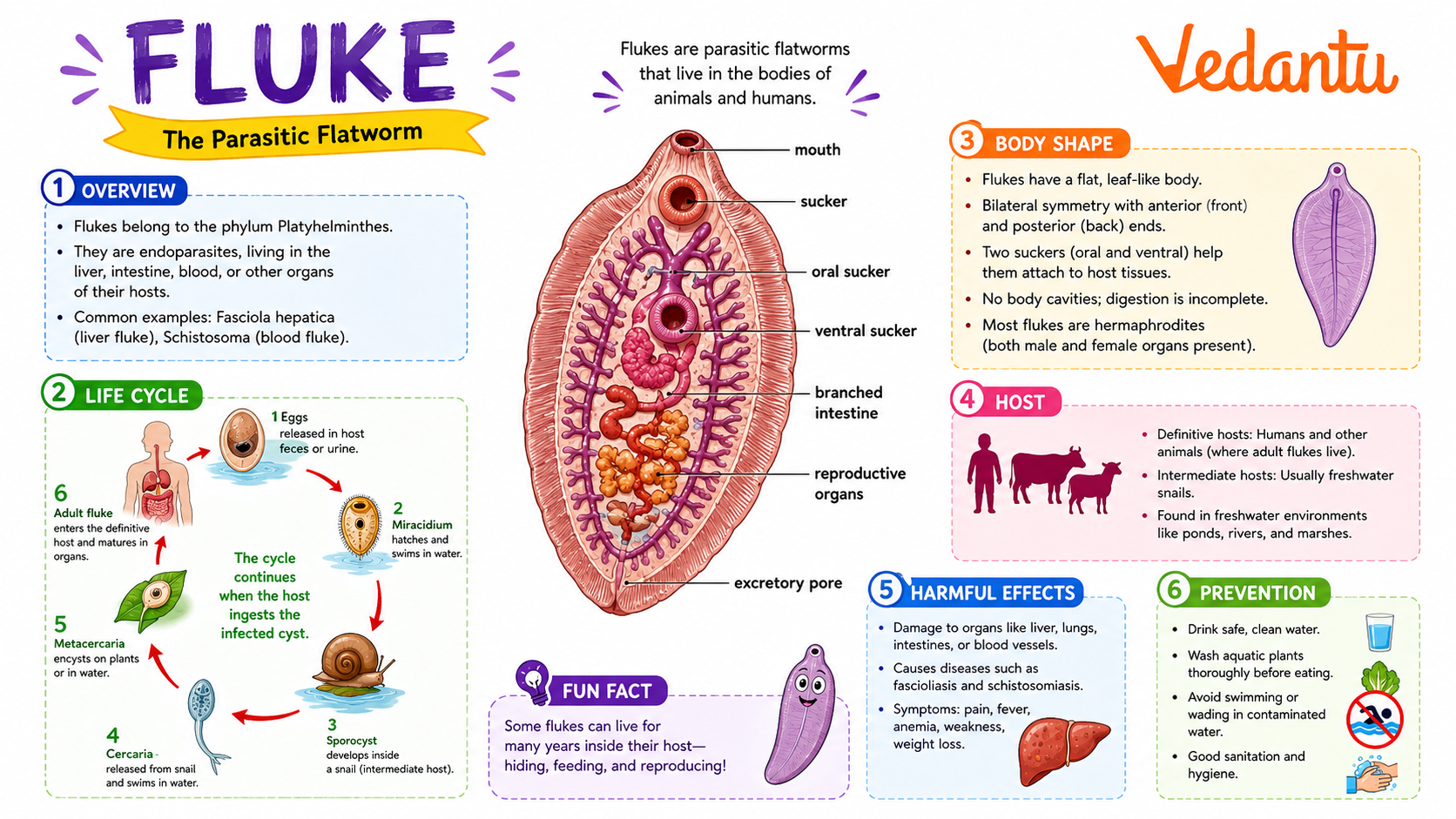
Appearance and Physical Characteristics
- Flat, soft, and leaf-shaped body.
- No body segments.
- Usually has two suckers – one for feeding and one for attachment.
- No separate breathing or circulatory system.
- Body is covered with a protective outer layer.
Fluke Habitat and Distribution
The fluke habitat changes during different stages of its life cycle.
- Inside the liver, blood, lungs, or intestines of animals and humans.
- Freshwater bodies like ponds, lakes, and rivers (during larval stages).
- Inside snails, which act as an intermediate host.
Flukes are found all over the world, especially in areas where sanitation is poor and freshwater snails are common.
Diet and Feeding Habits
The fluke diet depends on its host.
- Feeds on blood, tissue fluids, or nutrients from the host.
- Uses suckers to attach firmly inside the host’s body.
- Absorbs digested food directly through its body surface.
Behaviour and Lifestyle
Flukes live inside hosts and may cause diseases.
Use suckers to stay firmly attached inside organs.
Many flukes are hermaphrodites (have both male and female organs).
Produce many eggs to increase chances of survival.
Fluke Life Cycle
The fluke life cycle is complex and involves more than one host.
- Egg Stage: Eggs are released into water through the host’s waste.
- Larva Stage: Eggs hatch into larvae that infect freshwater snails.
- Inside Snail: The larva multiplies and develops further.
- Free-Swimming Stage: Leaves the snail and swims in water.
- Final Host: Infects humans or animals and becomes an adult fluke.
Types of Flukes
Lives in the liver of sheep, cattle, and sometimes humans.
Causes schistosomiasis in humans.
Infects lungs and may cause breathing problems.
Lives in the intestines of humans and animals.
Importance and Role in Nature
Part of the food web in freshwater systems.
Help control snail populations.
Help scientists understand parasitic diseases.
Teach importance of clean water and sanitation.
Amazing Fluke Facts
- Flukes are among the most common parasites in the world.
- Some species can live for many years inside a host.
- They produce thousands of eggs at once.
- They can infect fish, birds, mammals, and humans.
- Some flukes are visible to the naked eye.
- They have existed for millions of years.
Fun Facts for Kids
FAQs on Fluke Explained: Life Cycle, Types, and Health Risks
1. What is a fluke?
A fluke is a type of flat parasitic worm that lives inside animals or humans.
- It belongs to a group called flatworms (Trematodes).
- Flukes are usually very small and leaf-shaped.
- They are known as parasites because they live off a host.
- Common types include liver flukes and blood flukes.
2. Where do flukes live?
Flukes live inside the bodies of animals and sometimes humans.
- They can live in the liver, intestines, lungs, or blood.
- Many flukes start their life in freshwater snails.
- They are common in places with warm climates and unsafe water.
3. How do humans get infected by flukes?
Humans get infected by flukes through contaminated water or food.
- Drinking or swimming in infected freshwater.
- Eating raw or undercooked fish, crabs, or water plants.
- Poor hygiene and sanitation.
4. What are the symptoms of a fluke infection?
A fluke infection can cause different symptoms depending on the organ affected.
- Stomach pain or diarrhea.
- Fever and tiredness.
- Liver swelling in liver fluke cases.
- Blood in urine in blood fluke infections.
5. What is the life cycle of a fluke?
The life cycle of a fluke involves more than one host and several stages.
- Eggs leave the host through waste.
- Eggs hatch in water and infect a snail.
- The parasite grows inside the snail.
- It then leaves the snail and infects another host like a fish or human.
6. Are flukes dangerous?
Yes, flukes can be dangerous if not treated properly.
- They can damage important organs like the liver and lungs.
- Long-term infection may cause serious health problems.
- Early treatment helps prevent complications.
7. How are fluke infections treated?
Fluke infections are treated with special anti-parasitic medicines.
- Doctors may prescribe drugs like praziquantel.
- Proper diagnosis through lab tests is important.
- Follow-up care ensures the parasite is fully removed.
8. How can we prevent fluke infections?
Fluke infections can be prevented by practicing good hygiene and food safety.
- Avoid eating raw or undercooked seafood.
- Drink clean, safe water.
- Avoid swimming in contaminated freshwater.
- Wash hands regularly.
9. What is the difference between a fluke and a tapeworm?
A fluke and a tapeworm are both parasites, but they look and behave differently.
- Flukes are flat and leaf-shaped.
- Tapeworms are long and ribbon-like.
- Flukes often use snails in their life cycle.
- Tapeworms usually come from eating infected meat.
10. Why are flukes important to study in science?
Flukes are important in science because they help us understand parasites and diseases.
- They show how parasites adapt to different hosts.
- They teach us about life cycles in biology.
- Studying them helps doctors create better treatments.