


Introduction to Muscle Contraction
The capacity of muscle tissue to contract powerfully when activated by an action potential is referred to as contractility. When a muscle contracts, tension is generated while pulling on its attachment sites. If the created tension is sufficient to overcome the resistance of the item to be moved, the muscle shortens and movement occurs. This process is known as muscle contractility. In this article, the structure of muscles, types of muscles, and the mechanism of muscle contraction are discussed in detail.
What is Muscle Contraction?
Muscle is a specialised tissue that develops from the germ layer, the mesoderm. Myocytes are specialised cells that make up muscle tissue. These cells are linked by connective tissue to generate muscle tissue. It causes many forms of motion in both internal and exterior body components. A human body is made up of 639 muscles, each with its own set of characteristics such as contractibility, excitability, elasticity, and extensibility. They account for 40-50 percent of mature human body weight.
Myocytes are capable of contracting. They can shorten by $\dfrac{1}{3}$ to $\dfrac{1}{2}$ of their length owing to contraction and then return to their original length. Therefore, contractility refers to the trait of shortening and then returning to a relaxed condition of the muscle fibre. A stimulus, such as a nerve impulse, hormone, mechanical, thermal, or chemical stimulation, is necessary for muscle excitation, which results in muscular contraction. The stimulus received at one end of the muscle quickly travels to other portions as well as neighbouring myocytes.
Muscle Structure
A muscle is surrounded by a sheath of connective tissue known as the epimysium. A muscle has multiple muscular fibres organised in a bundle called fasciculi inside the epimysium. Each fascicle is covered in a sheath of connective tissue known as perimysium. The fasciculus is made up of parallel muscle fibres that are bordered by a connective tissue termed endomysium. The muscle bundle is further joined by a common collagenous sheath of connective tissue known as fascia.
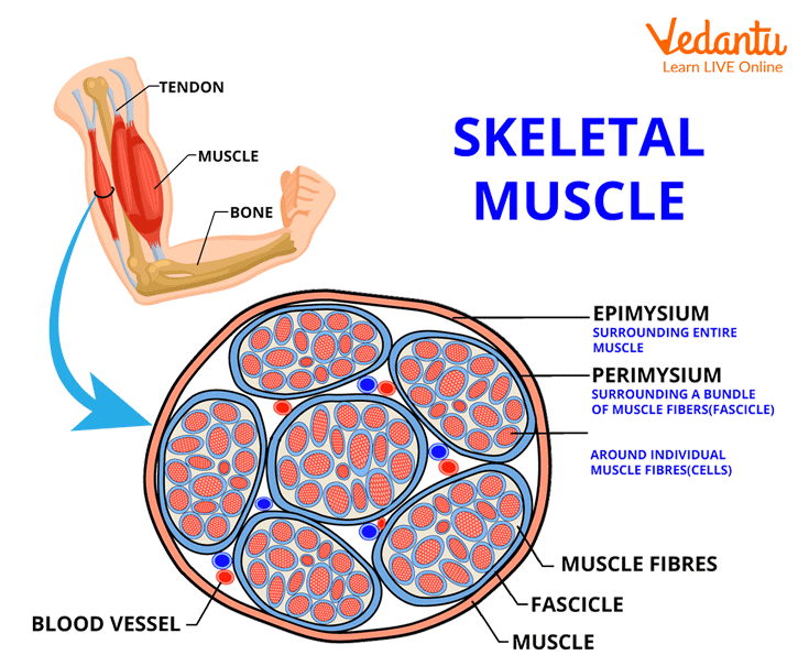
Showing Skeletal Muscle: Muscle structure
Types of Muscles
Muscles are classified based on a variety of factors, including their location, appearance, and the nature of the regulation of their activity. Muscles can be classified into the following types based on their location:
Skeletal muscle or voluntary muscle: These muscles are connected to the body's skeleton and are largely engaged in locomotory activities and changes in body posture. Under a microscope, alternate bright and dark bands may be seen on the muscle fibres. These muscles are known as striated or striped muscles because of the striated appearance of the muscular fibres. These muscles are voluntary since they are controlled by the animal's will or consciousness. For example, hindlimb muscles, body wall, mouth, throat, and the commencement of the oesophagus, among others.
Non-Striped or involuntary muscle: These muscle cells are elongated, spindle-shaped, wide in the middle, and taper at the ends. Since these muscles lack alternating light and dark stripes on their muscle fibres, they look smooth under a microscope. Their fibres are not laid out in parallel arrays.
These muscles border the hollow organs and are involuntary, such as the posterior section of the oesophagus, the stomach, the intestine, the lungs, the urine bladder, and the urogenital tract. The contraction of smooth muscles transports food through the digestive tract and gametes through the vaginal tract.
Cardiac muscle: As cardiac means "heart," muscles of the heart are referred to as cardiac muscles. The heart muscle cells form a branching pattern. These are striated in nature, which means that, under a microscope, alternate bright and dark bars may be seen on the muscle fibre. These are involuntary in nature since they provide their own impulse or excitement, allowing the heart to continue its pumping function of regular contraction and relaxation.
Muscle Contraction Mechanisms
A hypothesis known as the Sliding Filament Theory explains the process of muscle contraction. In 1954, two groups, Andrew Huxley and Ralph Niedergerke, and Hugh Huxley and Jean Hanson, presented this theory. According to this idea, muscle fibre contraction takes place when a thin actin filament glides across a thick myosin filament. The muscle contraction steps involved are listed below:
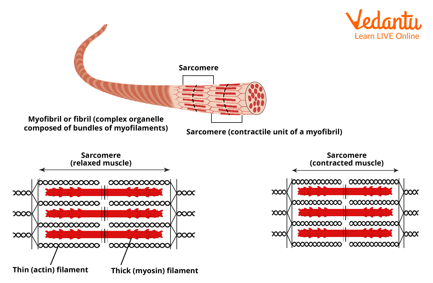
Sarcomere: The structural and functional unit of muscle fibres.
The signal supplied by the CNS via a motor neuron causes the muscle to contract. The CNS sends a nerve impulse through the motor neuron. When an impulse reaches an axon terminal or neuromuscular junction, neurotransmitter-containing vesicles fuse with the axon membrane. They release the neurotransmitter acetylcholine after fusing with the axon membrane, which passes via the synaptic cleft and generates action potentials in the sarcolemma.
The produced impulse or action potential then extends from the sarcolemma to the T-tubules. The impulse then activates the sarcoplasmic reticulum, causing calcium ions to be released into the sarcoplasm.
A rise in Ca2+ concentration in the sarcoplasm initiates filament sliding, whereas a decrease in concentration stops the process.
When Ca2+ concentration is low, then a muscle fibre is relaxed. This is due to the presence of Ca2+ active transport pumps in the sarcoplasmic reticulum (SR) membrane, which transfers Ca2+ from the sarcoplasm into the SR. Ca2+ is released as a muscle action potential travels over the sarcolemma and into the transverse tubule system.
As a result of this, Ca2+ floods the sarcoplasm surrounding the thick and thin filaments. Troponin changes its form as a result of the Ca2+ released from the sarcoplasmic reticulum. This change in shape shifts the troponin-tropomyosin complex away from actin myosin-binding sites.
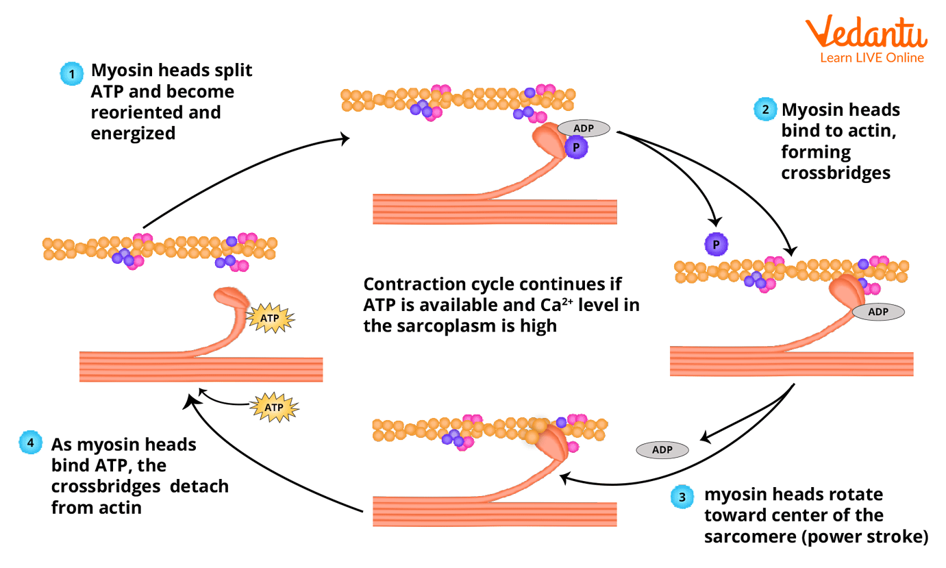
Myosin globular head functions as an ATPase, hydrolysing the ATP molecule. Myosin uses the energy released by ATP hydrolysis to bind to the exposed active site on the actin filament and form a cross bridge.
This moves the connected actin filament towards the centre of the A-band. The Z-line linked to these actions is likewise pulled inwards, causing the sarcomere to shorten or contract. The H-zone narrows as the thin myofilaments move past the thick myofilaments. This shortens the I-band while keeping the A-band the same length.
Myosin then releases ADP+Pi and returns to its relaxed state. ATP attaches to myosin once again, and the cross bridge between myosin and actin is broken. The ATP is hydrolysed again, and the cycle of cross-bridge production and breakdown is repeated, generating sliding. The procedure is repeated until the calcium ion is pumped back to the sarcoplasmic reticulum.
Muscle Contraction Mechanisms
Muscle Relaxation
Calcium returns to the sarcoplasmic reticulum following muscle contraction. As the quantity of calcium in the sarcoplasm diminishes, calcium does not bind to troponin C. Troponin changes form, and tropomyosin and troponin return to their original positions and states. This prevents myosin from binding to actin by blocking its active site. This induces muscular relaxation.
After a muscle fibre has contracted, two changes allow it to relax. First, an enzyme called acetylcholinesterase quickly degrades acetylcholine (AChE). When action potentials in the motor neuron halt, ACh release ceases, and AChE quickly degrades the ACh already present in the synaptic cleft. The formation of muscular action potentials is thereafter terminated, and the Ca release channels in the sarcoplasmic reticulum membrane shut.
Conclusions
Muscle is a specialised tissue that develops from the mesoderm. There are 639 muscles in the human body. Excitation of the muscle requires a nerve impulse, hormone, mechanical, thermal, or chemical stimulation. Skeletal muscles are striated and voluntary. The sarcoplasmic reticulum is a calcium-storage organ necessary during muscle contraction. Myosin makes up the thick myofilament. Actin is the main component of the thin myofilament. Myosin molecules have an ATP-binding site. Actin, tropomyosin, and troponin are the three proteins found in the thin myofilament. The contraction of muscles was explained by the Sliding-Filament theory. We have learnt about muscle relaxation and contraction.
FAQs on Mechanism of Muscle Contraction and Relaxation for NEET
1. What causes a muscle to contract?
When the neurological system sends a signal, muscle contraction begins. The signal, an impulse known as an action potential, goes via a motor neuron, a kind of nerve cell. The neuromuscular junction is the location where a motor neuron connects to a muscle cell. When a nervous system signal reaches the neuromuscular junction, the motor neuron sends a chemical message. Acetylcholine, a neurotransmitter, connects to receptors on the exterior of the muscle fibre to send the chemical message. This causes a chemical reaction to occur within the muscle.
2. What makes locomotion distinct from movement?
Locomotion is defined as the movement of a body from one location to another. Movement, on the other hand, is defined as the displacement of a body or a portion of a body from its initial location.
For example, Hydra employs tentacles for both catching prey/food and mobility. Amoeba employs pseudopodia for motility and food movement while Paramoecium uses cilia. Humans use their limbs to adjust their body position and move about. This suggests that movement and locomotion are not different entities, rather they are said to be interdependent on each other.
3. What are the three kinds of contractions?
There are three different types of contractions. Muscle contraction may be classified into three types: concentric, isometric, and eccentric.
Concentric: Concentric muscle motions generate sufficient force to overcome external strain and shorten the muscle. Example: lifting a barbell in a bicep curl.
Eccentric muscular motions generate force while the muscle lengthens - this is the movement's resistance. Example: lowering the barbell from the bicep curl.
Isometric: Isometric muscular motions generate force but do not affect muscle length. Example: Holding the barbell in one position.