


ICSE Class 10 Biology Chapter 11 Selina Concise Solutions - Free PDF Download
A. Multiple Choice Type
(Select the most appropriate option in each case)
1. Which part of the eye is grafted in a needy patient from a donated eye?
a. Conjunctiva
b. Cornea
c. Choroid
d. Ciliary muscles
Ans: (b) Cornea
2. Which part of our ear is shaped like a snail shell?
a. Semicircular canals
b. Cochlea
c. Stapes
d. Eustachian Tube
Ans: (b) Cochlea
3. The three parts of the human ear contributing to hearing are
a. Cochlea, ear ossicles and tympanum
b. Semicircular canals, utriculus and sacculus
c. Eustachian tube, tympanum and utriculus
d. Perilymph, ear ossicles and semicircular canals
Ans: (a) Cochlea, ear ossicles and tympanum
4. The region in the eye where the rods and cones are located is the
a. Retina
b. Cornea
c. Choroid
d. Sclera
Ans: (a) retina
B. Very Short Answer Type
1. Name the following:
a) The photosensitive pigment present in the rods of the retina
Ans: Rhodopsin
b) The part which equalizes the air pressure in the middle and external ear
Ans: Equalizer tube
c) The ear ossicle attached to the tympanum
Ans: Malleus, incus, and stapes
d) The tube which connects the cavity of the middle ear with the throat
Ans: Eustachian tube
e) The part of the eye responsible for its shape
Ans: Ciliary body
f) The nerves which transmit impulse from ear to the brain.
Ans: Auditory nerves
g) The photoreceptors found in the retina of the eye.
Ans: Rods and cones
h) The eye defect caused due to shortening of the eye ball from front to back.
Ans: Hypermetropia
2. Note the relationship between the first two words and suggest the suitable word/ words for the fourth place.
a) Cones: Iodopsin: Rods: ____________
Ans: Rhodopsin
b) Sound: Eardrum: Dynamic balance: ____________
Ans: Semicircular canals
3. Which one or more of the expressions in column II are appropriate for the items listed in column I? Rewrite the correct matching pairs:
Ans:
C. Short Answer Type questions
1. State whether the following statement is true (T) or false (F). If false, correct it by changing one single word.
a) Deafness is caused due to rupturing of pinna
Ans: False, Deafness is caused due to rupturing of tympanum.
b) Semicircular canals are concerned with static (positional) balance.
Ans: True.
2. Where are the following located? State their main functions:
a. Yellow Spot
Ans: On the horizontal axis of the eyeball, the yellow spot is virtually in the centre at the back of the eye. It's the brightest part of your vision, as well as your colour vision.
b. Lacrimal Gland
Ans: The upper sideward portion of the eye orbit contains the lacrimal glands. They discharge the secretion in the form of tears, that act as a lubricant, antibacterial, and even clean the eyes of dust particles.
c. Organ of Corti
Ans: The Organ of Corti is located in the middle of the cochlear canal. It assists with hearing.
d. Semicircular canal
Ans: In the inner ear, semicircular canals can be found. These aid in maintaining the body's dynamic homeostasis.
e. Oval Window
Ans: The oval window is in the center of the ear. It aids in the vibration of the fluid in the cochlear canals.
f. Utriculus
Ans: The inner ear contains the utriculus. It connects the cochlea to the semi-circular canals. It also aids in regulating the body's static balance.
3. Given below are two sets (a) and (b) of five parts in each. Rewrite them in the correct sequence.
a. Cochlea, tympanum, auditory canal, ear ossicles, oval window
Ans: Auditory canal, tympanum, ear ossicles, oval window, cochlea
b. Conjuctiva, retina, cornea, optic nerve, lens
Ans: Conjunctiva, cornea, lens, retina, optic nerve
4. Given below are certain structures. Write against them their special functional activity.
a. Cochlea
Ans: The Cochlea includes the Corti organ, which includes hearing sensory cells. They use the auditory nerve to send sound signals to the brain.
b. Auditory nerve
Ans: This is the major nerve that originates from the sensory cells in the cochlea. Sound impulses travel from the inner ear to the brain by this nerve.
c. Retina
Ans: The retina is the light-sensitive innermost layer of the eye. The image of an object perceived by the eyes is formed by the retina.
d. Choroid
Ans: The choroid is the eyeball's main vascular layer. The eye receives nutrients from choroid.
e. Sacculus
Ans: It is a component of the inner ear's semicircular canals. It contains sensory cells known as macula, that aid in the body's static balance while it is in a stationary position.
5. Complete the following table by filling in the blank spaces.
Ans:
6. Name the following:
a. Two Pigments of the sensory cells
Ans: Rhodopsin and iodopsin.
b. Two types of adaptations
Ans: Dark adaptation and light adaptation.
c. Two kinds of accommodations
Ans: Distant vision accommodation and near vision accommodation.
d. Three layers of the eyeball
Ans: Sclera, choroid and retina.
7. Name the eye defects caused due to each of the following:
Ans:
D. Descriptive Type
1. Define the Following Terms:
(a) Conjunctiva:
Ans: The conjunctiva is a thin connective membrane that surrounds the surface of the eyeball (bulbar conjunctiva) and reflects back to form the eyelid's inner layer (palpebral conjunctiva). At the limbus, in which the sclera touches the cornea, this tissue is firmly attached to the sclera. The conjunctiva contains the auxiliary lacrimal glands (Krause and Wolfring), as well as goblet cells, which are responsible for maintaining the eye moist.
(b) Macula Lutea:
Ans: The macula, also known as the macula lutea, is an oval-shaped pigmented area at the centre of the human and animal retinas. The umbo, foveal avascular zone, foveola, fovea, parafovea, and perifovea areas of the macula in humans have a diameter of roughly 5.5 mm (0.22 in) and are subdivided into the foveola, umbo, fovea, foveal avascular zone, parafovea, and perifovea areas.
(c) Adaptation:
Ans: The ability of the retina of the eye to adjust to different levels of light is known as adaptation.
(d) Ampulla:
Ans: When the body is in motion, the ampulla is the inflated broad section of each semicircular canal that includes sensory cells called cristae that aid in dynamic equilibrium or dynamic balance.
2. Differentiate between members of each of the following pairs with reference to what is given in brackets.
a) Myopia and hyperopia (Cause of the defect)
Ans:
b) Rods and cones (Sensitivity)
Ans:
c) Semi-circular canal and cochlea (Senses perceived)
Ans:
d) Rod and cone cells (Pigment)
Ans:
e) Dynamic balance and static balance (Definition)
Ans:
3. Give Reason:
A. Sometimes medicines dropped into the eye come into the nose and even the throat.
Ans: As the nasolacrimal duct directs the secretion into the nasal cavity, drugs spilled into the eyes can often end up in the nose and throat.
B. Three small bones of ear ossicles are advantageous as compared to one single bone for hearing.
Ans: The vibrations received by the tympanum are transmitted and amplified by three little bones in the ear called ossicles. The vibrations received by the tympanum would not be increased if these were substituted by a single bone. As a result, three tiny ossicles of the ear are preferable than a single bone for hearing.
C. Blind spot is considered as 'area of no vision'.
Ans: Since there are no sensory cells in the blind spot, it is referred to as a 'region of no vision,' and no picture striking it can be detected.
4. Answer the Following:
(a) What is meant by power of accommodation of the eye? Name the muscles of the eye responsible for the same.
Ans: The power of accommodation refers to the ability to focus the eye at various distances. The ciliary muscles are in charge of accommodation power.
(b) Mention the characteristics of the image that falls on the retina of the eye.
Ans: The Characteristics of the image that gets formed on the retina is inverted and real.
5. Describe the mechanism of focusing the image of a distant object in your eye when you raise your head after reading a book.
Ans: While reading a book, the lens tends to become more convex or rounded because of the contraction of ciliary muscles and that is why the book is usually read from a short distance. When we elevate our heads to look at something far away, the ciliary muscles relax in order to increase the tension on the suspensory ligament, allowing the lens to stretch. We can focus on distant objects because of the shift in lens curvature.
6. By closing the eyes and gently pressing them with your palms, you may see some specs of brilliant light. How do you get this sensation while there is no light entering your eyes?
Ans: When we look at a bright object and thereafter close our eyes, the light impression lasts only a few seconds. This is referred to as the persistence image or the following image. It just lasts a tenth of a second. As a result, we can see some brilliant light specks by closing your eyes on firmly touching them with our palms.
7. Name the three ear ossicles. How do they contribute in the mechanism of hearing?
Ans: Malleus (hammer), Incus (anvil), and Stapes (stirr up) are the three ear ossicles. The outer ear is where hearing begins. Sound waves, or vibrations, travel through the external auditory canal and hit the eardrum when a sound is made outside the outer ear (tympanic membrane). Vibrations are felt in the eardrum. The vibrations are subsequently transmitted to the ossicles, which are three small bones in the middle ear. The sound is amplified by the ossicles. They transmit sound waves to the inner ear, where they are received by the fluid-filled hearing organ (cochlea). Sound waves are transformed to electrical impulses when they reach the inner ear. These impulses are sent to the brain via the auditory nerve. These electrical impulses are then translated into sound by the brain.
E. STRUCTURED/APPLICATION/SKILL TYPE
1. With reference to the functioning of the eye, answer the questions that follow:
a. What is the shape of the lens during (1) near vision (2) distant?
Ans: Shape of the lens:
Near vision - flattened
Distant - rounded or more convex
b. Name the two structures in the eye responsible for bringing about the change in the shape of the lens.
Ans: Ciliary muscles and suspensory ligament are the two structures in the eye responsible for bringing about the change in the shape of the lens.
c. Name the cells of the retina and their respective pigments which get activated (1) in the dark and (2) in the light.
Ans: The cells of the retina and their respective pigments which get activated:
In the dark: Cells - rod cells, Pigment - rhodopsin
In the light: Cells - cone cells, Pigment - iodopsin
2. With reference to the human ear, answer the questions that follow:
a. Give the technical term for the structure found in the inner ear.
Ans: The inner ear is made up of tubes and channels known as the Labyrinth, which is made up of two structures: the bony labyrinth and membranous labyrinth.
b. Name the part of the ear associated with (1) static balance (2) hearing (3) dynamic balance.
Ans: The malleus, incus, and stapes are three ossicles (or small bones) in the middle ear that are connected in a chain-like way. They are collectively known as Ear ossicles.
c. Name the nerve, which transmits messages from the ear to the brain.
Ans: Auditory nerve transmits messages from the ear to the brain.
3. The figure given below refers to the vertical section of the eye of a mammal. Study the figure carefully and answer the following questions.
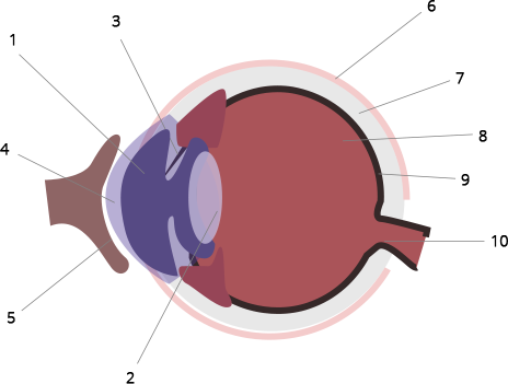
(a) Label the guidelines shown as 1 to 10.
Ans: 1 - Aqueous chamber,
2 - Lens,
3 - Iris,
4 - Cornea,
5 - Conjunctiva,
6 - Sclera,
7 - Choroid,
8 - Retina,
9 - Yellow spot,
10 - Optic nerve (Blind spot)
(b) Write one important role of parts shown as 3 and 7.
Ans: Part 3- It shows the Iris. Iris has radial and circular muscles that dilate and constrict the pupil, respectively.
Part 7- It shows Choroid. It's the eyeball's central layer, which is densely packed with blood vessels and feeds the eye.
(c) Write one structural difference between the parts shown as 9 and 10.
Ans:
(d) Mention one functional difference between the parts shown as 6 and 8.
Ans:
4. Given below is a diagram depicting a defect of the human eye? Study the same and answer the questions that follow:
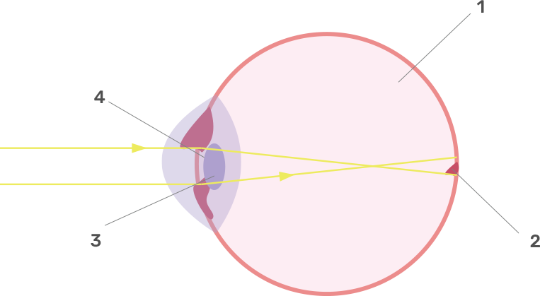
(a) Name the defect shown in the diagram.
Ans: Myopia is the defect shown in the diagram.
(b) Give two possible reasons for this defect.
Ans: The two possible reasons for this defect are:-
(i) The lens of the eye becomes convex or curved.
(ii) The eyeball's depth is excessive, i.e. the eyeball is stretched from front to rear.
(c) Name the parts labeled 1 to 4.
Ans: Part 1 - vitreous humour,
Part 2 - blind spot,
Part 3-lens,
Part 4-pupil.
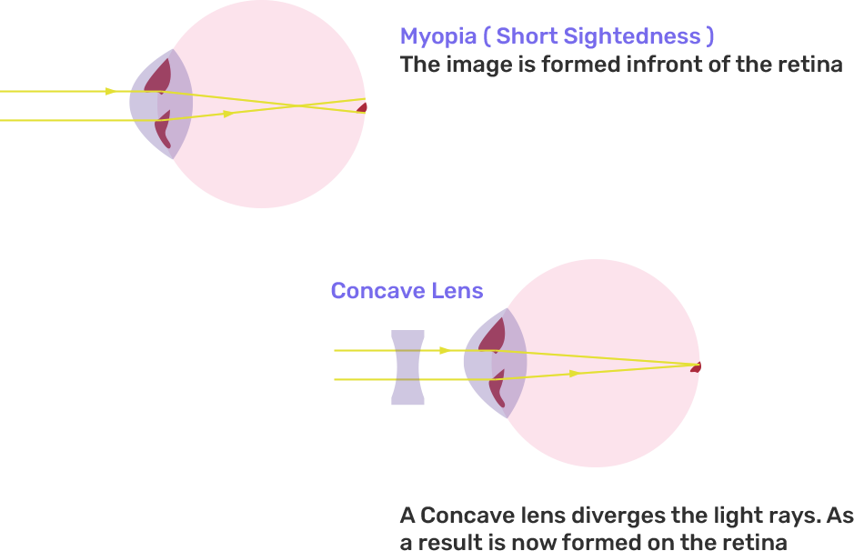
(d) Name the type of lens used to correct this eye defect.
Ans: Concave lens the type used to correct this eye defect.
(e) Draw a labeled diagram to show how the above mentioned defect is rectified using the lens named above.
Ans:
5. The figure below is the sectional view of a part of the skull showing a sense organ:
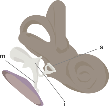
(i) Name the sense organ.
Ans: Ear
(ii) What are the parts labeled 'm', 'i' and 's'? What do these parts constitute collectively?
Ans: m - malleus,
i - incus and
s - stapes
These are collectively called ear ossicles.
(iii) What do you call the part shown in the form of a spiral? What is its function?
Ans: Cochlea. Vibrations in the hair of the sense cells in the cochlea transfer the hearing impulse to the brain through the auditory nerve.
(iv) Name the part labeled 'tm'. What is its function?
Ans: Membrane of the tympanic cavity. In the process of hearing, it vibrates and then sets the ear ossicles vibrating.
6. Given below is a diagram of a part of the human ear. Study the same and answer the questions that follow:
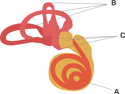
(i) Give the collective biological term for Malleus, Incus and Stapes.
Ans: Ear ossicles is the collective biological term for Malleus, Incus and stapes.
(ii) Name the parts labeled A, B and C in the diagram.
Ans: A - Cochlea,
B - Semicircular canals,
C - Ear ossicles.
(iii) State the functions of the parts labeled 'A' and 'B'.
Ans: Part A- The cochlea aids in the transmission of impulses from the auditory nerve to the brain.
Part B- The body's dynamic balance is maintained via semicircular canals.
(iv) Name the audio receptor region present in the part labeled 'A'.
Ans: Organ of Corti is the audio receptor region present in the part labeled 'A'.
7. Draw a labeled diagram of the inner ear. Name the part of the inner ear that is responsible for static balance in human beings.
Ans:
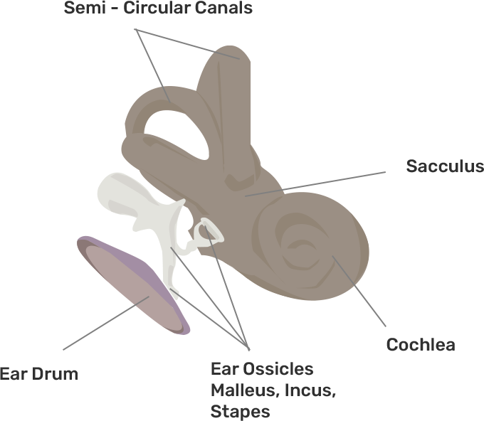
Utriculus and Sacculus are responsible for maintaining static balance in human beings.
8. Have a look at the posture of this woman who is reading a book and answer the questions which follow:

(a) What problem is she facing? Name the problem.
Ans: Myopia is the problem that she is facing.
(b) What are the two conditions shown in sections A and B of the eye as applicable to her?
Ans: A-Normal eye, B-Myopia
(c) What kind of looking glasses she needs?
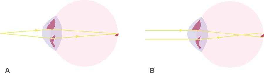
Ans: Here, concave-lens looking glasses are required.
9. The figure given below shows the principal parts of a human ear. Study the diagram and answer the following questions.
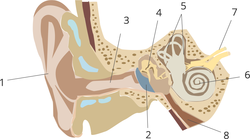
(a) Label the parts 1 to 8.
Ans: 1 - External ear (pinna),
2 - Ear drum (tympanum),
3 - Auditory canal,
4 - Malleus,
5 - Semicircular canals,
6 - Cochlea,
7 - Auditory nerve,
8 - Eustachian tube.
(b) State the role of parts 6, 7 and 8.
Ans: The role of:
Part 6 (Cochlea) - Cochlea is responsible for carrying the sensory cells for hearing.
Part 7 (Auditory nerve) - Auditory Nerve is responsible for transmitting the impulse of hearing to the brain.
Part 8 (Eustachian tube) - Eustachian tube is responsible for equalizing air pressure on both the sides of the tympanum.
(c) Why is it harmful to use a sharp object to remove ear wax? Mention the number and name of the part involved.
Ans: Using a sharp tool to remove ear wax is dangerous since it can rupture the ear drum. The part involved is part 2 - Ear drum (tympanum).
The sense organs are specialized organs that help us to perceive the world around us. They are an integral part of our lives and are the only way to perceive our environment. The sense organs provide the data needed for their interpretation through various organs and a network of nerves in response to a particular physical phenomenon. These senses govern our association and our interaction with the environment.
We have five sense organs, namely:
Eyes
Ears
Nose
Tongue
Skin
These five sense organs contain receptors that transmit information through sensory neurons to the right places in the nervous system.


FAQs on Concise Biology Class 10 ICSE Solutions for Chapter 11 - Sense Organs
1. What are the key features of Concise Biology Class 10 ICSE Solutions for Chapter 11 - Sense Organs?
The Selina solutions have been curated by some of our teachers who hold expertise in this particular subject. The solutions have a detailed explanation in simple and easy language that can be understood by every student. The chapter deals with the features of sense organs, their types, and their importance. It also deals with the defect that can arise in each sense organ. The solution book is so well crafted that the students can easily follow the book and get a good score in the examination. The pointers used in the books to explain the points are a great way to promote learning.
2. Why should we refer to Selina Solutions Concise Biology Class 10 book for Sense Organ Chapter?
The Selina solution book has all the questions with their solution and detailed explanation, which makes it easy for the students to follow. The detailed explanation helps the students in clearing their doubts and improving their understanding skills while they are preparing for their board exams. The line-by-line explanation helps the student understand the concept better and also helps in building a strong foundation. The solutions are so well crafted that the students would not require any extra help to understand the chapter.
3. Why should we follow Vedantu for Concise Biology Class 10 ICSE Solutions for Chapter 11 - Sense Organs?
Vedantu is an online platform providing the students with materials for their preparation in PDF format for free. On Vedantu we can find the solutions for the concise biology book in such a manner that it can easily be grasped by the students. The solutions help the students in their self-study and are a perfect guide to the students. The Vedantu website is easy to use and the offline PDF can also be used later on by the students when they do not have access to the internet.
4. Where can we get Concise Biology Class 10 ICSE Solutions for Chapter 11 - Sense Organs?
The Selina solutions are easily available online for the students to download and understand the chapter. They are available in two forms, that is they can either be viewed online or can be downloaded. The solutions are also available on the Vedantu website which provides accurate solutions keeping the best interest of the student in mind. These solutions help the students in understanding the question and help them solve the question in an easy and faster method keeping the understanding of the student in mind.
5. Can we download Concise Biology Class 10 ICSE Solutions for Chapter 11 - Sense Organs online?
The Selina publication Biology book cannot be downloaded online. Though these books can be bought online from different online stores, it isn't possible to download them online. These books can also be brought from the nearest book store easily. Though the book cannot be downloaded for free from online websites, the solution to these books can easily be downloaded without any difficulties from the Vedantu website. The students can download the solutions for free from the website in PDF format.