


Overview of Microscope
A microscope helps us to see the details that our eyes miss. As the word suggests, Micro means “tiny”, and scope means “to look at”. The entire universe is made up of millions and billions of little things. The microscope is used to magnify small objects for the study. The evolution of the microscope has had a great impact. Before its invention, we had very little information on many organisms like bacteria and protozoa as they can not be seen with the naked eye. To view these organisms, you must place your eye over the microscope.
The Microscope
What is a Microscope?
A microscope is an instrument used to observe small objects, including cells. Two Dutch spectacle-makers and a father-and-son team, Hans and Zacharias Janssen, create the first microscope.
When light reflects from an object being viewed under the microscope and passes through the lens, it bends towards the eye, making the object look bigger than its actual size.
This allows scientists to see the shape of the cell, its nucleus, and other organelles.
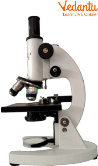
Different Parts of Microscope
The Eyepiece or Ocular Lens: Eyepiece lens magnifies the image of the specimen. This part is also known as ocular. Most school microscopes have an eyepiece with 10X magnification.
Eyepiece Tube or Body Tube: The tube holds the eyepiece.
Nosepiece: Nosepiece holds the objective lenses and is sometimes called a revolving turret. You choose the objective lens by rotating to the specific lens you want.
Objective Lenses: Most compound microscopes come with three or four objective lenses around the nosepiece. The most common objective lenses have the power of 4X, 10X and 40X. Combined with the eyepiece's magnification, the resulting magnification is 40X, 100X and 400X magnification. Total magnification is calculated by multiplying the eyepiece's power by the objective lens's power. (10X Eyepiece AND 40X Objective = 400X Total Magnification) Some more advanced microscopes have an additional objective lens with 100X power. This results in 1,000X magnification.
Arm: The Arm connects the base to the nosepiece and eyepiece. It is the structural part that is also used to carry the microscope.
Stage: The stage is where the specimen is placed. This place is for observation.
Diaphragm (sometimes Called the Iris): The diaphragm controls the amount of light passing through the slide. It is located below the stage and is usually controlled by a round dial.
Coarse Focus: Coarse focus moves the stage to provide general focus on the specimen. The course dial is the first used when bringing a specimen into focus.
Fine Focus: Fine focus moves the stage in smaller increments to provide a clear view of the specimen. The fine focus dial is the second used when bringing a specimen into focus.
Base: The base is the main support of the microscope. The bottom is where all the other parts of the microscope stand.
Importance of Microscopes
The importance of microscopes is as under:
Microscopes have opened up a broader aspect of science. With the help of microscopes, scientists can identify the existence of microorganisms, study the structure of cells, and see the smallest parts of plants, animals, and fungi.
Microscopes are used in hospitals to diagnose diseases and are used all over the world. They magnify the blood cells so that it gets easier for the doctors to identify the illness.
Scanning electron microscopes have magnifications up to several million times for viewing molecules, viruses, and nano-particles. Corrective software is used to increase the magnification and resolutions of the images.
Electron or light microscopes are the oldest and the most commonly used, where the light is passed through the machine, and the sample is used to magnify it. A specialised camera is used to produce a film or a digital image.
What Can You See Through a Microscope?
The microscope view gives a clear picture of what is on the slide. There are a large number of fascinating things under the microscope that can be viewed. Some of them include the following:
Amoeba
Amoeba is a unicellular organisms that can be seen under a microscope. This organism can be observed directly without staining or even after staining and fixing it with a particular dye. If observed directly, the amoeba appears like a jelly-like structure that shows the crawling movement of the organism.
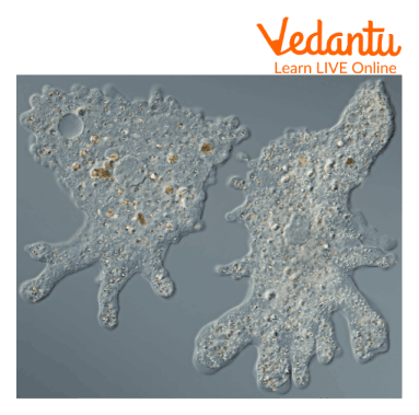
Amoeba Under a Microscope
Algae
Algae are usually found in freshwaters or marine sources, and most of them are provided with pigments that assist the organisms in producing food or oxygen. Algae are classified into various groups and can be observed under a microscope.
Animal Cells
Animal cells can be observed under a microscope; they appear different based on the cell type. Although, the internal structure of the organelles is almost similar. They are usually transparent, and the thickness of the cell differs throughout the cytoplasm.

Animal Cells Under a Microscope
Butterfly Scales
Butterfly scales can be observed under a microscope and appear like feathers or a woven basket when observed. This shows the interlaced structure of a fragile butterfly.
Yeast Cells
Yeast is oval-shaped single-celled fungi that play an important role in daily life. When seen under a microscope, these oval-shaped can be seen dividing or doubling themselves. Yeast is used in our daily life in the form of bread, beer, and other fermented food products.
Yeast cells under a microscope
Eggshell Membrane
A thin film is attached to the inside wall of an eggshell, known as the Eggshell membrane, and it can be observed under a microscope. Not only one, but it has two membranes that defend against bacterial invasion. These layers are strong enough and are made up of keratin.
Facts for Kids
Some facts about Microscope include the following:
The first microscope was used to study insects and was named ‘Flea Glass’.
With the help of an electronic microscope, an object can be viewed millions of times when it is being observed.
The smallest sample observed with the help of a light microscope was 500 nanometers long.
Antony van Leeuwenhoek invented the single-lens microscope in the 1660s.
Summary
Microscopes have established a new dimension in science and technology. To view, we have to place our eye over the microscope. In today’s time, it has been of great help to scientists and in many other spheres like hospitals, laboratories, and manufacturing plants to study and discover things that our eyes cannot identify. Over time, the advancements in microscopes have made them easier to use and improved the quality of the images. We hope you enjoyed reading this article; in case of any other doubts, feel free to ask in the comments.
FAQs on What can you See Through a Microscope?
1. What are some common things you can see through a microscope?
A microscope helps us see a hidden world of tiny things that are invisible to our naked eyes. As per the 2025-2026 EVS syllabus, with a microscope, you can discover things like:
- Living microorganisms: Tiny creatures in pond water like amoeba and paramecium.
- Plant cells: The building blocks of plants, clearly visible in an onion peel slide.
- Parts of insects: The detailed structure of a fly's wing or an ant's leg.
- Fibres: The individual threads in a piece of cloth or paper.
- Crystals: Beautiful shapes of salt or sugar crystals.
2. Which part of a microscope do you use to see the object?
You look through the eyepiece, which is the lens at the very top of the microscope. The object you want to see is placed on a glass slide on the stage below. The objective lenses, which are the rotating lenses closer to the object, perform the first and most powerful step of magnification.
3. What are some everyday items from home that look amazing under a microscope?
Many things around your house look completely different and fascinating under a microscope. Some great examples to try are:
- A pinch of salt or sugar to see their unique crystal shapes.
- A single strand of your hair to see its texture.
- The fibres in a small piece of cotton or wool.
- The coloured dots that make up a picture in a newspaper or magazine.
- A small, thin piece of an onion skin (the see-through layer).
4. Can you see germs like bacteria with a simple school microscope?
You can see some larger microorganisms like amoeba from pond water with a good school microscope. However, most bacteria and all viruses are far too small to be seen with a basic light microscope found in schools. Scientists need extremely powerful tools called electron microscopes to see these much smaller forms of life.
5. Why is it important for scientists to use microscopes?
Microscopes are one of the most important tools in science and medicine. They are crucial because they allow scientists and doctors to:
- Discover microorganisms: To understand tiny living things that can cause diseases or are beneficial to our environment.
- Study cells: To learn how plants, animals, and humans are built and how their bodies function.
- Develop medicines: To observe how medicines affect germs and cells in order to find new cures.
- Analyse materials: To check the purity of water or the structure of new materials.
6. What is the main difference between what a magnifying glass and a microscope can show you?
The main difference is their power of magnification. A magnifying glass has only one lens and typically makes things look 5 to 10 times bigger. A microscope, on the other hand, uses multiple lenses (the eyepiece and objective lenses) to make things look hundreds or even thousands of times bigger. This allows a microscope to reveal details a magnifying glass cannot, such as a single plant cell or bacteria.
7. Why do things sometimes appear upside down when viewed through a microscope?
This is a fascinating effect caused by the way light travels through the lenses. A standard compound microscope uses at least two lenses, and the combination of them bends the light in a way that inverts the image. The image is flipped both upside down and back-to-front. So, when you move the slide to the right, the image you see through the eyepiece appears to move to the left.
8. How do microscopes actually make tiny things look bigger?
A microscope works by using a system of curved glass lenses and a light source. Light from a lamp or mirror at the bottom shines through the thin specimen on the slide. This light then enters the objective lens, which creates a magnified, inverted image inside the microscope tube. Finally, the eyepiece lens acts like a magnifying glass to enlarge that first image, making the object appear big enough for your eye to see in detail.