


Microtubules take part in
(a) Formation of spindle fibres
(b) Movement of cilia and flagella
(c) Both A and B
(d) Cyclosis
Answer
612.3k+ views
Hint: The cytoskeletal structure of eukaryotic cells that forms during cell division to separate sister chromatids between daughter cells. Prokaryotic cell locomotion results from sliding of outer microtubules doublets relative to one another by motor activity of axonemal dynein.
Complete step by step answer:
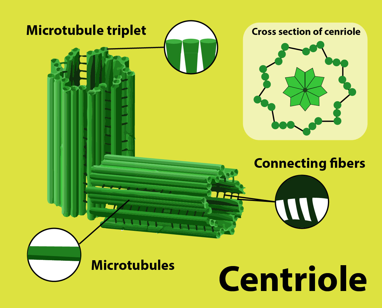
In animal cells, the centrosome features a pair of centrioles, each with nine triplets of microtubules arranged during a ring. The centrioles are surrounded by an electron- rich pericentriolar matrix. Microtubules are major components of spindle fibre wont to pull apart chromosomes during cellular division.
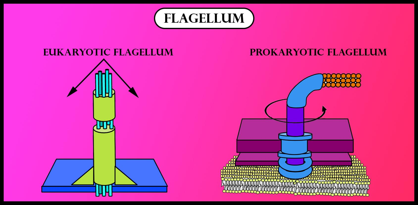
Microtubules are the central structure which supports cilia and flagella. Both can move unicellular and little multicellular organisms by propelling water past the organism. If these structures are anchored during a large structure, they move fluid over a surface.
So, the correct answer is, ‘Both A and B.’
Additional Information:
Spindle fiber:
- It is mentioned because the mitotic spindle during mitosis, a process that produces genetically identical daughter cells, or the meiotic spindle during meiosis, a process that produces gametes with half the amount of chromosomes of the parent cell.
- Astral microtubules develop within the actin skeleton and interact with the cell cortex to assist in spindle orientation.
- The turn- over rate of this population of microtubules is above the other population.
- The role of astral microtubules is assisted by dyneins specific to the present role.
- The globular chains plan to move towards the centrosome, but as they're sure to the cell wall, this leads to pulling the centrosomes towards the membrane, thus assisting cytokinesis.
- Kinetochore microtubules directly connect to the kinetochores.
- Each chromosome has two chromatids, and every chromatid features a kinetochore.
- The two kinetochores are associated with a region of the chromosome called the centromere.
Cilia and flagella:
- Cilia or flagella is composed of microtubules that are encased in a microtubule that is encased in a plasma membrane.
- This bundle of microtubules is called the axoneme.
- These are called the outer microtubules are called the outer microtubule doublets.
- The outer microtubules are connected to every other during a ring with cross- links.
- The outer microtubules also hook up with the middle structure with radial spokes.
- These outer microtubules surround another pair of central microtubules, another pair of central microtubules, which are not connected.
- The $9+2$ pattern continues throughout the entire organelle until the base.
- It is the foundation of the cilia or foundation of the cilia or flagella and is embedded flagella and is embedded in the cell membrane.
Note:
- Each protofilament is assembled from dimeric building blocks consisting of 1 alpha and one beta subunits.
- The protofilament is asymmetric with alpha subunit on one end and beta on the opposite
- One end of the protofilament is known as the plus end is terminated by a row of beta- tubulin units and the minus end is terminated by the alpha- tubulin units.
Complete step by step answer:
In animal cells, the centrosome features a pair of centrioles, each with nine triplets of microtubules arranged during a ring. The centrioles are surrounded by an electron- rich pericentriolar matrix. Microtubules are major components of spindle fibre wont to pull apart chromosomes during cellular division.
Microtubules are the central structure which supports cilia and flagella. Both can move unicellular and little multicellular organisms by propelling water past the organism. If these structures are anchored during a large structure, they move fluid over a surface.
So, the correct answer is, ‘Both A and B.’
Additional Information:
Spindle fiber:
- It is mentioned because the mitotic spindle during mitosis, a process that produces genetically identical daughter cells, or the meiotic spindle during meiosis, a process that produces gametes with half the amount of chromosomes of the parent cell.
- Astral microtubules develop within the actin skeleton and interact with the cell cortex to assist in spindle orientation.
- The turn- over rate of this population of microtubules is above the other population.
- The role of astral microtubules is assisted by dyneins specific to the present role.
- The globular chains plan to move towards the centrosome, but as they're sure to the cell wall, this leads to pulling the centrosomes towards the membrane, thus assisting cytokinesis.
- Kinetochore microtubules directly connect to the kinetochores.
- Each chromosome has two chromatids, and every chromatid features a kinetochore.
- The two kinetochores are associated with a region of the chromosome called the centromere.
Cilia and flagella:
- Cilia or flagella is composed of microtubules that are encased in a microtubule that is encased in a plasma membrane.
- This bundle of microtubules is called the axoneme.
- These are called the outer microtubules are called the outer microtubule doublets.
- The outer microtubules are connected to every other during a ring with cross- links.
- The outer microtubules also hook up with the middle structure with radial spokes.
- These outer microtubules surround another pair of central microtubules, another pair of central microtubules, which are not connected.
- The $9+2$ pattern continues throughout the entire organelle until the base.
- It is the foundation of the cilia or foundation of the cilia or flagella and is embedded flagella and is embedded in the cell membrane.
Note:
- Each protofilament is assembled from dimeric building blocks consisting of 1 alpha and one beta subunits.
- The protofilament is asymmetric with alpha subunit on one end and beta on the opposite
- One end of the protofilament is known as the plus end is terminated by a row of beta- tubulin units and the minus end is terminated by the alpha- tubulin units.
Recently Updated Pages
Master Class 11 Social Science: Engaging Questions & Answers for Success
Master Class 11 Chemistry: Engaging Questions & Answers for Success
Master Class 9 General Knowledge: Engaging Questions & Answers for Success
Master Class 9 Maths: Engaging Questions & Answers for Success
Master Class 9 Science: Engaging Questions & Answers for Success
Master Class 9 English: Engaging Questions & Answers for Success
Trending doubts
One Metric ton is equal to kg A 10000 B 1000 C 100 class 11 physics CBSE
Difference Between Prokaryotic Cells and Eukaryotic Cells
Two of the body parts which do not appear in MRI are class 11 biology CBSE
1 ton equals to A 100 kg B 1000 kg C 10 kg D 10000 class 11 physics CBSE
Explain zero factorial class 11 maths CBSE
10 examples of friction in our daily life