


What separates the heart chambers?
Answer
545.1k+ views
Hint: Human heart is mesodermal in origin and is located between the lungs. It has four chambers and each chamber is separated to prevent the mixing of oxygenated and deoxygenated blood and this separation created a depression called fossa ovalis.
Complete Answer:
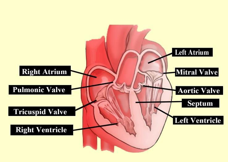
Internally the wall present in the heart is called the septum and valves which separates the heart chambers. It separates the right and left ventricle and also the right and left atria. The right and left atria get separated by the interatrial septum and this septum creates an oval depression called fossa ovalis. The right atrium receives an opening of the superior vena cava, inferior vena cava and coronary sinus. The opening of the vena cava gets supported by the eustachian valve towards the inferior side whereas the coronary sinus get supported by the coronary and the left atrium received the four openings of the pulmonary vein whereas the two ventricles get separated by the interventricular septum.
Externally the left and right atria of the heart get separated by the interatrial groove and the atria and ventricle get separated by the coronary sulcus. The groove present on the ventricle is called the anterior interventricular sulcus and posterior interventricular sulcus and these sulci consist of coronary arteries by which the heart receives the blood. The ascending aorta on the left ventricles helps in the systemic circulation of the body.
Note:
The passage of constriction waves through the heart chamber takes place with the help of cardiac muscle fibre, connective tissue, blood vessels and the net-like arrangement of the muscle fibres which branch and cross-connect with each other.
Complete Answer:
Internally the wall present in the heart is called the septum and valves which separates the heart chambers. It separates the right and left ventricle and also the right and left atria. The right and left atria get separated by the interatrial septum and this septum creates an oval depression called fossa ovalis. The right atrium receives an opening of the superior vena cava, inferior vena cava and coronary sinus. The opening of the vena cava gets supported by the eustachian valve towards the inferior side whereas the coronary sinus get supported by the coronary and the left atrium received the four openings of the pulmonary vein whereas the two ventricles get separated by the interventricular septum.
Externally the left and right atria of the heart get separated by the interatrial groove and the atria and ventricle get separated by the coronary sulcus. The groove present on the ventricle is called the anterior interventricular sulcus and posterior interventricular sulcus and these sulci consist of coronary arteries by which the heart receives the blood. The ascending aorta on the left ventricles helps in the systemic circulation of the body.
Note:
The passage of constriction waves through the heart chamber takes place with the help of cardiac muscle fibre, connective tissue, blood vessels and the net-like arrangement of the muscle fibres which branch and cross-connect with each other.
Recently Updated Pages
Master Class 11 English: Engaging Questions & Answers for Success
Master Class 11 Social Science: Engaging Questions & Answers for Success
Master Class 11 Maths: Engaging Questions & Answers for Success
Master Class 11 Biology: Engaging Questions & Answers for Success
Master Class 11 Physics: Engaging Questions & Answers for Success
Master Class 11 Chemistry: Engaging Questions & Answers for Success
Trending doubts
One Metric ton is equal to kg A 10000 B 1000 C 100 class 11 physics CBSE
Difference Between Prokaryotic Cells and Eukaryotic Cells
Find the value of the expression given below sin 30circ class 11 maths CBSE
Difference between physical and chemical change class 11 chemistry CBSE
Two of the body parts which do not appear in MRI are class 11 biology CBSE
Draw a diagram of a plant cell and label at least eight class 11 biology CBSE