


Draw the well labelled diagram of the internal structure of the human heart.
Answer
614.4k+ views
Hint:Heart is the main organ of the circulatory system. It is the main organ which supplies oxygenated blood to the whole body.
Complete answer:
Human heart is a muscular organ made up of cardiac muscles situated between the two lungs. It is well protected by a membrane called the pericardium.
Four chambers of heart- The left and right atrium (upper chamber) and the left and right ventricles (lower chamber). The two atriums are the receiving chambers for the blood entering the heart. The ventricles are thicker as they have to pump the blood away from the heart. In between the left ventricle and atrium, two cusp valves are present called the mitral valve and in between the right atrium and ventricle, three cusp tricuspid valves are present which prevents the backflow of the blood.
>Aorta- It is the main artery that carries oxygenated blood away from the heart to the entire body. It is guarded by aortic valves.
>Pulmonary artery- It carries deoxygenated blood from right ventricles to the lungs for oxygenation.
>Pulmonary vein- It carries oxygenated blood from lungs to the heart.
>Superior vena cava- It carries the deoxygenated blood from the upper half of the body to the right atrium of the heart.
>Inferior vena cava- It is a vein that carries deoxygenated blood from the lower end of the body to the right atrium of the heart.
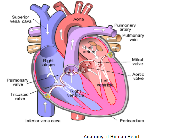
Note: Human heart is a four chambered organ which pumps blood throughout the body. The right side of the heart deals with deoxygenated blood (blue colour in the above fig.) and the left side deals with oxygenated blood (red colour in the fig.)
Complete answer:
Human heart is a muscular organ made up of cardiac muscles situated between the two lungs. It is well protected by a membrane called the pericardium.
Four chambers of heart- The left and right atrium (upper chamber) and the left and right ventricles (lower chamber). The two atriums are the receiving chambers for the blood entering the heart. The ventricles are thicker as they have to pump the blood away from the heart. In between the left ventricle and atrium, two cusp valves are present called the mitral valve and in between the right atrium and ventricle, three cusp tricuspid valves are present which prevents the backflow of the blood.
>Aorta- It is the main artery that carries oxygenated blood away from the heart to the entire body. It is guarded by aortic valves.
>Pulmonary artery- It carries deoxygenated blood from right ventricles to the lungs for oxygenation.
>Pulmonary vein- It carries oxygenated blood from lungs to the heart.
>Superior vena cava- It carries the deoxygenated blood from the upper half of the body to the right atrium of the heart.
>Inferior vena cava- It is a vein that carries deoxygenated blood from the lower end of the body to the right atrium of the heart.
Note: Human heart is a four chambered organ which pumps blood throughout the body. The right side of the heart deals with deoxygenated blood (blue colour in the above fig.) and the left side deals with oxygenated blood (red colour in the fig.)
Recently Updated Pages
Master Class 12 Business Studies: Engaging Questions & Answers for Success
Master Class 12 Biology: Engaging Questions & Answers for Success
Master Class 12 Chemistry: Engaging Questions & Answers for Success
Class 12 Question and Answer - Your Ultimate Solutions Guide
Master Class 11 Social Science: Engaging Questions & Answers for Success
Master Class 11 English: Engaging Questions & Answers for Success
Trending doubts
Which are the Top 10 Largest Countries of the World?
Draw a labelled sketch of the human eye class 12 physics CBSE
Differentiate between homogeneous and heterogeneous class 12 chemistry CBSE
Sulphuric acid is known as the king of acids State class 12 chemistry CBSE
Why is the cell called the structural and functional class 12 biology CBSE
Which is the correct genotypic ratio of mendel dihybrid class 12 biology CBSE