


Draw a labelled diagram of a female reproductive system.
Answer
622.2k+ views
Hint: The reproductive system of a female is immature at birth and develops at maturity which is made up of the internal and external sex organs that have a role in the reproduction of new offspring.
Complete answer:
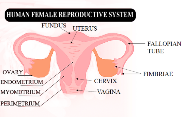
The reproductive system has a pair of ovaries, uterus, fallopian tubes, cervix, and vagina.
Ovary- It is located in the upper pelvic cavity and releases one egg cell once a month simultaneously. There is one pair of ovaries shaped like an almond of size around 2 cm to 3.5 cm wide and 1 cm thick. The ovary also secretes female hormones i.e. estrogen and progesterone.
Fimbriae- these are connected to the ovary and are small, finger-like projections at the end of the fallopian tubes, through which eggs move from the ovaries to the uterus.
Fallopian tubes- or uterine tubes or salpinges (singular salpinx), are tubes that stretch from the uterus to the ovaries and contains 3 parts: isthmus (closest to the uterus), ampulla (increases in diameter and is the most common site for fertilization), and final segment infundibulum. The fertilized egg passes through the tubes from the ovaries of female mammals to the uterus.
Uterus- it is a muscular organ above the vagina and between the bladder and rectum. The fetus or unborn child develops here and the walls of the uterus contract to push out the fetus during childbirth.
Endometrium and myometrium- The innermost layer is the endometrium which thickens each month for the preparation of an embryo and sheds in the menstrual period if fertilization does not occur. The middle layer of the uterine wall, consisting mainly of uterine smooth muscle cells is myometrium, it helps in supporting stromal and vascular tissue, and mainly induces uterine contractions.
Cervix and fundus- Cervix is a canal connecting the vagina and uterus and is the neck of the uterus that extends downward to the vagina. The sperm or the infant passes through here. The fundus is the top portion that is opposite of the cervix. Fundal height is usually measured during pregnancy from the top of the pubic bone to determine growth rates.
Vagina- is an elastic and muscular canal and the place where sperm are usually ejaculated during sexual intercourse. The vagina is the birth canal from where the developed baby is delivered out of the female.
Note: The external female reproductive organs are known as the vulva and are the openings for the urethra and vagina. It includes the clitoris and two pairs of labia. The vulva is lubricated by the Secretions from Bartholin’s glands near the vagina opening.
Complete answer:
The reproductive system has a pair of ovaries, uterus, fallopian tubes, cervix, and vagina.
Ovary- It is located in the upper pelvic cavity and releases one egg cell once a month simultaneously. There is one pair of ovaries shaped like an almond of size around 2 cm to 3.5 cm wide and 1 cm thick. The ovary also secretes female hormones i.e. estrogen and progesterone.
Fimbriae- these are connected to the ovary and are small, finger-like projections at the end of the fallopian tubes, through which eggs move from the ovaries to the uterus.
Fallopian tubes- or uterine tubes or salpinges (singular salpinx), are tubes that stretch from the uterus to the ovaries and contains 3 parts: isthmus (closest to the uterus), ampulla (increases in diameter and is the most common site for fertilization), and final segment infundibulum. The fertilized egg passes through the tubes from the ovaries of female mammals to the uterus.
Uterus- it is a muscular organ above the vagina and between the bladder and rectum. The fetus or unborn child develops here and the walls of the uterus contract to push out the fetus during childbirth.
Endometrium and myometrium- The innermost layer is the endometrium which thickens each month for the preparation of an embryo and sheds in the menstrual period if fertilization does not occur. The middle layer of the uterine wall, consisting mainly of uterine smooth muscle cells is myometrium, it helps in supporting stromal and vascular tissue, and mainly induces uterine contractions.
Cervix and fundus- Cervix is a canal connecting the vagina and uterus and is the neck of the uterus that extends downward to the vagina. The sperm or the infant passes through here. The fundus is the top portion that is opposite of the cervix. Fundal height is usually measured during pregnancy from the top of the pubic bone to determine growth rates.
Vagina- is an elastic and muscular canal and the place where sperm are usually ejaculated during sexual intercourse. The vagina is the birth canal from where the developed baby is delivered out of the female.
Note: The external female reproductive organs are known as the vulva and are the openings for the urethra and vagina. It includes the clitoris and two pairs of labia. The vulva is lubricated by the Secretions from Bartholin’s glands near the vagina opening.
Recently Updated Pages
Master Class 12 Business Studies: Engaging Questions & Answers for Success
Master Class 12 Biology: Engaging Questions & Answers for Success
Master Class 12 Chemistry: Engaging Questions & Answers for Success
Class 12 Question and Answer - Your Ultimate Solutions Guide
Master Class 11 English: Engaging Questions & Answers for Success
Master Class 11 Social Science: Engaging Questions & Answers for Success
Trending doubts
Which are the Top 10 Largest Countries of the World?
Draw a labelled sketch of the human eye class 12 physics CBSE
Differentiate between homogeneous and heterogeneous class 12 chemistry CBSE
Why is the cell called the structural and functional class 12 biology CBSE
Draw ray diagrams each showing i myopic eye and ii class 12 physics CBSE
Which is the correct genotypic ratio of mendel dihybrid class 12 biology CBSE